INTRODUCTION
Autism is a neuropsychiatric condition of child-hood characterized by impairment in personal rela-tionships, communication difficulties, and restricted and repetitive interests and activities. Autism is now
widely considered a neurobiologic disorder. Several studies aiming to identify the etiology of autism, have indicated involvement of limbic system struc-tures including amygdala, hippocampus, and ante-rior cingulate cortex, as well as prefronatal lobe, temporal lobe, and cerebellum (1-4). However, the neural basis of autism remains poorly understood.
Neuropsychologically, impairments in Theory of Mind (ToM), executive function, central coherence, and recognition of emotions have been suggested in autism. The frontal lobe particitates importantly in executive function and ToM. It is recognized that
ORIGINAL
Function of the frontal lobe in autistic individuals
: a proton magnetic resonance spectroscopic study
Emiko Fujii
1, Kenji Mori
1, Masahito Miyazaki
1, Toshiaki Hashimoto
2,
Masafumi Harada
3, and Shoji Kagami
1 1Department of Pediatrics, Institute of Health Biosciences, the University of Tokushima Graduate School ;2
Department of Pediatrics, Tokushima Red Cross Hinomine General Nursing Center ; and 3
Department of Radiologic Technology, School of Health Sciences, the University of Tokushima Graduate School, Tokushima, Japan
Abstract : Purpose. In this investigation, we studied differences in chemical metabolites in certain brain regions between autistic patients and normal control subjects. Methods. Pro-ton magnetic resonance spectroscopy (1H-MRS) was used to evaluate functional activity
in these regions. Specific regions studied were right and left dorsolateral prefrontal cor-tex(DLPFC) and the anterior cingulated cortex(ACC). Results. In the ACC, the N - acety-laspartate(NAA)/creatine/phosphocreatine(Cr) ratio in autistic patients (n=31) was signifi-cantly lower than that in control subjects (n=28). The decrease in the NAA/Cr ratio for the ACC was much greater in the group with worst social ability. NAA/Cr for the left DLPFC and social ability of autistic patients also correlated well. Furthermore, NAA/Cr for the left DLPFC in the group with intelligence quotient (IQ) below 50 was significantly less than in controls. NAA/Cr for the right DLPFC in autistic patients was not decreased compared to controls, and did not correlate with IQ or social ability. Conclusions. These findings suggest neuronal dysfunction in the ACC and left DLPFC in autism, and also a relation-ship between social disability and metabolic dysfunction in these regions. Dysfunction in the ACC and the left DLPFC may contribute to the pathogenesis of autism. J. Med. In-vest. 57 : 35-44, February, 2010
Keywords : autism, proton magnetic resonance spectroscopy (1H-MRS), dorsolateral prefrontal cortex(DLPFC), anterior cingulated cortex(ACC)
Received for publication May 29, 2009 ; accepted August 20, 2009.
Address correspondence and reprint requests to Emiko Fujii, Department of Pediatrics, Institute of Health Biosciences, the University of Tokushima Graduate School, Kuramoto - cho, Tokushima 770 - 8503, Japan and Fax : + 81 - 88 - 631 - 8697.
the portion of the brain that plays a major role in ex-ecutive function is the dorsolateral prefrontal cortex (DLPFC : Brodmann’s area 46/9) (5-7). The ante-rior cingulate cortex (ACC) not only participates in executive function but also in emotion and ToM (7-10).
Recently, proton magnetic resonance spectros-copy (1H-MRS) has been used to examine brain
me-tabolism in patients. The main metabolites that can be assessed using this technique are N-acetylas-partate (NAA), creatine/phosphocreatine (Cr), and choline-containing compounds (Cho). The NAA sig-nal, the most prominent1H spectral peak, is present
at high concentrations in neurons, and might be related to mitochondrial function. Therefore, NAA often is used to assess neuronal density (11-13). The Cho signal, which might indicate glial cell den-sity (13-15), increases in intenden-sity with increased membrane synthesis and turnover (16, 17). The Cr signal might reflect glial or overall (neurons plus glia) cell density (13, 14) ; phosphocreatine repre-sents a key component of high-energy phosphate metabolism (18).1H-MRS studies often use Cr as an
internal intensity reference for other peaks, on the assumption that its concentration is relatively con-stant. We previously investigated brain function in autistic patients using1H-MRS, and reported that
the concentrations of NAA were decreased in the amygdala, Wernicke’s area, and the cerebellum (19, 20). We consider1H-MRS to be a valuable tool for
detection of neuronal impairment in autistic brain. In the present study, we used1H-MRS to
inves-tigate metabolism in the DLPFC and ACC of sub-jects with autism and healthy control subsub-jects, com-paring amounts and patterns of chemical metabo-lites. We also related the metabolic findings to intel-lectual and social abilities in subjects with autism.
2 PATIENTS AND METHODS
2-1 Patients2-1-1 Anterior cingulate cortex (ACC)
The study group included 31 autistic patients (2 to 13 years old ; mean age 6.1 ; 25 boys and 6 girls). These individuals were recruited from among outpatients at the Department of Pediatrics of Tokushima University. All subjects in this group were diagnosed with autistic disorder by two expe-rienced pediatric neurologists according to the cri-teria of the Diagnostic and Statistical Manual of Mental Disorders IV(DSM-IV). The intelligence
quotient (IQ) determined by the Tanaka-Binet in-telligence scale was over 85 in 4 patients ; 71 to 84 in no patients ; 50 to 70 in 7 patients ; 35 to 49 in 10 patients ; and 20 to 34 in 10 patients. Social ability of the autistic patients was evaluated with a social ma-turity scale (Nihon Bunka Kagakusha) ; (S-M scale). The social quotient (SQ) by the S-M scale was over 85 in 3 patients ; 71 to 84 in 2 patients ; 50 to 70 in 7 patients ; 35 to 49 in 15 patients ; and 20 to 34 in 4 patients.
The control subjects were 28 children underwent an MRI examination because of headache or head trauma. They showed no developmental or behav-ioral abnormality. Ages and genders (2 to 15 years old ; mean, 6.8 ; 21 boys and 5 girls) were matched with those of autistic patients.
Informed consent was obtained from the parents of all subjects. Informed consent was also obtained from the subjects who could understand the content and purpose of this syudy.
2-1-2 Bilateral dorsolateral prefrontal cortex (DLPFC)
This group consisted of 20 individuals (2 to 13 years old ; mean, 7.5 ; 17 boys and 3 girls). These individuals were a subgroup of the subjects in the ACC examination. We first examined1H-MRS in
the ACC and next in the DLPFC. We performed1
H-MRS in the ACC only when patients could not lie still until all1H-MRS measurements were
com-pleted. IQ was over 85 in 4 patients who underwent
1H-MRS in both areas ; 71 to 84 in 0 patients ; 50 to
70 in 4 patients ; 35 to 49 in 6 patients ; and 20 to 34 in 6 patients. SQ was over 85 in 3 patients ; 71 to 84 in 2 patients ; 50 to 70 in 4 patients ; 35 to 49 in 9 pa-tients ; and 20 to 34 in 2 papa-tients. Control subjects were 18 children (2 to 15 years old ; mean, 7.8 ; 16 boys and 2 girls), who showed no developmental or behavioral abnormalities.
2-2 1H-MRS measurement
All1H-MRS studies were performed with a
1.5-tesla clinical MRI system (Signa Horizon, GE, Mil-waukee, WI) with a standard head coil, using a point-resolved spectroscopic (PRESS) sequence, with TR of 1300 and TE of 135 ms, and 256 free-induction decays (FID). Gradient map shimming was con-ducted in the location of measurement by a high-order shim program ; the full width of the half maxi-mum of the water peak was less than 8 Hz. We ac-quired T1 and T2 MRI images in axial and coronal views before the1H-MRS examination, and placed
(VOI) in each DLPFC (Brodmann’s area 46/9. Fig. 1). In the present study, the VOI was placed mainly in Brodmann’s area 46, which is considered to play a leading role in central executive (21). Spectra were processed using SA/GE software. FIDs were zero-filled to 4096 data-points, and Fourier transforma-tion was performed. After DC offset correctransforma-tion, we calculated peak areas, i.e. signal intensities of NAA, Cr, and Cho, with curvefitting using a Gaussian func-tion. Criteria for selecting reliable metabolite signals were based on the S.D. of the fit for each metabo-lite ; only results with S.D. below 20% were included in the analysis. Signal-intensity ratios for NAA/Cr, NAA/Cho, and Cho/Cr were compared with those of control subjects. Furthermore, LCModel ver. 5.6, a fully automated program for analyzing metabolic products was used to estimate NAA, Cr, and Cho concentrations. We placed a single 4.5 ml (2.0
!
1.5!
1.5 cm) VOI in the ACC, where we measuredthe above metabolites using the same method (Fig. 1). This study was approved by the Institutional Re-view Board of our institution.
2-3 Statistical analysis
We compared autistic patients with control indi-viduals with respect to the signal ratios for NAA/ Cr, NAA/Cho, and Cho/Cr. Furthermore, we also compared concentrations of NAA, Cho, and Cr. We used Student’s t test to determine statistically sig-nificant differences between autistic patients and the control group, and used a paired Student’s t test to compare left vs, right side among several groups. A value of p below 0.05 was considered statistically significant. Furthermore, we examined the correla-tion between the signal ratio of NAA/Cr and IQ and SQ in autistic patients using Pearson’s correlation coefficient.
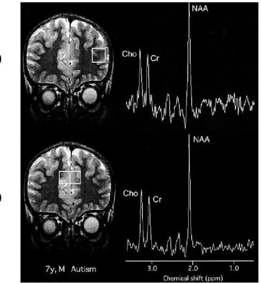
Fig. 1 Measurement locations for1H- MRS and representative spectra obtained.
(A) : Left dorsolateral prefrontal cortex (DLPFC). (B) : Anterior cingulate cortex (ACC). NAA, N- acetylaspartate ; Cr, creatine/phos-phocreatine ; Cho, choline - containing compounds.
control autism control autism control autism
RESULTS
The baseline MRI was evaluated individually by two experienced pediatric neurologists and one neuroradiologist. No abnormal signal or distinct atro-phy was detected in frontal lobes of autistic patients or control individuals at the time when1H-MRS
studies were performed.
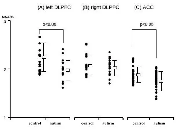
The NAA/Cr ratio for the ACC and the left DLPFC in autistic patients was significantly decreased
compared with the control group (p!0.05). There was no significant difference in this value in the right DLPFC (Fig. 2, Table 1). The NAA/Cho ratio in the ACC was significantly decreased compared with the control group (p!0.01). There was no significant difference in this value in left or right DLPFC (Ta-ble 1).
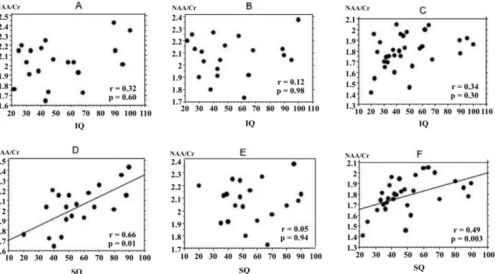
We investigated the relationship between IQ and NAA/Cr in autistic patients. They were poorly cor-related, as indicated by low correlation coefficients
Table 1 Metabolite concentrations and ratios in autistic and control subjects
NAA (mM) Cho (mM) Cr (mM) NAA/Cr NAA/Cho Cho/Cr
ACC autism 5.91!0.93 1.60!0.21 4.30!0.90 1.81*!0.17 1.52**!0.20 1.17!0.13 control 6.03!0.89 1.51!0.19 4.10!0.71 1.90!0.15 1.64!0.12 1.16!0.13 left DLPFC autism 6.98!1.60 1.50!0.41 4.70!1.30 2.01*!0.21 2.02!0.36 1.03!0.19 control 7.51!2.43 1.40!0.30 4.11!0.89 2.23!0.30 2.17!0.45 1.05!0.16 right DLPFC autism 7.49!1.42 1.50!0.20 4.70!0.89 2.07!0.16 1.92!0.31 1.09!0.24 control 7.68!1.30 1.43!0.31 4.40!1.10 2.14!0.22 2.20!0.41 1.10!0.15 Values are the mean!SD. * p!0.05 ; ** p!0.01. ACC, anterior cingulate cortex ; DLPFC, dorsolateral prefrontal cortex ; NAA, N-acetylaspartate ; Cho, choline - containing compounds ; Cr, creatine/phosphocreatine.
Fig. 2 NAA/Cr ratio for left and right DLPFC and the right ACC in control subjects and autistic patients. NAA/Cr ratio for the left DLPFC and the ACC in autistic patients is significantly decreased compared with control subjects (p!0.05).
control IQ≧50 IQ<50 control IQ≧50 IQ<50 control IQ≧50 IQ<50 1
3
2 NAA/Cr
in each area (r=0.32 for left DLPFC, r=0.12 for right DLPFC, r=0.34 for ACC) ; Fig. 3A, B, and C. How-ever, NAA/Cr for the left DLPFC in the group
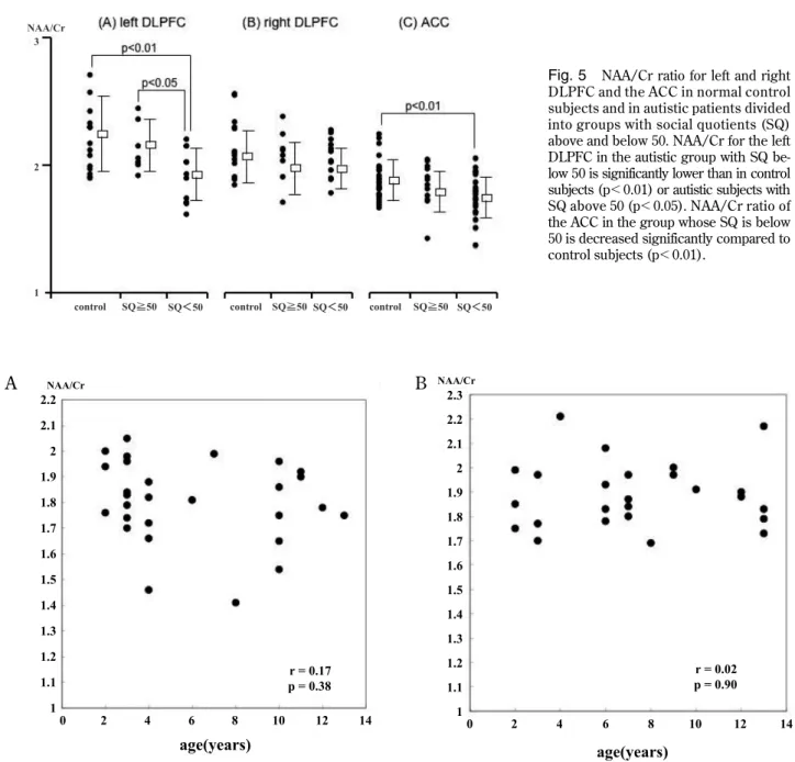
whose IQ was below 50 was significantly lower than in the control group (p!0.05, Fig. 4A).
We investigated the relationship between SQ and
Fig. 4 NAA/Cr ratio for left and right DLPFC and the ACC in control subjects and in autistic patients divided into groups with in-telligence quotients (IQ) above and below 50. NAA/Cr ratio for the left DLPFC in the autistic group with IQ below 50 is significantly lower than in control subjects (p!0.05).
Fig. 3 Correlation between IQ, SQ, and NAA/Cr in autistic patients. No correlation is evident between intelligence quotient (IQ) and NAA/Cr in any area studied (A, B, C), but a significant correlation is confirmed between social quotient (SQ) and NAA/Cr in the left DLPFC and the ACC (D, F). A, left DLPFC ; B, right DLPFC ; C, ACC ; D, left DLPFC ; E, right DLPFC ; F, ACC.
control SQ≧50 SQ<50 control SQ≧50SQ<50 control SQ≧50 SQ<50 1
3
2 NAA/Cr
NAA/Cr in autistic patients. NAA/Cr for the left DLPFC in autism was correlated with SQ. The cor-relation coefficient was 0.66 (p=0.01 ; Fig. 3D). NAA/Cr for the left DLPFC in the subgroup whose SQ was below 50 was significantly low compared to the control group (p!0.01) and the subgroup whose SQ was above 50(p!0.05, Fig. 5A). NAA/Cr for the ACC in the autism group also correlated with SQ. The correlation coefficient was 0.49 (p=0.003 ; Fig. 3F). NAA/Cr for the ACC in the subgroup, whose SQ was below 50 was decreased significantly com-pared to the control group (p!0.01 Fig. 5C). NAA/ Cr and SQ were poorly correlated in the right DLPFC Fig. 3E and 5B).
There was no significant difference in concentra-tions of NAA, Cho, and Cr in any region between the autism and control groups (Table 1). When we evaluated laterality of metabolite concentrations in the DLPFC, there was no significant difference in either the control or autism group.
There was no significant difference in Cho/Cr ratio for any region between autism and control groups (Table 1).
The distributions of NAA/Cr ratio for the ACC according to age in auristic patients and control sub-jects are shown in Fig. 6. The correlation coefficients for NAA/Cr ratio and age were not statistically sig-nifficant in the ACC of autistic patients and control
Fig. 5 NAA/Cr ratio for left and right DLPFC and the ACC in normal control subjects and in autistic patients divided into groups with social quotients (SQ) above and below 50. NAA/Cr for the left DLPFC in the autistic group with SQ be-low 50 is significantly be-lower than in control subjects (p!0.01) or autistic subjects with SQ above 50 (p!0.05). NAA/Cr ratio of the ACC in the group whose SQ is below 50 is decreased significantly compared to control subjects (p!0.01).
A
A BB
Fig. 6 The distributions of NAA/Cr ration for the ACC according to age in autistic patients (A) and control subjects (B). No corre-lation is evident between NAA/Cr and age in autistic patients and control subjects.
subjects. No correlation was also observed in left and right DLPFC of autistic patients and control sub-jects.
DISCUSSION
It is recognized that executive function deficits are frequently seen in individuals who have sustained damage to the frontal lobes. They include repetitive, aimless movements or speech, difficulty inhibiting familiar or obvious responses, inappropriate repeti-tion of previous thoughts or acrepeti-tions, and diminished capacity for planning. Stuss described several ad-ditional information-processing deficits resulting from frontal lobe pathology, including a tendency to focus on one aspect of information, difficulty re-lating or integrating isolated details, problems man-aging simultaneous or multiple sources of informa-tion, and impaired ability to act on or apply knowl-edge in a meaningful manner (22, 23). Recently, it was recognized that the portion of the brain that plays a major role in executive function is the DLPFC (5, 6).
Some features of autism are reminiscent of the executive function deficits that follow frontal injury. The behavior of autistic people often appears rigid and inflexible ; many children with autism become distressed over trivial changes in the environment and insist on following routines in precise detail. They are often very perseverative, focusing on one narrow interest or repetitively engaging in one stereotyped behavior. They may be impulsive, hav-ing trouble delayhav-ing or inhibithav-ing responses. Some individuals with autism possess a large store of in-formation, but seem to have trouble applying or us-ing this knowledge meanus-ingfully. Finally, autistic people often seem narrowly focused on details and have difficulty in “seeing the big picture”. Thus, there appear to be similarities between autism and executive function deficits at both descriptive and behavioral levels (24).
In recent years, neuroradiologic studies have re-vealed disorders in autistic patients’ frontal function. Hashimoto (25) and Ohnishi, et al. (26) used single-photon emission computed tomography (SPECT) to examine cerebral blood flow of autistic patients, reporting that blood flow was decreased in the fron-tal region (27). Chugani, et al. used positron emis-sion tomography (PET) to examine serotonin me-tabolism, finding a decline in synthesis of serotonin in the left frontal lobe and left thalamus in autistic
patients.
This study used1H-MRS to demonstrate that
NAA/Cr for the left DRPFC of autistic patients cor-related well with SQ ; autistic persons whose SQ scores were below 50 had a significant decrease in NAA/Cr in the DLPFC. However, those whose SQ scores were above 50 had no such decrease in the left DLPFC. NAA is present in neurons, and reduced NAA reflects decreases in the number of neurons, lowered neuron activity, and/or disorders of neuro-nal development. These findings suggest that disor-ders of neurons of the left DLPFC participate in dis-turbances in social orienting in autism. Highly devel-oped executive function is considered to be neces-sary in order to adapt to variable environments, and to maintain normal social interactions. Ozonoff, et al. (24, 28) also hypothesized that alterations in execu-tive function could explain the social impairments of autism. Furthermore, we found that left DLPFC NAA/Cr was decreased significantly in patients whose IQ was below 50. It is possible that dysfunc-tion of neurons in the left DLPFC participates in dis-ordered communication through language, which contributes to lower IQ scores in patients with autism.
The ACC shapes a part of the executive function neuronal network together with the DLPFC, and participates in the cognitive control of attention (7, 8). The ACC also has close anatomic connections to the amygdala, and orbitofrontal cortex, and partici-pates in emotional expression. Monkeys in whom the ACC is destroyed have poor vocal and facial ex-pression, showing decreased tendency to approach other monkeys and decreased vocalization to mon-keys approaching them (29). These monmon-keys also engage in playing with plastic toys for prolonged periods of time while neglecting other monkeys. Humans with injury to the ACC have been noted to have emotional changes such as decreased feeling, hypalgesia, and emotional instability (30). Gallagher, et al. (9) used functional magnetic resonance im-aging (fMRI) to examine brain metabolic activity in response to both verbal ToM stories and non-verbal ToM tasks that involved the processing of visually presented cartoons. They observed brain activation associated with both tasks, specifically in the parac-ingulate area of the dorsomedial frontal cortex. Brunet, et al. (10) used PET to examine processing of comic strips depicting stories either involving the attribution of intention to characters or understand-ing physical causal sequences involvunderstand-ing the charac-ters. Comparison of these conditions suggests that
the former is associated with regional cerebral blood flow increases in the right dorsomedial frontal cortex and in the left and right ACC. These findings sug-gest that the ACC is likely to be responsible for an important part of social recognitive function and emotional expression.
Autistic patients have impairments of ToM, and frequently have emotional and sensory disturbances. Haznedar, et al. (31) reported decreased glucose metabolism in the ACC of autistic patients using18F
fluoro-deoxyglucose PET during verbal learning by the California method. Furthermore, MRI studies have shown decreased volume of the ACC in autis-tic patients. Histologic abnormalities in the ACC, in-cluding decreased neuron size, have been seen in the brains of autistic patients (32). Our examination of NAA/Cr in the ACC of autistic patients showed a significant decrease compared to the control group. This was particularly true for patients whose SQ scores were decreased. These findings are consis-tent with histologic findings reported in autistic pa-tients, and present understanding of the functions of the ACC.
In the present study, NAA/Cr ratio was signifi-cantly decreased for many regions in autistic pa-tients, but we could not detect any significant differ-ences between concentrations of NAA, Cho, and Cr in any region between the autistic groups. This dis-crepancy might have resulted from small sample size in this study. The NAA concentration in each re-gion in autistic patients was slightly but not signifi-cantly decreased from that in the control group. On the other hand, concentrations of Cho and Cr in each region were slightly increased in autistic pa-tients compared with the control group. Conse-quently, the NAA/Cr and NAA/Cho ratios were sig-nificantly decreased for many regions in autistic pa-tients. The increased concentrations of Cr and Cho might reflect glial activation and increased mem-brane turnover in autistic brain (33, 34).
Some reports have described biochemical differ-ences in the brain related to age and/or gender. Kadota, et al. (35) determined that white matter NAA/Cho ratios showed rapid growth during the first decade and reached a maximum value in the second or early third decade, followed by a steady decline starting in the latter half of the third decade. Furthermore, they found that the growth spurt and age-related decline of the white matter NAA/Cho were steeper in male than in female subjects. In this study, we used age- and gender-matched control subjects. Furthermore, no correlation was evident
between NAA/Cr and age in any region of autistic patients and control subjects. Therefore, our findings should not be greatly influenced by age or gender. Evidence of neuronal dysfunction was found in the DLPFC and ACC of autistic brains in this study. There is some possibility that dysfunction of the DLPFC and ACC is responsible for the pathogenesis of autism. However, as described in previous reports (19-21, 36, 37), many other regions, including the temporal lobe, amygdala, hippocampus, brainstem, and cerebellum, have been suggested to be involved in autism. We previously found decreased concen-trations of NAA in the amygdaloid-hippocampal re-gion, Wernicke’s area, and the cerebellum. The amygdala has a close anatomic connection to the ACC, and is considered part of the social brain. Pvious functional MRI studies in autism reported re-duced activation in the amygdala when making emo-tional judgments concerning eyes, processing facial emotion, and performing a face perception task (1, 38, 39). These findings suggest that the amygdala is important in autistic symptoms such as emotional and social impairment. Neuronal impairment or dys-function in Wernick’s area may correlate with the language disorder in autistic patients. The cerebel-lum has close anatomic connections with the fron-tal lobe, involving not only motor functions but also cognitive functions. Many patients with autism show clumsiness. Moreover, a postmortem study detected a significant decrease in number of Purkinje cells in the cerebellar cortex of autistic brains (2). Fu-ture studies will help to clarify the relationship be-tween above mentioned many regions of the autis-tic brain.
REFERENCES
1. Baron-Cohen S, Ring HA, Wheelwright S, Bullmore ET, Brammer MJ, Simmons A, Williams SC : Social intelligence in the normal and autistic brain : An fMRI study. Eur J Neu-rosci 11 : 1891-8, 1999
2. Bauman ML, Kemper TL : Structural brain anat-omy in autism : What is the evidence? The Neu-robiology of Autism, 2nded. Baltimore,
Mary-land : Johns Hopkins University Press : 121-35, 2005
3. Palmen SJ, Van Engeland H, Hof PR, Schmitz C : Neuropathological findings in autism. Brain 127 : 2572-83, 2004
in autistic spectrum disorder (ASD). J Neuroi-maging 14 : 8-15, 2004
5. Owen AM, Evans AC, Petrides M : Evidence for a two-stage model of spatial working mem-ory processing within the lateral frontal cortex : A positron emission tomography study. Cere-bral Cortex 6 : 31-8, 1996
6. Berman KF, Ostrem JL, Randolph C, Gold J, Goldberg TE, Coppola R, et al. : Physiological activation of a cortical network during perform-ance of the Wisconsin Card Sorting Test : A positron emission tomography study. Neuropsy-chologia 33 : 1027-46, 1995
7. Osaka N, Osaka M, Kondo H, Morishita M, Fukuyama H, Shibasaki H : The neural basis of executive function in working memory. An fMRI study based on individual differences. Neuroimage 21 : 623-31, 2004
8. Cohen JD, Botvinick M, Carter CS : Anterior cingulated and prefrontal cortex : who’s in con-trol? Nat Neurosci 3 : 421-3, 2000
9. Gallagher HL, Happé F, Brunswick N, Fletcher PC, Frith U, Frith CD : Reading the mind in cartoons and stories : an fMRI study of ‘theory of mind’ in verbal and nonverbal tasks. Neu-ropsychologia 38 : 11-21, 2000
10. Brunet E, Sarfati Y, Hardy-Baylé MC, Decety J : A PET investigation of the attribution of in-tentions with a nonverbal task. Neuroimage 11 : 157-66, 2000
11. Birken DL, Oldendorf WH : N-acetyl-L-aspartic acid : A literature review of a compound promi-nent in 1H-NMR spectroscopic studies of brain. Neurosci Biobehav Rev 13 : 23-31, 1989 12. Clark JB : N-acetyl aspartate : a marker for
neuronal loss or mitochondrial dysfunction. Dev Neurosci 20 : 271-6, 1998
13. Urenjak J, Williams SR, Gardian DG, Noble M : Proton nuclear magnetic resonance spectros-copy unambiguously identifies different neural cell types. J Neurosci 13 : 981-9, 1993
14. Brand A, Richter-Landsberg C, Leibfritz D : Multinuclear NMR studies on the energy me-tabolism of glial and neural cells. Dev Neurosci 15 : 289-98, 1993
15. Gupta RK, Cloughesy TF, Sinha U, Garakian J, Lazareff J, Rubino G, Rubino L, Becker DP, Vinters HV, Alger JR : Relationships between choline magnetic resonance spectroscopy, ap-parent diffusion coefficient and quantitative histopathology in human glioma. J Neurooncol 50 : 215-26, 2000
16. Gill SS, Thomas DG, Van Bruggen N, Gadian DG, Peden CJ, Bell JD, Cox IJ, Menon DK, Iles RA, Bryant DJ : Proton MR spectroscopy of in-tracranial tumors : In vivo and in vitro studies. J Comp Assist Tomogr 14 : 497-504, 1990 17. Speck O, Thiel T, Hennig J : Grading and
ther-apy monitoring of astrocytomas with 1H-spec-troscopy : preliminary study. Anticancer Res 16 : 1581-5, 1996
18. Tedeschi G, Bertolino A, Righini A, Campbell G, Raman R, Duyn JH, Moonen CT, Alger JR, Di Chiro G : Brain regional distribution pattern of metabolite signal intensities in young adults by proton magnetic resonance spectroscopic imaging. Neurology 45 : 1384-91, 1995
19. Otsuka H, Harada M, Mori K, Hisaoka S, Nishitani H : Brain metabolites in the hippocam-pus-amygdala region and cerebellum in autism : an 1H-MR spectroscopy study. Neuroradiology 41 : 517-9, 1999
20. Hisaoka S, Harada M, Nishitani H, Mori K : Regional magnetic resonance spectroscopy of the brain in autistic individuals. Neuroradiology 43 : 496-8, 2001
21. Hashimoto T, Tayama M, Miyazaki M, Murakawa K, Kuroda Y : Brainstem and cere-bellar vermis involvement in autistic children. J Child Neurol 8 : 149-53, 1993
22. Stuss DT, Benson D : The frontal lobes. New York : Raven Press, 1986
23. Mateer CA, Williams D : Effects of frontal lobe injury in childhood. Devel Neuropsychol 7 : 359-76, 1991
24. Ozonoff S, Pennington BF, Rogers SJ : Execu-tive function deficits in high functioning autis-tic individuals : Relationship to theory of mind. Child Psychol Psychiatry 32 : 1081-105, 1991 25. Hashimoto T, Sasaki M, Fukumizu M : Single
emission computed tomography of the brain in autism : effect of the developmental level. Pe-diatr Neurol 23 : 416-20, 2000
26. Ohnishi T, Matsuda H, Hashimoto T : Abnor-mal regional cerebral blood flow in childhood autism. Brain 123 : 1838-44, 2000
27. Chugani DC, Muzik O, Rothermel R, Behen M, Chakraborty P, Mangner T, Silva EA, Chugani HT : Altered serotonin synthesis in the dentatothalamocortical pathway in autistic boys. Ann Neurol 42 : 666-9, 1997
28. Ozonoff S, Griffith EM : Neuropsychological function and the external validity of Asperger syndrome. In : Klin A, Volkmar FR, Sparrow
SS editors. Asperger syndrome. Guilford Press, New York, 2000, PP. 72-96
29. Hadland KA, Rushworth MF, Gaffan D, Passingham RE : The effect of cingulate lesions on social behavior and emotion. Neuropsy-chologia 41 : 919-31, 2003
30. Vogt BA, Finch DM, Olson CR : Functional het-erogeneity in cingulated cortex : the anterior executive and posterior evaluative regions. Cereb Cortex 2 : 435-43, 1992
31. Haznedar MM, Buchsbaum MS, Metzger M, Solimando A, Spiegel-Cohen J, Hollander E : Anterior cingulate gyrus volume and glucose metabolism in autistic disorder. Am J Psychia-try 154 : 1047-50, 1997
32. Kemper TL, Bauman M : Neuropathology of in-fantile autism. J Neuropathol Exp Neurol 57 : 645-52, 1998
33. Vargas DL, Nascimbene CN, Krishnan C, Zimmerman AW, Parao CA : Neuroglial activa-tion and neuroinflammaactiva-tion in the brain of pa-tients with autism. Ann Neurol 57 : 67-81, 2005 34. Sokol DK, Dunn DW, Edwards-Brown M, Feinberg J : Hydrogen proton magnetic reso-nance spectroscopy in autism : preliminary evi-dence of elevated choline/creatine ratio. J Child
Neurol 17 : 245-9, 2002
35. Kadota T, Horinouchi T, Kuroda C : Develop-ment and aging of the cerebrum : assessDevelop-ment with proton MR spectroscopy. AJNR Am J Neuroradiol 22 : 128-35, 2001
36. Courchesne E, Saitoh O, Yeung-Courchesne R, Press GA, Lincoln AJ, Haas RH, Schreibman L : Abnormality of cerebellar vermian lobules VI and VII in patients with infantile autism : iden-tification of hypoplastic and hyperplastic sub-groups with MR imaging. AJR 162 : 123-30, 1994
37. Frith U, Frith CD : Development and neuro-physiology of mentalizing. Philos Trans R Soc Lond B Biol Sci 358 : 459-73, 2003
38. Critchley HD, Daly EM, Bullmore ET, Williams SC, Van Amelsvoort T, Robertson DM, Rowe A, Phillips M, McAlonan G, Howlin P, Murphy DG : The functional neuroanatomy of social be-haviour ; Changes in cerebral blood flow when people with autistic disorder process facial ex-pressions. Brain 123 : 2203-12, 2000
39. Pierce K, Muller RA, Ambrose J, Allen G, Courchesne E : Face processing occurs outside the fusiform ‘face area’ in autism : Evidence from functional MRI. Brain 124 : 2059-73, 2001