I. INTRODUCTION
Brain metastases constitute the most frequent ma-lignant disease in the central nervous system (CNS), outnumbering primary brain tumor cases 10-fold
(1). Up to 20-40% of patients with adult systemic malignancies will develop brain metastases in the course of their disease (2). A variety of systemic malignancies can metastasize to the CNS, although the majority of the lesions come from lung cancer
REVIEW
The molecular biology of lung cancer brain metastasis :
an overview of current comprehensions and future
perspectives
Masaki Hanibuchi
1, Sun-Jin Kim
2, Isaiah J. Fidler
2, and Yasuhiko Nishioka
1 1Department of Respiratory Medicine and Rheumatology, Institute of Health Biosciences, The Univer-sity of Tokushima Graduate School, Tokushima, Japan, 2
Department of Cancer Biology, The University of Texas M. D. Anderson Cancer Center, Houston, USA
Abstract : Brain metastases occur in 20-40%% of patients with advanced malignancies and lung cancer is one of the most common causes of brain metastases. The occurrence of brain metastases is associated with poor prognosis and high morbidity in patients with advanced lung cancer, even after intensive multimodal therapy. Progress in treating brain metas-tases has been hampered by a lack of model systems, a lack of human tissue samples, and the exclusion of brain metastatic patients from many clinical trials. While the biology of brain metastasis is still poorly understood, it is encouraging to see more efforts are be-ginning to be directed toward the study of brain metastasis. During the multi-step process of metastasis, functional significance of gene expressions, changes in brain vasculature, abnormal secretion of soluble factors and activation of autocrine/paracrine signaling are considered to contribute to the brain metastasis development. A better understanding of the mechanism of this disease will help us to identify the appropriate therapeutic strate-gies, which leads to circumvent brain metastases. Recent findings on the biology of lung cancer brain metastases and translational leads identified by molecular studies are dis-cussed in this review. J. Med. Invest. 61 : 241-253, August, 2014
Keywords :brain metastasis, lung cancer, molecular biology
Abbreviation used : CNS, central nervous system ; QOL, quality of life ; NSCLC, non - small cell lung cancer ; SCLC, small cell lung cancer ; WBRT, whole brain radiation therapy ; BBB, blood - brain barrier ; TJ, tight junction ; ECM, extracellular matrix ; ICAM, intercellular adhesion molecule ; VCAM, vascular - cell adhesion molecule ; PECAM, platelet- endothelial - cell adhesion molecule ; EMT, epithelial - mesenchymal transition ; VEGF, vascular endo-thelial growth factor ; PlGF, placental growth factor ; MMP, ma-trix metalloproteinase ; ADAM, a disintegrin and metalloprotease ; EGFR, epidermal growth factor receptor ; ALK, anaplastic lym-phoma kinase ; ROCK, Rho kinase ; HGF, hepatocyte growth fac-tor ; PI3K, phosphoinositide 3 - kinase ; ET, endothelin ; miRNA, microRNA ; SNP, single nucleotide polymorphism ; TGF -β, transforming growth factor -β ; CEA, carcinoembronic antigen ;
ProGRP, pro - gastrin - releasing peptide ; PCI, prophylactic cranial irradiation ; IL, interleukin ; TNF -α, tumor necrosis factor-α ; EGFR- TKI, epidermal growth factor receptor - tyrosine kinase in-hibitor ; CSF, cerebrospinal fluid ; EML4, microtubule - associated protein - like 4
Received for publication May 27, 2014 ; accepted June 23, 2014. Address correspondence and reprint requests to Yasuhiko Nishioka, Department of Respiratory Medicine and Rheumatol-ogy, Institute of Health Biosciences, the University of Tokushima Graduate School, 3 - 18 - 15 Kuramoto - cho Tokushima, 770 - 8503, Japan and Fax : +81 - 88 - 633 - 2134.
(40-50%) followed by breast cancer (20-30%), mela-noma (5-10%), lymphoma, and various other pri-mary sites like the gastrointestinal tract (4-6%) and prostate (3, 4). Brain metastasis represents a signifi-cant healthcare problem and has an adverse impact on patient morbidity and outcome (5). In addition, tumors in the CNS strongly affect patients’ quality of life (QOL), impairing sensory and motor neural functions and inducing headache, nausea, vomiting, and seizures (6). While conventional treatment regi-mens provide marginal survival benefits (7), the prognosis for patients with brain metastases is dis-mal. With an increasing incidence (8), and a fre-quent occurrence in patients whose extracranial cancer has been controlled, brain metastasis is be-coming a major limiting factor for cancer patients’ QOL and survival.
Lung cancer, including non-small cell lung cancer (NSCLC) and small cell lung cancer (SCLC), is the leading cause of malignancy-related death world-wide. Lung cancer is the malignancy that most com-monly gives rise to brain metastases. Approximately 10-25% of lung cancer patients have brain metasta-ses at initial diagnosis and about 40-50% of them develop brain metastases during the course of dis-ease (9). Treatment options are limited and standard of care is generally whole brain radiation therapy (WBRT) with corticosteroids to alleviate edema and overall survival is 3-6 months after diagnosis of CNS disease. Favorable prognostic factors that af-fect survival include Karnofsky’s performance status, patient age (!65 years), control of primary tumor, and absence of extracranial metastatic disease (10).
To overcome lung cancer brain metastases, an un-derstanding of the molecular biology of brain metas-tasis is crucial. The current review gives a broad overview about the accumulating recent knowledge of lung cancer brain metastasis and the latest devel-opments in the field.
II. BIOLOGY OF BRAIN METASTASIS
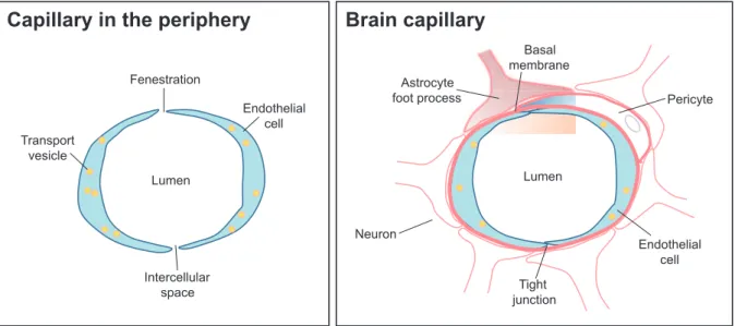
Similar to the metastatic process in other organs, brain metastasis formation is a highly selective, multi-step process, involving complex interactions between tumor and host cells, which is clearly ex-plained by the “seed-and-soil” hypothesis (11, 12). The brain is considered as a “sanctuary” site for metastatic cancer cells because many of the therapeutic agents can not cross the blood-brain barrier (BBB) (13). The BBB is a network posed of both endothelial cells and supporting com-ponents that protects the CNS microenvironment. In contrast to the periphery, the endothelium of brain micro-vessels is characterized by continuous tight junctions (TJs), decreased pinocytosis activity, very low pinocytic activity, and overexpressed efflux pumps (14) (Figure 1). With the reinforcement of the surrounding extracellular matrix (ECM), basal membrane, pericytes, and the end-feet of astrocytes, the BBB effectively prevents the free exchange of substances between the blood and the interstitial fluid of the brain (15). Therefore, the brain forms a special challenge for tumor cells because of BBB and all other steps have to be successfully completed
Figure 1. Schematic comparison of a brain capillary with a capillary in the periphery. Schematic structure of the blood - brain barrier. Cerebral endothelial cells, coming in contact with pericytes and astrocytes, form the morphological basis of the blood - brain barrier. Endothelial cells are interconnected by a continuous line of tight junctions (Cited from reference number 14 and revised).
for tumor cells to invade through BBB and sur-vive.
When metastatic cancer cells enter the brain circulation, they might arrest in sites of slow flow within the capillary bed at vascular branch points. Then, interactions between cancer cells and brain endothelial cells or transendothelial migration are mediated by interaction of tumor cell-surface recep-tors and endothelial cell adhesion molecules (16). After overcoming the BBB, tumor cells are con-fronted with components of the local microenviron-ment including the extracellular matrix (ECM), resi-dent brain parenchymal cells (astrocytes, microglia), and paracrine signaling molecules including cy-tokines and growth factors (16). Survival and suc-cessful tumor growth require adaption to and inter-action with these factors. The brain also establishes an adequate blood supply via angiogenesis, angio-ectasia, vasculogenesis, vasculogenic mimicry, mak-ing brain metastases grow and proliferate better (17). During the above process, functional signifi-cance of gene expressions, changes in brain vas-culature, abnormal secretion of soluble factors and activation of autocrine/paracrine signaling contribute to the brain metastasis development.
III. MOLECULAR PATHWAYS MEDIATING
LUNG CANCER BRAIN METASTASIS
1. Gene expression analyses
In order to identify pathways specific to the de-velopment of brain metastasis and novel molecular targets/translational leads, several groups have un-dertaken gene expression studies in animal models and human tissue cohorts.
Kikuchi et al. compared gene expression of pri-mary lung adenocarcinoma with brain metastases originating from these tumors. Of 23,040 genes tested, 244 showed a different expression level, in-cluding genes coding for plasma membrane pro-teins, cellular antigens and cytoskeletal propro-teins, which may modulate cell-cell interactions (18). Brain metastases of lung adenocarcinoma were evaluated in another study as well, by comparing the gene expression profile of metastatic brain tumors originating from lung adenocarcinoma with that of healthy lung. Zohrabian et al. have found 1,561 dif-ferently expressed genes by using cDNA microar-ray. The overexpression of genes associated with invasion and metastasis, adhesion, angiogenesis and cell migration was validated by real-time PCR (19).
In a cohort of primary tumors from lung cancer patients who developed brain metastases, Grinberg-Rashi et al. found the expression levels of three genes, CDH2, KIFC1, and FALZ, was highly pre-dictive of brain metastases (20). N-cadherin, which is coded by CDH2 gene, is involved in multiple proc-esses including inducing invasion, migration, pro-moting survival of cancer cells, regulating adhesion and neurite outgrowth (21). Clinically, N-cadherin overexpression has been shown to be associated with decreased survival in poorly differentiated SCLCs (22).
DCUN1D1, a squamous cell carcinoma-related oncogene, is associated with tumor progression and poor outcomes in NSCLC. Recently, the over-expression of DCUN1D1 has been found to be use-ful in identifying patients who are at higher risk for brain metastases. Yoo et al. showed that 14 of 16 DCUN1D1-positive NSCLC patients (87.5%) re-sulted in brain metastases (23).
However, as previously reported in other primary and metastatic clinical or experimental tumors (24-26), the genetic heterogeneity would limit the trans-lation of the gene profiles into the clinical trials.
2. Cell surface molecules
1) Integrins
Integrins are a family of cell surface receptors that mediate cell adhesion and signal transduction. Integrins interact with ECM components such as collagen, laminin, and fibronectin and play crucial roles in cell migration. Integrins can also activate signaling cascades to mediate cell survival (27). The expression of integrinα3β1 has been associated with lung cancer brain metastasis. Compared with their parental cell line and bone-metastasizing coun-terparts, tumor cells that preferably metastasize to the brain highly expressedα3β1 integrin (28). Moreover, inhibiting α3β1 integrin function de-creased brain metastases formation in nude mice (28). It has been posited that the interaction of the α3β1 integrin with laminin, which promotes tumor cell migration and invasion, may be critical to this effect (28).
2) Immunoglobulin superfamily of cell adhesion mole-cules
Endothelial cells express several adhesion mole-cules belonging to the immunoglobulin superfamily, including members of the intercellular adhesion molecules (ICAMs), vascular-cell adhesion molecule (VCAM) and platelet-endothelial-cell adhesion mole-cule (PECAM) (29). These are essential in immune
response and inflammation, but some of them were also shown to be involved in the interaction of vas-cular endothelial and tumor cells and formation of metastases. Recently, ICAM-1 and VCAM-1 were shown to play a crucial role in polychlorinated bi-phenyl-mediated enhancement of brain metastasis formation of lung carcinoma cells (30).
3) Cadherins
Cadherin dysfunction is involved in tumor pro-gression and metastasis formation. Loss of expres-sion of E-cadherin induces epithelial-mesenchymal transition (EMT) in carcinoma cells, which initi-ates an increase in cell motility and metastasis for-mation. In metastatic lesions, a re-expression of E-cadherin has been observed, which plays an im-portant role in the proliferation of tumor cells at the metastatic site. Correspondingly, metastatic brain tumors were shown to express high levels of E-cadherin (31), while low expression of E-E-cadherin in primary NSCLC was shown to correlate with in-creased risk for the development of brain metastasis (32). In addition, the expression level of N-cadherin was observed to be highly predictive of brain me-tastasis formation in NSCLC (20).
3. Soluble factors
1) Vascular endothelial growth factor (VEGF) Angiogenesis is an important aspect of tumor metastasis. Recent studies have examined the roles of VEGF, which influences both angiogenesis and blood vessel permeability. VEGF signaling and func-tion in brain metastasis has also been extensively characterized in preclinical models. Measurement of VEGF levels in the culturing media of cells grow-ing in vitro has shown that VEGF is secreted by tumor cells with high brain metastatic activity (33). Increased VEGF secretion has also been detected in brain metastasis xenografts in nude mouse mod-els (33). Antisense VEGF transfectants of PC14-PE6 lung adenocarcinoma cells exhibited a decreased incidence of experimental brain metastases, suggest-ing that VEGF is necessary for brain tumor initiation and growth (33). Jubb et al. compared VEGF ex-pression, proliferation, microvessel density, vascular pattern and vascular maturity in matched primary NSCLC and brain metastases (34). They found that brain metastases are characterized by a significantly higher proliferation rate and vascular maturity than their matched primaries. These findings are impor-tant because if the vasculature of brain metastasis is mature, then patients with cerebral secondaries may be less likely to respond to anti-VEGF treatment,
even though patients with primary NSCLC might benefit from such therapy (34).
2) Placental growth factor (PlGF)
Li et al. (35) recently found PlGF, a member of VEGF subfamily, may be associated with SCLC brain metastasis. PlGF in the serum of SCLC pa-tients with brain metastases was significantly higher than that without brain metastases and normal speci-mens. In addition, SCLC cell-derived PlGF activates the VEGFR1/Rho/ERK signaling pathway in cere-bral endothelial cells, resulting in the disassembly of TJs and promoting transendothelial migration (35). Moreover, the down-regulation of PlGF sup-pressed SCLC cell metastasis to the brain in an ex-perimental brain metastasis model.
3) Chemokines
Chemokines play an important role in cell migra-tion, invasion and tumor angiogenesis (36). Emerg-ing data support the putative involvement of the CXCR4/CXCL12 signaling axis in NSCLC brain me-tastasis (37). High CXCL12 expression within sites of lung cancer metastasis has been demonstrated in mouse xenograft studies (38). Hartmann et al. showed that the CXCR4 chemokine receptor closely co-operates with integrins to promote adhesion and chemoresistance of SCLC cells (39). Overexpres-sion of CXCR4 and CXCL12 has been described in histolopathological specimens of brain metastases and correlated with brain-specific metastasis in a cohort of NSCLC patients (40). In a recent study, Paratore et al. (41) have investigated the expression of CXCR4/CXCL12 in primary NSCLC specimens of patients with and without brain metastases. CXCL12 and CXCR4 immunoreactivities in metastatic NSCLC samples were significantly higher than that in paired non-metastatic NSCLC.
4. Proteases
1) Matrix metalloproteinases (MMPs)
Different proteolytic enzymes have been impli-cated in brain metastasis formation and migration of tumor cells through BBB endothelial cells. MMPs might have special importance in the process of transendothelial migration of tumor cells through the BBB, because TJ proteins can be targets of MMP degradation. MMP-induced disruption of TJs was shown to promote invasion of tumor cells into the CNS (42). MMP-9 was reported to be overex-pressed by brain metastatic lung adenocarcinoma cells (43).
2) A disintegrin and metalloprotease 9 (ADAM9) ADAM-9 is a membrane-tethered protease and
belongs to a member of the “a disintegrin and met-alloprotease” family. ADAM9 is overexpressed in brain metastatic lung cancer cells. Shintani et al. found that the expression of ADAM9 up-regulated integrinα3β1 and facilitated brain metastasis for-mation (44).
5. Driver mutations
1) Epidermal growth factor receptor (EGFR) mutations It is found that driver mutations in NSCLC, at least in part, would be associated with brain metas-tases. In East Asian patients, Matsumoto et al. (45) and Gow et al. (46) have found EGFR mutations in 63 and 44% of brain metastases, respectively. This prevalence is similar to that reported in primary tu-mors of the same population, varying from 30 to 50% (47, 48). Among the 110 patients enrolled, Li et al. found the frequencies of EGFR mutations were 64% and 31% in the patients with and without brain me-tastases, respectively (49). Eichler et al. showed that the numbers of brain metastases were significantly higher in patients with EGFR-mutated NSCLC com-pared to those with wild-type (50). Sekine et al. demonstrated that NSCLC patients with the exon 19 deletions had more multiple and smaller brain metastases with smaller peritumoral brain edema than those without any mutations (51). Moreover, Heron et al. (52) reported that tumors with exon 19 deletions showed a higher incidence of CNS involve-ment as compared with tumors bearing a L858R mutation (21% vs. 3%). While published data are limited to draw any definite conclusions, brain me-tastases would be more frequent in NSCLC patients with EGFR mutations.
2) Anaplastic lymphoma kinase (ALK) translocations More recently, ALK translocations have been found to be another ‘‘druggable’’ alteration besides activating EGFR mutations in NSCLC (53). ALK translocations appear to be constant between pri-mary tumors and brain metastases (54), and ALK-positive tumors may predispose to brain metastasis formation (55). In contrast, Doebele et al. found pa-tients with ALK translocations were predisposed to liver, but not adrenal, bone, or brain metastases, compared to ALK-negative cohort (56). Further studies should be required to clarify the significance of ALK translocations in NSCLC patients with brain metastases.
6. Growth factors and signaling pathways
1) Wnt signaling
The activation of canonical Wnt/TCF pathway has
also been identified as playing a role in lung cancer spread to the brain. Treatment of a brain-seeking lung cancer cell line H2030-BrM3 with Wnt3a sig-nificantly increased the expressions of the WNT/ TCF target genes, LEF1 and HOXB9. Confirming that LEF1 and HOXB9 are involved in metastasis, overexpression of the two genes led to an increase in brain metastases whereas knockdown of each gene decreased metastatic incidence (57). Support-ing the hypothesis that TCF4 may play an important role in lung cancer development, Xu et al. found in-creased TCF4 overexpression in lung cancer pa-tients with advanced stages (stage III-IV) compared with early stages (stage I-II) (58). Furthermore, ex-pression of Wnt3a in a four-gene signature pre-dicted increased mortality rates in lung cancer pa-tients (59).
2) Rho/Rho kinase (ROCK) signaling
During invasion of tissues and migration through vessel walls and ECM components, metastasizing tumor cells require increased motility, which is de-pendent on the remodeling of the cytoskeleton. Rho/ROCK pathways had been proposed to be in-volved in the regulation of paracellular permeability and junctional dynamics in endothelial cells (60). While little is known about the behavior of tumor cells during transmigration through the BBB, it has been shown that inhibition of ROCK decreases the migration of SCLC cells through the brain endo-thelium (61).
3) Hepatocyte growth factor (HGF)/Met signaling The receptor tyrosine kinase Met and its ligand HGF promoted metastatic spread to the lung, liver and brain in an experimental model using the NCI-H460 lung cancer cell line (62). A study of matched lung cancer brain metastases and primary tumors from the same patients identified increased expres-sion of total and phosphorylated c-Met in the brain metastases. The expression and activation of c-Met in the primary lung tumors also correlated with the development of brain metastases (63).
4) Phosphoinositide 3-kinase (PI3K)-Akt signaling The PI3K-Akt pathway is a crucial regulator of cell survival and proliferation, and increased PI3K activity has been reported in several cancer types. Recently, a PI3K inhibitor was found to effectively control metastatic growth of HER2-positive breast cancer cells in multiple organs, including brain me-tastases (64). However, inhibition of PI3K had no effect on the transmigration of SCLC cells through brain endothelial cells (61).
5) Endothelin (ET)/ET receptor signaling
The binding of ET to the ET receptors exerts pleiotropic biological effects that influence cell sur-vival, proliferation, invasion, metastasis, as well as angiogenesis (65). In a preclinical model of sponta-neous melanoma brain metastasis, Cruz-Muñoz et al. have identified the alterations in the expression of ET receptor as a potential factor that influences brain metastatic potential. Induced overexpression of this gene mediated enhanced overall metastatic disease, and resulted in an increased incidence of spontaneous brain metastases (66). We recently demonstrated that the blockade of ET receptor sig-nificantly inhibited experimental brain metastases of human NSCLC cells (67).
7. MicroRNA (miRNA)
Mounting evidence indicates that miRNA may be key players in the regulation of tumor cell invasion and metastasis. Chen et al. (68) found miR-378 was significantly differentially expressed in the matched NSCLC surgical specimens from 8 patients with brain metastases and 21 without brain metastases. Arora et al. (69) reported that miR-328 had a role in conferring migratory potential to NSCLC cells, which might be incorporated into clinical treatment decision making to stratify NSCLC patients at higher risk for brain metastases.
8. Single nucleotide polymorphisms (SNPs)
Studying multiple SNPs in signaling pathways may be useful for pinpointing the genes and poly-morphisms involved in conferring risk of brain me-tastases (70). Multivariate analyses of 33 SNPs from 13 genes in the transforming growth factor-β (TGF-β) signaling pathway have revealed that the GG genotype of SMAD6 : rs12913975 and TT genotype of INHBC : rs4760259 were associated with a sig-nificantly higher incidence of brain metastasis in patients with NSCLC at 24 months follow-up, com-pared with the GA or CT/CC genotypes, respec-tively (71). In melanoma, TGF-β2 was reported to be crucial, since its expression is indispensable for the metastasis formation in the brain parenchyma (72).
9. Circulating markers
1) Tumor markers
Tumor markers may be helpful in the prediction of brain metastases. Among them, carcinoembronic antigen (CEA) is the most widely studied. Lee et al. found that the pretreatment serum CEA level was
significantly correlated with brain metastases in 227 advanced NSCLC patients (73). Arrieta et al. also reported that high serum CEA level at diagnosis is an independent prognostic factor of CNS metastasis development and survival in patients with advanced NSCLC (74). They considered that surface expres-sion of CEA in tumor cells could be a mechanism of invasion to CNS through immunoglobulin-related transport in BBB.
Pro-gastrin-releasing peptide (ProGRP) is a widely used tumor marker for the screening of SCLC. Yonemori et al. retrospectively analyzed the characteristics of the first failure event due to brain metastasis in SCLC patients treated with prophylactic cranial irradiation (PCI). Elevation of ProGRP level before PCI was found to be a significant predictive factor for brain metastasis on multivariate analysis (75).
2) Indicators of CNS injury in the blood
Elevated levels of certain proteins or neurotrans-mitters in the blood may be indicators of CNS dam-age caused by invasion of brain metastases (70). S100β is a nervous system specific cytoplasmic pro-tein found in astrocytes and is released into serum when the BBB is breached (76). However, a con-founding factor was the presence of BBB changes due to cerebrovascular disease. Therefore, patients who are found to have evidence of chronic cere-brovascular disease will likely receive no further benefit from routine screening of their serum S100β level. More recently, proapolipoprotein A1, the pre-cursor of the cholesterol-binding protein apolipo-protein A1, was reported to be significantly in-creased in patients with CNS disease compared with those affected only by vascular diseases (77).
10. Role of astrocytes in brain metastasis formation
Astrocytes have an indispensable role in the main-tenance of BBB properties of cerebral endothelial cells. Therefore, they support endothelial cells in impeding tumor cells from penetrating into the brain. On the other hand, astrocytes have a pro-tective role for brain metastases. Reactive astrocytes induce the protection of tumor cells from chemo-therapy through sequestration of calcium from the cytoplasm of tumor cells and by up-regulating sur-vival genes in tumor cells (78, 79). Moreover, astro-cytes secrete soluble factors that stimulate the pro-liferation of tumor cells in the brain microenviron-ment. In addition, astrocytes were shown to induce proliferation of lung and breast cancer cells by pro-ducing interleukin (IL)-1β, tumor necrosis factor-α
(TNF-α) and/or IL-6 (80, 81). These proinflamma-tory cytokines secreted by astrocytes might not only induce proliferation of tumor cells, but also support the transendothelial migration and formation of new metastatic colonies in the brain.
IV. TARGETED THERAPIES FOR LUNG
CANCER BRAIN METASTASIS
In the study by Kienast et al., the VEGF-A inhibi-tor bevacizumab blocked angiogenesis and resulted in dormancy of brain metastasis derived from lung cancer cells (82), indicating that anti-angiogenic agents might be promising to inhibit brain metas-tasis. In early clinical studies, hemorrhagic episodes were reported after treatment with bevacizumab in patients with metastatic spread, including brain me-tastasis (83). In consequence, guidelines prohibited the use of bevacizumab in this cohort and patients with brain metastasis have been excluded from par-ticipating in clinical trials that investigated anti-angiogenic drugs (84, 85). However, recent large meta-analyses performed in over 10,000 patients that received anti-angiogenic agents revealed that these drugs do not increase the risk of intracranial bleed-ing compared to the untreated population with brain metastasis (0.8-3.3% bleeding risk) (85). These re-ports have prompted to change the guidelines and allow administration of bevacizumab in patients with brain metastasis from non-squamous NSCLC (84). Based on the new guidelines, the decision to use anti-angiogenics in this patient cohort should be made upon careful assessment of the potential bene-fits and risks for individual patients (85).
The EGFR tyrosine kinase inhibitors (EGFR-TKIs), gefitinib and erlotinib, have been tested in patients with NSCLC and brain metastasis (50, 86, 87). Similar to primary tumors, the response of brain metastasis to EGFR inhibitors is better in patients with activating EGFR mutations while the activity of these drugs in individuals with wild-type EGFR metastatic disease is very modest (50, 86, 87). In-terestingly, the response of chemotherapy-naïve, never-smoker patients with brain metastases after treatment with erlotinib and gefitinib was 74%, and maybe inhibition of EGFR in this patient subgroup is more effective compared to other cohorts (88). The BBB penetrability of erlotinib might be better than gefitinib as small but measurable penetration of erlotinib into cerebrospinal fluid (CSF) has been documented (89). Notably, the incidence of CNS
progression after treatment with gefitinib or erlotinib was lower in NSCLC patients with EGFR mutations compared to patients with wild-type EGFR and therefore these targeted agents might also have a value as prophylactic agents (52). Furthermore, the resistance EGFR mutation T790M occurs also in CNS metastasis. Whether afatinib, a second gen-eration EGFR inhibitor that inhibits T790M also has activity in CNS metastasis with this mutation remains to be determined. In one report, encour-agingly, dose escalation of afatinib resulted in re-mission of a brain metastasis (90). The question whether erlotinib or gefitinib should be combined with other WBRT or stereotactic radiosurgery is subject of current studies.
Activation of ALK oncogene leads to fusion of ALK and the echinoderm microtubule-associated protein-like 4 (EML4) that is encountered in ap-proximately 4% of patients with NSCLC. The tar-geted agent crizotinib inhibits this oncogenic fusion and can lead to effective local tumor control (91). In a recent case report of a patient with EML4-ALK fusion, though, brain metastasis developed despite control of extracerebral metastases (92). Very low levels of crizotinib were detected in plasma and CSF of the patient, suggesting insufficient BBB penetra-bility for this agent (92). However, another recent case report demonstrated a twelve-month progres-sion-free survival after treatment with crizotinib in a patient with lung adenocarcinoma with EML4-ALK fusion that developed metachronous miliary lung metastases and brain metastasis (93).
V. CONCLUSION
Brain metastasis has become an increasingly chal-lenging clinical problem, largely due to the recently improved clinical control of systemic metastatic dis-eases. While the biology of brain metastasis is still poorly understood, it is encouraging to see more ef-forts are beginning to be directed toward the study of brain metastasis.
The biomarkers mentioned in this review would be promising tools for the prediction of brain metas-tases. However, currently none of them can predict occurrence of brain metastases alone and much challenge remain for their translation into practice. In this article, the mechanistic basis of lung cancer metastasis to the brain is described (Table 1). The better understanding of molecular biology of lung cancer brain metastasis, including heterogeneous
genetic profiles, is essential to find appropriate tar-gets of prevention of brain metastasis formation. In addition, prospective randomized clinical studies are
needed to further assess the utility of these biologi-cal markers.
It is indisputable that the microenvironment cells Table 1. Risk factors for the development of lung cancer brain metastasis.
Risk factors Authors Reference No. Genes
high CDH2 expression Grinberg - Rashi H et al. 20 high KIFC1 expression Grinberg - Rashi H et al. 20 low FALZ gene expression Grinberg - Rashi H et al. 20 high DCUN1D1 expression Yoo et al. 23 Cell surface molecules
highα3β1 integrin expression Yoshimasu T et al. 28 high ICAM- 1 expression Sipos E et al. 30 high VCAM- 1 expression Sipos E et al. 30 low E- cadherin expression Yoo JY et al. 32 Saad AG et al. 9 high N- cadherin expression Grinberg - Rashi H et al. 20 Soluble factors
high VEGF expression Saad AG et al. 9 Yano S et al. 33 Hu L et al. 43 Kienast Y et al. 82 high PlGF expression Li B et al. 35 high CXCR4 expression Salmaggi A et al. 40 Paratore S et al. 41 high CXCL12 expression Salmaggi A et al. 40 Paratore S et al. 41 Proteases
high MMP - 9 expression Hu L et al. 43 high ADAM9 expression Shintani Y et al. 44 Driver mutations
EGFR mutation Li Z et al. 49 Eichler AF et al. 50 ALK translocation Yang P et al. 55 Signaling pathways
activation of WNT/TCF signaling Nguyen DX et al. 57 activation of Rho/ROCK signaling Li B et al. 61 activation of HGF/Met signaling Navab R et al. 62 Benedettini E et al. 63 Circulating markers
CEA elavation Lee DS et al. 73 Arrieta O et al. 74 ProGRP elavation Yonemori K et al. 75 Others
high Amphiregulin expression Sun M et al. low Caspase - 3 expression Saad AG et al. high Caveolin - 1 expression Cassoni P et al. low CD44 expression Kergi HA et al. high EGF expression Sun M et al. high ERCC1 expression Gomez - Roca C et al. high IGF - 1 expression Hwang CC et al. high Ki - 67 levels Saad AG et al. low Neuregulin1 expression Sun M et al. inactivating mutation of PTEN Hahn M et al. high S100A7 expression Zhang H et al. high phosphorylated Her3 expression Sun M et al. high phospho - S6 expression McDonald JM et al. low TGF -α expression Sun M et al.
in the tumor stroma contribute significantly to the outgrowth of cancer cells both at the primary site and in distant metastatic organs. The occurrence of brain metastasis reflects the culmination of such tumor-microenvironment interactions. Particularly, the specialized physiology of the brain not only con-tributes to the colonization of metastatic tumor le-sions but also significantly affects the efficacy and outcome of therapeutic interventions. Future clinical interventions to treat patients with brain metastasis must take into consideration the impact of these im-portant microenvironmental determinants.
DISCLOSURE OF CONFLICT OF
INTER-ESTS
All authors have no conflict of interests.
REFERENCES
1. Weil R, Palmieri D, Bronder J, Stark A, Steeg PS : Breast cancer metastasis to the central nervous system. Am J Pathol 167 : 913-920, 2005
2. Nathoo N, Chahlavi A, Barnett GH, Toms SA : Pathobiology of brain metastases. J Clin Pathol 58 : 237-242, 2005
3. Wen PY, Loeffler JS : Brain metastases. Curr Treat Options Oncol 1 : 447-458, 2000
4. Kim SH, Chao ST, Toms SA, Vogelbaum MA, Barnett GH, Suh JH, Weil RJ : Sereotactic ra-diosurgical treatment of parenchymal brain me-tastases from prostate adenocarcinoma. Surg Neurol 69 : 641-646, 2008
5. Soffietti R, Ruda R, Trevisan E : Brain metas-tases : current management and new develop-ments. Curr Opin Oncol 20 : 676-684, 2008 6. Gril B, Evans L, Palmieri D, Steeg PS :
Trans-lational research in brain metastasis is identi-fying molecular pathways that may lead to the development of new therapeutic strategies. Eur J Cancer 46 : 1204-1210, 2010
7. Eichler AF, Loeffler JS : Multidisciplinary man-agement of brain metastases. Oncologist 12 : 884-898, 2007
8. Smedby KE, Brandt L, Backlund ML, Blomqvist P : Brain metastases admissions in Sweden be-tween 1987 and 2006. Br J Cancer 101 : 1919-1924, 2009
9. Saad AG, Yeap BY, Thunnissen FB, Pinkus GS,
Pinkus JL, Loda M, Sugarbaker DJ, Johnson BE, Chirieac LR : Immunohistochemical mark-ers associated with brain metastases in patients with non-small cell lung carcinoma. Cancer 113 : 2129-2138, 2008
10. Schwer AL, Gaspar LE : Update in the treat-ment of brain metastases from lung cancer. Clin Lung Cancer 8 : 180-186, 2006
11. Paget S : The distribution of secondary growths in cancer of the breast. 1889. Cancer Metastasis Rev 8 : 98-101, 1989
12. Fidler IJ : The role of the organ microenviron-ment in brain metastasis. Semin Cancer Biol 21 : 107-112, 2011
13. Palmieri D, Chambers AF, Felding-Habermann B, Huang S, Steeg PS : The biology of metas-tasis to a sanctuary site. Clin Cancer Res 13 : 1656-1662, 2007
14. Deeken JF, Löscher W : The blood-brain bar-rier and cancer : transporters, treatment, and Trojan horses. Clin Cancer Res 13 : 1663-1674, 2007
15. Abbott NJ, Ronnback L, Hansson E : Astrocyte-endothelial interactions at the blood-brain bar-rier. Nat Rev Neurosci 7 : 41-53, 2006
16. Preusser M, Capper D, Ilhan-Mutlu A, Berghoff AS, Birner P, Bartsch R, Marosi C, Zielinski C, Mehta MP, Winkler F, Wick W, von Deimling A : Brain metastases : pathobiology and emerg-ing targeted therapies. Acta Neuropathol 123 : 205-222, 2012
17. Eichler AF, Chung E, Kodack DP, Loeffler JS, Fukumura D, Jain RK : The biology of brain meta-stases-translation to new therapies. Nat Rev Clin Oncol 8 : 344-356, 2011
18. Kikuchi T, Daigo Y, Ishikawa N, Katagiri T, Tsunoda T, Yoshida S, Nakamura Y : Expres-sion profiles of metastatic brain tumor from lung adenocarcinomas on cDNA microarray. Int J Oncol 28 : 799-805, 2006
19. Zohrabian VM, Nandu H, Gulati N, Khitrov G, Zhao C, Mohan A, Demattia J, Braun A, Das K, Murali R, Jhanwar-Uniyal M : Gene expression profiling of metastatic brain cancer. Oncol Rep 18 : 321-328, 2007
20. Grinberg-Rashi H, Ofek E, Perelman M, Skarda J, Yaron P, Hajdúch M, Jacob-Hirsch J, Amariglio N, Krupsky M, Simansky DA, Ram Z, Pfeffer R, Galernter I, Steinberg DM, Ben-Dov I, Rechavi G, Izraeli S : The expression of three genes in primary non-small cell lung can-cer is associated with metastatic spread to the
brain. Clin Cancer Res 15 : 1755-1761, 2009 21. Derycke LD, Bracke ME : N-cadherin in the
spotlight of cell-cell adhesion, differentiation, embryogenesis, invasion and signalling. Int J Dev Biol 48 : 463-476, 2004
22. Nakashima T, Huang C, Liu D, Kameyama K, Masuya D, Kobayashi S, Kinoshita M, Yokomise H : Neural-cadherin expression associated with angiogenesis in non-small-cell lung cancer pa-tients. Brit J Cancer 88 : 1727-1733, 2003 23. Yoo J, Lee SH, Lym KI, Park SY, Yang SH, Yoo
CY, Jung JH, Kang SJ, Kang CS : Immunohisto-chemical expression of DCUN1D1 in non-small cell lung carcinoma : its relation to brain metas-tasis. Cancer Res Treat 44 : 57-62, 2012 24. Ricketts CJ, Linehan WM : Intratumoral
hetero-geneity in kidney cancer. Nat Genet 46 : 214-215, 2014
25. Wu JM, Fackler MJ, Halushka MK, Molavi DW, Taylor ME, Teo WW, Griffin C, Fetting J, Davidson NE, De Marzo AM, Hicks JL, Chitale D, Ladanyi M, Sukumar S, Argani P : Hetero-geneity of breast cancer metastases : compari-son of therapeutic target expression and pro-moter methylation between primary tumors and their multifocal metastases. Clin Cancer Res 14 : 1938-1946, 2008
26. Nakamura T, Kuwai T, Kitadai Y, Sasaki T, Fan D, Coombes KR, Kim SJ, Fidler IJ : Zonal heterogeneity for gene expression in human pancreatic carcinoma. Cancer Res 67 : 7597-7604, 2007
27. Desgrosellier JS, Cheresh DA : Integrins in can-cer : biological implications and therapeutic op-portunities. Nat Rev Cancer 10 : 9-22, 2010 28. Yoshimasu T, Sakurai T, Oura S, Hirai I, Tanino
H, Kokawa Y, Naito Y, Okamura Y, Ota I, Tani N, Matsuura N : Increased expression of in-tegrinα3β1 in highly brain metastatic subclone of a human non-small cell lung cancer cell line. Cancer Sci 95 : 142-148, 2004
29. Wilhelm I, Molnár J, Fazakas C, Haskó J, Krizbai IA : Role of the blood-brain barrier in the formation of brain metastases. Int J Mol Sci 14 : 1383-1411, 2013
30. Sipos E, Chen L, Andras IE, Wrobel J, Zhang B, Pu H, Park M, Eum SY, Toborek M : Proin-flammatory adhesion molecules facilitate poly-chlorinated biphenyl-mediated enhancement of brain metastasis formation. Toxicol Sci 126 : 362-371, 2012
31. Shabani HK, Kitange G, Tsunoda K, Anda T,
Tokunaga Y, Shibata S, Kaminogo M, Hayashi T, Ayabe H, Iseki M : Immunohistochemical expression of E-cadherin in metastatic brain tumors. Brain Tumor Pathol 20 : 7-12, 2003 32. Yoo JY, Yang SH, Lee JE, Cho DG, Kim HK,
Kim SH, Kim IS, Hong JT, Sung JH, Son BC, Lee SW : E-cadherin as a predictive marker of brain metastasis in non-small-cell lung cancer, and its regulation by pioglitazone in a preclini-cal model. J Neurooncol 109 : 219-227, 2012 33. Yano S, Shinohara H, Herbst RS, Kuniyasu H,
Bucana CD, Ellis LM, Davis DW, McConkey DJ, Fidler IJ : Expression of vascular endothelial growth factor is necessary but not sufficient for production and growth of brain metastasis. Cancer Res 60 : 4959-4967, 2000
34. Jubb AM, Cesario A, Ferguson M, Congedo MT, Gatter KC, Lococo F, Mule A, Pezzella F : Vascular phenotypes in primary non-small cell lung carcinomas and matched brain metastases. Br J Cancer 104 : 1877-1881, 2011
35. Li B, Wang C, Zhang Y, Zhao XY, Huang B, Wu PF, Li Q, Li H, Liu YS, Cao LY, Dai WM, Fang WG, Shang DS, Cao L, Zhao WD, Chen YH : Elevated PLGF contributes to small-cell lung cancer brain metastasis. Oncogene 32 : 2952-2962, 2013
36. Keeley EC, Mehrad B, Strieter RM : CXC chemokines in cancer angiogenesis and metas-tases. Adv Cancer Res 106 : 91-111, 2010 37. Fokas E, Steinbach JP, Rödel C : Biology of
brain metastases and novel targeted therapies : Time to translate the research. Biochim Bio-phys Acta 1835 : 61-75, 2013
38. Belperio JA, Phillips RJ, Burdick MD, Lutz M, Keane M, Strieter R : The SDF-1/CXCL12/ CXCR4 biological axis in non-small cell lung cancer metastases. Chest 125 : 156S, 2004 39. Hartmann TN, Burger JA, Glodek A, Fujii N,
Burger M : CXCR4 chemokine receptor and in-tegrin signaling co-operate in mediating adhe-sion and chemoresistance in small cell lung cancer (SCLC) cells. Oncogene 24 : 4462-4471, 2005
40. Salmaggi A, Maderna E, Calatozzolo C, Gaviani P, Canazza A, Milanesi I, Silvani A, DiMeco F, Carbone A, Pollo B : CXCL12, CXCR4 and CXCR7 expression in brain metastases. Cancer Biol Ther 8 : 1608-1614, 2009
41. Paratore S, Banna GL, D’Arrigo M, Saita S, Iemmolo R, Lucenti L, Bellia D, Lipari H, Buscarino C, Cunsolo R, Cavallaro S : CXCR4
and CXCL12 immunoreactivities differentiate primary non-small-cell lung cancer with or without brain metastases. Cancer Biomark 10 : 79-89, 2011
42. Feng S, Cen J, Huang Y, Shen H, Yao L, Wang Y, Chen Z : Matrix metalloproteinase-2 and -9 secreted by leukemic cells increase the perme-ability of blood-brain barrier by disrupting tight junction proteins. PLoS One 6 : e20599, 2011 43. Hu L, Zhang J, Zhu H, Min J, Feng Y, Zhang
H : Biological characteristics of a specific brain metastatic cell line derived from human lung adenocarcinoma. Med Oncol 27 : 708-714, 2010 44. Shintani Y, Higashiyama S, Ohta M, Hirabayashi H, Yamamoto S, Yoshimasu T, Matsuda H, Matsuura N : Overexpression of ADAM9 in non-small cell lung cancer correlates with brain metastasis. Cancer Res 64 : 4190-4196, 2004 45. Matsumoto S, Takahashi K, Iwakawa R,
Matsuno Y, Nakanishi Y, Kohno T, Shimizu E, Yokota J : Frequent EGFR mutations in brain metastases of lung adenocarcinoma. Int J Can-cer 119 : 1491-1494, 2006
46. Gow CH, Chang YL, Hsu YC, Tsai MF, Wu CT, Yu CJ, Yang CH, Lee YC, Yang PC, Shih JY : Comparison of epidermal growth factor recep-tor mutations between primary and correspond-ing metastatic tumors in tyrosine kinase inhibi-tor-naive non-small-cell lung cancer. Ann On-col 4 : 696-702, 2009
47. Huang SF, Liu HP, Li LH, Ku YC, Fu YN, Tsai HY, Chen YT, Lin YF, Chang WC, Kuo HP, Wu YC, Chen YR, Tsai SF : High frequency of epidermal growth factor receptor mutations with complex patterns in non-small cell lung cancers related to gefitinib responsiveness in Taiwan. Clin Cancer Res 24 : 8195-8203, 2004 48. Wu YL, Zhong WZ, Li LY, Zhang XT, Zhang L,
Zhou CC, Liu W, Jiang B, Mu XL, Lin JY, Zhou Q, Xu CR, Wang Z, Zhang GC, Mok T : Epider-mal growth factor receptor mutations and their correlation with gefitinib therapy in patients with non-small cell lung cancer : a meta-analy-sis based on updated individual patient data from six medical centers in mainland China. J Thorac Oncol 5 : 430-439, 2007
49. Li Z, Guo H : The retrospective analysis of the frequency of EGFR mutations and efficacy of gefitinib in NSCLC patients with brain metas-tases. J Clin Oncol 29 : e18065, 2011
50. Eichler AF, Kahle KT, Wang DL, Joshi VA, Willers H, Engelman JA, Lynch TJ, Sequist LV :
EGFR mutation status and survival after diag-nosis of brain metastasis in nonsmall cell lung cancer. Neuro Oncol 11 : 1193-1199, 2010 51. Sekine A, Kato T, Hagiwara E, Shinohara T,
Komagata T, Iwasawa T, Satoh H, Tamura K, Kasamatsu T, Hayashihara K, Saito T, Takahashi H, Ogura T : Metastatic brain tu-mors from non-small cell lung cancer with EGFR mutations : distinguishing influence of exon 19 deletion on radiographic features. Lung Cancer 77 : 64-69, 2012
52. Heon S, Yeap BY, Britt GJ, Costa DB, Rabin MS, Jackman DM, Johnson BE : Development of central nervous system metastases in pa-tients with advanced non-small cell lung can-cer and somatic EGFR mutations treated with gefitinib or erlotinib. Clin Cancer Res 16 : 5873-5882, 2010
53. Soda M, Choi YL, Enomoto M, Takada S, Yamashita Y, Ishikawa S, Fujiwara S, Watanabe H, Kurashina K, Hatanaka H, Bando M, Ohno S, Ishikawa Y, Aburatani H, Niki T, Sohara Y, Sugiyama Y, Mano H : Identification of the transforming EML4-ALK fusion gene in non-small-cell lung cancer. Nature 448 : 561-566, 2007
54. Preusser M, Berghoff AS, Ilhan-Mutlu A, Magerle M, Dinhof C, Widhalm G, Dieckmann K, Marosi C, Wöhrer A, Hackl M, Zöchbauer-Müller S, von Deimling A, Schoppmann SF, Zielinski CC, Streubel B, Birner P : ALK gene translocations and amplifications in brain me-tastases of non-small cell lung cancer. Lung Cancer 80 : 278-283, 2013
55. Yang P, Kulig K, Boland JM, Erickson-Johnson MR, Oliveira AM, Wampfler J, Jatoi A, Deschamps C, Marks R, Fortner C, Stoddard S, Nichols F, Molina J, Aubry MC, Tang H, Yi ES : Worse disease-free survival in never-smokers with ALK+lung adenocarcinoma. J Thorac On-col 7 : 90-97, 2012
56. Doebele RC, Lu X, Sumey C, Maxson DA, Weickhardt AJ, Oton AB, Bunn PA Jr, Barón AE, Franklin WA, Aisner DL, Varella-Garcia M, Camidge DR : Oncogene status predicts patterns of metastatic spread in treatment-naïve non-small cell lung cancer. Cancer 118 : 4502-4511, 2012
57. Nguyen DX, Chiang AC, Zhang XH, Kim JY, Kris MG, Ladanyi M, Gerald WL, Massagué J : WNT/TCF signaling through LEF1 and HOXB9 mediates lung adenocarcinoma metastasis. Cell
138 : 51-62, 2009
58. Xu HT, Wei Q, Liu Y, Yang LH, Dai SD, Han Y, Yu JH, Liu N, Wang EH : Overexpression of axin downregulates TCF-4 and inhibits the de-velopment of lung cancer. Ann Surg Oncol 14 : 3251-3259, 2007
59. Raz DJ, Ray MR, Kim JY, He B, Taron M, Skrzypski M, Segal M, Gandara DR, Rosell R, Jablons DM : A multigene assay is prognostic of survival in patients with early-stage lung ade-nocarcinoma. Clin Cancer Res 14 : 5565-5570, 2008
60. Wojciak-Stothard B, Ridley AJ : Rho GTPase and the regulation of endothelial permeability. Vascul Pharmacol 39 : 187-199, 2003
61. Li B, Zhao WD, Tan ZM, Fang WG, Zhu L, Chen YH : Involvement of Rho/ROCK signal-ling in small cell lung cancer migration through human brain microvascular endothelial cells. FEBS Lett 580 : 4252-4260, 2006
62. Navab R, Liu J, Seiden-Long I, Shih W, Li M, Bandarchi B, Chen Y, Lau D, Zu YF, Cescon D, Zhu CQ, Organ S, Ibrahimov E, Ohanessian D, Tsao MS : Co-overexpression of Met and hepa-tocyte growth factor promotes systemic metas-tasis in NCI-H460 non-small cell lung carci-noma cells. Neoplasia 11 : 1292-1300, 2009 63. Benedettini E, Sholl LM, Peyton M, Reilly J,
Ware C, Davis L, Vena N, Bailey D, Yeap BY, Fiorentino M, Ligon AH, Pan B, Richon V, Minna JD, Gazdar AF, Draetta G, Bosari S, Chirieac LR, Lutterbach B, Loda M : Met acti-vation in non-small cell lung cancer is associ-ated with de novo resistance to EGFR inhibitors and the development of brain metastasis. Am J Pathol 177 : 415-423, 2010
64. Nanni P, Nicoletti G, Palladini A, Croci S, Murgo A, Ianzano ML, Grosso V, Stivani V, Antognoli A, Lamolinara A, Landuzzi L, di Tomaso E, Iezzi M, De Giovanni C, Lollini PL : Multiorgan metastasis of human HER-2+breast cancer in Rag2-/-; Il2rg-/-mice and treatment with PI3K inhibitor. PLoS One 7 : e39626, 2012 65. Bagnato A, Rosanò L : The endothelin axis in cancer. Int J Biochem Cell Biol 40 : 1443-1451, 2008
66. Cruz-Muñoz W, Jaramillo ML, Man S, Xu P, Banville M, Collins C, Nantel A, Francia G, Morgan SS, Cranmer LD, O’Connor-McCourt MD, Kerbel RS : Roles for endothelin receptor B and BCL2A1 in spontaneous CNS metasta-sis of melanoma. Cancer Res 72 : 4909-4919,
2012
67. Hanibuchi M, Kim SJ, Otsuka K, Mitsuhashi A, Goto H, Nishioka Y, Fidler IJ : Therapeutic effi-cacy of endothelin receptor blockade on experi-mental brain metastases of human non-small cell lung cancer. 18th Congress of the Asian Pacific Society of Respirology, 2013
68. Chen LT, Xu SD, Xu H, Zhang JF, Ning JF, Wang SF : MicroRNA-378 is associated with non-small cell lung cancer brain metastasis by promoting cell migration : invasion and tumor angiogenesis. Med Oncol 29 : 1673-1680, 2012 69. Arora S, Ranade AR, Tran NL, Nasser S, Sridhar S, Korn RL, Ross JT, Dhruv H, Foss KM, Sibenaller Z, Ryken T, Gotway MB, Kim S, Weiss GJ : MicroRNA-328 is associated with (non-small) cell lung cancer (NSCLC) brain metastasis and mediates NSCLC migration. Int J Cancer 129 : 2621-2631, 2011
70. Huang Q, Ouyang X : Predictive biochemical-markers for the development of brain metas-tases from lung cancer : Clinical evidence and future directions Cancer Epidemiol 37 : 703-707, 2013
71. Li Q, Wu H, Chen B, Hu G, Huang L, Qin K, Chen Y, Yuan X, Liao Z : SNPs in the TGF-β signaling pathway are associated with increased risk of brain metastasis in patients with non-small-cell lung cancer. PLoS One 7 : e51713, 2012
72. Zhang C, Zhang F, Tsan R, Fidler IJ : Trans-forming growth factor-beta2 is a molecular de-terminant for site-specific melanoma metastasis in the brain. Cancer Res 69 : 828-835, 2009 73. Lee DS, Kim YS, Jung SL, Lee KY, Kang JH,
Park S, Kim YK, Yoo IeR, Choi BO, Jang HS, Yoon SC : The relevance of serum carcinoem-bryonic antigen as an indicator of brain metas-tasis detection in advanced non-small cell lung cancer. Tumour Biol 33 : 1065-1073, 2012 74. Arrieta O, Saavedra-Perez D, Kuri R,
Aviles-Salas A, Martinez L, Mendoza-Posada D, Castillo P, Astorga A, Guzman E, De la Garza J : Brain metastasis development and poor sur-vival associated with carcinoembryonic antigen (CEA) level in advanced non-small cell lung cancer : a prospective analysis. BMC Cancer 9 : 119, 2009
75. Yonemori K, Sumi M, Fujimoto N, Ito Y, Imai A, Kagami Y, Ikeda H : Pro-gastrin-releasing peptide as a factor predicting the incidence of brain metastasis in patients with small cell lung
carcinoma with limited disease receiving pro-phylactic cranial irradiation. Cancer 104 : 811-816, 2005
76. Kanner AA, Marchi N, Fazio V, Mayberg MR, Koltz MT, Siomin V, Stevens GH, Masaryk T, Aumayr B, Vogelbaum MA, Barnett GH, Janigro D : Serum S100beta : a noninvasive marker of blood-brain barrier function and brain lesions. Cancer 97 : 2806-2813, 2003
77. Marchi N, Mazzone P, Fazio V, Mekhail T, Masaryk T, Janigro D : ProApolipoprotein A1 : a serum marker of brain metastases in lung cancer patients. Cancer 112 : 1313-1324, 2008 78. Kim SJ, Kim JS, Park ES, Lee JS, Lin Q,
Langley RR, Maya M, He J, Kim SW, Weihua Z, Balasubramanian K, Fan D, Mills GB, Hung MC, Fidler IJ : Astrocytes upregulate survival genes in tumor cells and induce protection from chemotherapy. Neoplasia 13 : 286-298, 2011 79. Lin Q, Balasubramanian K, Fan D, Kim SJ, Guo
L, Wang H, Bar-Eli M, Aldape KD, Fidler IJ : Reactive astrocytes protect melanoma cells from chemotherapy by sequestering intracellu-lar calcium through gap junction communica-tion channels. Neoplasia 12 : 748-754, 2010 80. Seike T, Fujita K, Yamakawa Y, Kido MA,
Takiguchi S, Teramoto N, Iguchi H, Noda M : Interaction between lung cancer cells and as-trocytes via specific inflammatory cytokines in the microenvironment of brain metastasis. Clin Exp Metastasis 28 : 13-25, 2011
81. Sierra A, Price JE, Garcia-Ramirez M, Mendez O, Lopez L, Fabra A : Astrocyte-derived cytoki-nes contribute to the metastatic brain specific-ity of breast cancer cells. Lab Invest 77 : 357-368, 1997
82. Kienast Y, von Baumgarten L, Fuhrmann M, Klinkert WE, Goldbrunner R, Herms J, Winkler F : Real-time imaging reveals the single steps of brainmetastasis formation. Nat Med 16 : 116-122, 2010
83. Carden CP, Larkin JM, Rosenthal MA : What is the risk of intracranial bleeding during anti-VEGF therapy? Neuro Oncol 10 : 624-630, 2008 84. Besse B, Lasserre SF, Compton P, Huang J, Augustus S, Rohr UP : Bevacizumab safety in patients with central nervous system metasta-ses. Clin Cancer Res 16 : 269-278, 2010 85. De Braganca KC, Janjigian YY, Azzoli CG,
Kris MG, Pietanza MC, Nolan CP, Omuro AM,
Holodny AI, Lassman AB : Efficacy and safety of bevacizumab in active brain metastases from non-small cell lung cancer. J Neurooncol 100 : 443-447, 2010
86. Jamal-Hanjani M, Spicer J : Epidermal growth factor receptor tyrosine kinase inhibitors in the treatment of epidermal growth factor receptor-mutant non-small cell lung cancer metastatic to the brain. Clin Cancer Res 18 : 938-944, 2012 87. Wu C, Li YL, Wang ZM, Li Z, Zhang TX, Wei
Z : Gefitinib as palliative therapy for lung ade-nocarcinoma metastatic to the brain, Lung Can-cer 57 : 359-364, 2007
88. Kim JE, Lee DH, Choi Y, Yoon DH, Kim SW, Suh C, Lee JS : Epidermal growth factor recep-tor tyrosine kinase inhibirecep-tors as a first-line therapy for never-smokers with adenocarci-noma of the lung having asymptomatic syn-chronous brain metastasis. Lung Cancer 65 : 351-354, 2009
89. Masuda T, Hattori N, Hamada A, Iwamoto H, Ohshimo S, Kanehara M, Ishikawa N, Fujitaka K, Haruta Y, Murai H, Kohno N : Erlotinib effi-cacy and cerebrospinal fluid concentration in patients with lung adenocarcinoma developing leptomeningeal metastases during gefitinib therapy. Cancer Chemother Pharmacol 67 : 1465-1469, 2011
90. Yap TA, Vidal L, Adam J, Stephens P, Spicer J, Shaw H, Ang J, Temple G, Bell S, Shahidi M, Uttenreuther-Fischer M, Stopfer P, Futreal A, Calvert H, de Bono JS, Plummer R : Phase I trial of the irreversible EGFR and HER2 kinase inhibitor BIBW 2992 in patients with advanced solid tumors. J Clin Oncol 28 : 3965-3972, 2010 91. Gandhi L, Jänne PA : Crizotinib for ALK-rear-ranged non-small cell lung cancer : a new tar-geted therapy for a new target. Clin Cancer Res 18 : 3737-3742, 2012
92. Costa DB, Kobayashi S, Pandya SS, Yeo WL, Shen Z, Tan W, Wilner KD : CSF concentration of the anaplastic lymphoma kinase inhibitor crizotinib. J Clin Oncol 29 : e443-e445, 2011 93. Falk AT, Poudenx M, Otto J, Ghalloussi H,
Barriere J : Adenocarcinoma of the lung with miliary brain and pulmonary metastases with echinoderm microtubule-associated protein like 4-anaplastic lymphoma kinase translocation treated with crizotinib : a case report. Lung Can-cer 78 : 282-284, 2012