Posted at the Institutional Resources for Unique Collection and Academic Archives at Tokyo Dental College,
Title
Simple and rapid detection of Rothia mucilaginosa
by loop-mediated isothermal amplification method
Author(s)
Keisuke, Nakai; Hiroshi, Maeda; Hideaki, Ikenaga;
Norimasa, Tsuji
Journal
日本口腔検査学会雑誌, 10(1): 44-52
URL
http://hdl.handle.net/10130/4543
Right
Description
Keisuke Nakai, Hiroshi Maeda*, Hideaki Ikenaga, Norimasa Tsuji
Department of Endodontics, Osaka Dental University 8-1 Kuzuhahanazonocho Hirakata, Osaka 573-1121, Japan *: 8-1 Kuzuhahanazonocho Hirakata, Osaka 573-1121, Japan Tel: 81-72-864-3043 Fax: 81-72-864-3143 e-mail: [email protected]Simple and rapid detection of
Rothia mucilaginosa
by loop-mediated isothermal amplification method
Abstract
Aim: Rothia mucilaginosa is an emerging opportunistic pathogen in immunocompromised hosts. The aim of this study was to develop a rapid and simple examination method for this pathogen. Methods: The loop-mediated isothermal amplification (LAMP) method was applied. Two primer sets (FIP, BIP, F3, B3 and two loop primers) were designed including species-specific sequences of the 16S ribosomal RNA gene. The LAMP reactions were performed under isothermal conditions at 64°C for 20 or 50 minutes. Results: The target gene was specifically amplified by LAMP with one primer set. Unexpectedly, the other primer set showed nonspecific amplification for other Rothia species. The amplicons were visualized by agarose gel electrophoresis with detection limits of 10 and 100 cells/tube for the 50-minute and 20-minute LAMP methods, respectively. By addition of SYBR-Green I, naked eye inspection was possible with the same detection limit. To further simplify the method, intact cells were subjected to the LAMP assay, and the amplicons were detected with a limit of 10 cells in the 50-minute reaction. Conclusion: Using LAMP, we developed a simple and rapid detection assay for R. mucilaginosa. This assay may be useful for microbiological diagnosis.
Key words: Rothia mucilaginosa, loop-mediated isothermal amplification (LAMP) method, naked eye inspection, opportunistic pathogens, bacteremia
Recieved; December 5th 2017 Accepted: December 11st, 2017
Introduction
Rothia mucilaginosa is a gram-positive coccobacillus, previously known as Stomatococcus mucilaginosus. This microorganism is a member of the oral microflora and is usually not associated with systemic infections in healthy hosts. Recently, R. mucilaginosa and other
Rothia species have been recognized as emerging opportunistic pathogens associated with serious infections in immunocompromised hosts1). Rothia infections have been reported in endocarditis2), meningitis3), pneumonia4), bacteremia5), and endophthalmitis5). Immunocompromised hosts, such
JJ S E D P Vol. 10 No. 1: , 2018
as patients with hematological malignancies under chemotherapy or neutropenic patients, have been reported as targets of this microorganism6).
The source of R. mucilaginosa infection is presumed to be gut translocation, mucositis, or catheter-related infection6). In addition, it is necessary to pay attention to oral infectious diseases. Periapical periodontitis is a disease caused by polymicrobial infection of oral bacteria in the tooth root canal. R. mucilaginosa has been reported to be a pathogen of persistent periapical periodontitis7). Root canals connected to alveolar bone are known as a source of dental focal infection, and therefore bacterial examination of the root canal is critical in dental treatment, especially for immunocompromised hosts.
The loop-mediated isothermal amplification (LAMP) method, developed by Notomi et al.8), relies on autocycling strand displacement DNA synthesis by the Bst DNA polymerase large fragment. The reaction can be conducted under isothermal conditions ranging from 60°C to 65°C. The specificity is attributable to four primers that recognize six distinct sequences. Continuous amplification under isothermal conditions produces large amounts of target DNA in a short time. Therefore, the method has high sensitivity, rapidity, and enables simple visual (naked eye) judgment of DNA amplification through a color change of the reaction mixture with SYBR Green I9). LAMP methods have been developed for bacterial examination in many fields10-12),
Table 1 Reference strains and results of LAMP and conventional RCR for each strain
Strains LAMP conventional
Primer set A Primer set B PCR Rothia mucilaginosa DY-18 + + + Rothia mucilaginosa ATCC 25296 + + + Rothia aeria JCM 11412 – + – Rothia dentocariosa ATCC 17931 – + – Actinomyces viscosus ATCC 15987 – – – Actinomyces oralis MG-1 – – – Escherichia coli ATCC 25922 – – – Staphylococcus aureus NBRC 14462 – – – Staphylococcus epidermidis ATCC 14990 – – – Enterococcus faecalis ATCC 19433 – – – Streptococcus sanguinis ATCC 10556 – – – Streptococcus salivarius JCM 5707 – – – Streptococcus mutans ATCC 700610 – – – Pseudomonas aeruginosa 13275 – – – Tannerella forsythia ATCC 43037 – – – Campylobacter rectus ATCC 33238 – – – Porphyromonas gingivalis FDC 381 – – – Aggregatibacter actinomycetemcomitans Y4 – – – Prevotella intermedia ATCC 25611 – – – Treponema denticola ATCC 35405 – – – Fusobacterium nucleatum ATCC 25586 – – – Eikenella corrodens ATCC 23834 – – – 44 - 52
including examination of antibiotic resistance13). As the method does not require special apparatus, such as a thermal cycler, it is practical to use for chair-side microbiological diagnosis in dental offices. In the current study, we aimed to develop a new LAMP method for R. mucilaginosa. For the purpose, two sets of LAMP primers were designed and the performance of the methods were described.
Materials and Methods
1. Bacterial strains and cell preparation
The bacterial strains used in this study are listed in Table 1. Rothia species, including Rothia mucilaginosa, Rothia aeria, and Rothia dentocariosa, were cultivated on blood agar plates (BBLTM Microbiology Systems: BD, Franklin Lakes, NJ, USA). Bacterial colonies on the agar plates were suspended in phosphate-buffered saline (PBS) (Thermo Fisher Scientific K.K., Tokyo, Japan). The cell suspensions were subjected to DNA extraction prior to use for LAMP reaction, or used directly as templates. Cell numbers in the suspensions were estimated by real-time PCR using universal primers for the 16S ribosomal RNA gene (16S rDNA) as described previously14). For
determination of the detection limits, tenfold serial dilution samples were prepared. In addition to the Rothia species, cell suspensions were prepared from 18 reference strains (Table 1) as described previously11,13,14).
2. DNA extraction
InstaGeneTM Matrix (Bio-Rad, Hercules, CA, USA) was used for DNA extraction from cultivated strains according to the manufacturer’s instructions. Briefly, bacterial cells suspended in PBS were pelleted by centrifugation (14000 rpm for 10 minutes) and resuspended in 200 µl of InstaGeneTM Matrix. The suspensions were incubated at 56°C for 30 minutes and then 100°C for 8 minutes. After incubation, the suspensions were centrifuged and 2 µl aliquots of the resulting supernatants were used for LAMP and conventional PCR. 3. Primers for LAMP Primers for LAMP were designed to target the 16S rDNA of R. mucilaginosa using Primer Explorer version 5 (Fujitsu, Tokyo, Japan) at the Net Laboratory website (https://primerexplorer.jp/). Two sets of primer set A Primer Sequence F3 GCCTTCGGGTTGTAAACCTC B3 GTTAAGCMCCGGCCTTTC LF TTTCTCTGCAGGTACCGTCA LB GAGCGTTGTCCGGAATTATTGG FIP GCTGCTGGCACGTAGTTAGCC-TAGCAGGGAAGAAGAGARAT BIP CGCGGTAATACGTAGGGCGC-ACAGCAGACGCGACAAAC primer set B primer Sequence F3 CGAGCGTTGTCCGGAATT B3 AGTTACAGCCCAGAGACCTG LF GCAGACGCGACAAACCG LB ATTCCTGGTGTAGCGGTGGA FIP TACACGGAGTTAAGCYCCGG-GGGCGTAAAGAGCTTGTAGG BIP AGAGTGCAGTAGGGGAGACTGG-CCTTCGCCATCGGTGTTC Table 2 Nucleotide sequences of LAMP primers
JJ S E D P Vol. 10 No. 1: , 2018 four primers, a forward inner primer (FIP), a reverse inner primer (BIP), and two outer primers (F3 and B3)8), were selected from the candidates to include R. mucilaginosa-specific sequences. Prior to selection, R. mucilaginosa-specific regions on the 16S rDNA were identified by alignment of the published sequence data of 16S rDNA of other bacterial species, including R. aeria and R. dentocariosa, obtained from the National Center for Biotechnology Information website and human oral microbiome database. Alignments were performed in Genetyx® ver. 8 (Genetyx Co., Ltd., Tokyo, Japan). Two loop primers (LF, LB)15) were designed for each primer set and used to accelerate the reactions.
4. LAMP reaction
The LAMP reaction was carried out using a Loopamp® DNA Amplification Kit (Eiken Chemical
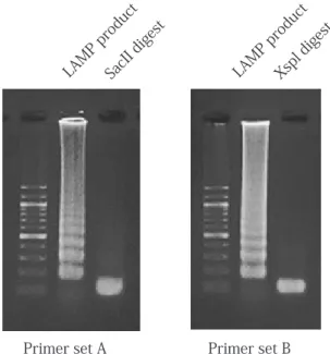
Fig. 1. Target sequence of 16S rDNA and primer locations. The nucleotide sequence of the 16S rDNA of R. mucilaginosa (accession NO. AP011540) was used to design the primers, and the target region is shown with the sequences of R. aeria (accession NO. AB071952) and R. dentocariosa (accession NO. M59055). A forward inner primer (FIP), a backward inner primer (BIP), and two outer primers (F3 and B3c) were used for a basic LAMP. Two sets of primers (A and B) were designed in this study, and their locations are indicated by lines or broken lines. The FIP consisted of a sequence of F1c and F2 (5'-F1c-F2-3'). The BIP consisted of a sequence of B1 and B2c (5'-B1-B2c-3'). The numbers on the right indicate the base position of the 16S rDNA. The restriction enzymes SacII and XspI were used for digestion of LAMP products with primer sets A and B, respectively. Their recognition sites are indicated by boxes.
Fig. 2. Electrophoresis pattern of LAMP products. The LAMP reactions were carried out with primer set A or B and extracted
R. mucilaginosa DNA template. The LAMP products and the digestion products with SacII and XspI were electrophoresed on agarose gels. Primer set A Primer set B LAMP product LAMP product SacII digest Xspl digest mucilaginosa:TGGGGAATATTGCACAATGGGCGCAAGCCTGATGCAGCGACGCCGCGTGAGGGATGACGG 415 aeria :TGGGGAATATTGCACAATGGGCGCAAG-CTGATGCAGCGACGCCGCGTGAGGGATGACGG dentocariosa:TGGGGAATATTGCACAATGGGCGCAAGCCTGATGCAGCGACGCCGCGTGAGGGATGACGG mucilaginosa:CCTTCGGGTTGTAAACCTCTGTTAGCAGGGAAGAAGAGAGATTGACGGTACCTGCAGAGA 475 aeria :CCTTCGGGTTGTAAACCTCTGTTAGCATCGAAGAAGCGAAAGTGACGGTAGGTGCAGAGA dentocariosa:CCTTCGGGTTGTAAACCTCTGTTAGCATCGAAGAAGCGAAAGTGACGGTAGGTGCAGAGA mucilaginosa:AAGCGCCGGCTAACTACGTGCCAGCAGCCGCGGTAATACGTAGGGCGCGAGCGTTGTCCG 535 aeria :AAGCGCCGGCTAACTACGTGCCAGCAGCCGCGGTAATACGTAGGGCGCGAGCGTTGTCCG dentocariosa:AAGCGCCGGCTAACTACGTGCCAGCAGCCGCGGTAATACGTAGGGCGCGAGCGTTGTCCG mucilaginosa:GAATTATTGGGCGTAAAGAGCTTGTAGGCGGTTTGTCGCGTCTGCTGTGAAAGGCCGGGG 595 aeria :GAATTATTGGGCGTAAAGAGCTTGTAGGCGGTTGGTCGCGTCTGCTGTGAAAGGCCGGGG dentocariosa:GAATTATTGGGCGTAAAGAGCTTGTAGGCGGTTGGTCGCGTCTGCTGTGAAAGGCTGGGG mucilaginosa:CTTAACTCCGTGTATTGCAGTGGGTACGGGCAGACTAGAGTGCAGTAGGGGAGACTGGAA 655 aeria :CTTAACTCC-GGTTTTGCAGTGGGTACGGGCTAACTAGAGTGCAGTAGGGGAGACTGGAA dentocariosa:CTTAAC-CCTGGTTTTGCAGTGGGTACGGGCTAACTAGAGTGCAGTAGGGGAGACTGGAA mucilaginosa:TTCCTGGTGTAGCGGTGGAATGCGCAGATATCAGGAGGAACACCGATGGCGAAGGCAGGT 715 aeria :TTCCTGGTGTAGCGGTGGAATGCACAGATATCAGGAGGAACACCGATGGCGAAGGCAGGT dentocariosa:TTCCTGGTGTAGCGGTGGAATGCGCAGATATCAGGAGGAACACCGATGGCGAAGGCAGGT mucilaginosa:CTCTGGGCTGTAACTGACGCTGAGAAGCGAAAGCATGGGGAGCGAACAGGATTAGATACC 775 aeria :CTCTGGGCTGTAACTGACGCTGAGAAGCGAAAGCATGGGGAGCGAACAGGATTAGATACC dentocariosa:CTCTGGGCTGTAACTGACGCTGAGAAGCGAAAGCATGGGGAGCGAACAGGATTAGATACC primer set B-B3 primer set B-B2 Primer set B-B1 Primer set B-F1 Primer set B-F2 Primer set B-F3 Primer set A-F3 Primer set A-F2
Primer set A-F1 Primer set A-B1
Primer set A-B2 Primer set A-B3
XspI
SacII
Co. Ltd., Tokyo, Japan). The 25-µl reaction mixtures contained 40 pmol each of FIP and BIP, 5 pmol each of F3 and B3, 20 pmol of LF and LB primers, 2 µl of template, 1 µl of Bst DNA polymerase (8 units), and 12.5 µl of reaction mix supplied with the kit. The reaction mixtures were incubated under isothermal conditions at 64°C for 20 or 50 minutes. After incubation, the reaction was terminated by heating at 80°C for 2 minutes.
5. Detection of LAMP products
The LAMP product was detected by agarose gel electrophoresis or naked eye inspection. For electrophoresis, aliquots of 2 µl of the reaction mixture were loaded onto 2% agarose gels, which were stained with ethidium bromide (50 µg/ml), and examined under UV illumination (302 nm). For naked eye detection, 1.0 µl of 10–1-diluted SYBR Green I (Takara Bio, Shiga, Japan) was added to the reaction mixture, and the color change was observed under natural light. 6. Conventional PCR method
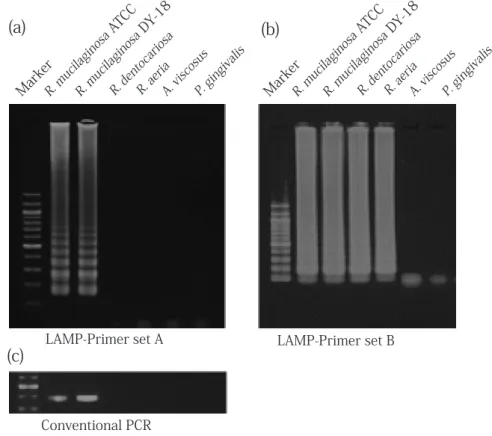
Conventional PCR16) for detection of R. mucilaginosa was performed simultaneously with the LAMP m e t h o d s . T h e s e q u e n c e s o f f o r w a r d a n d reverse PCR primers targeting the 16S rDNA w e r e 5 ' - G C C TAG C T T G C TAG G T G G AT- 3 ' a n d 5'-GCAGGTACCGTCAATCTCTC-3', respectively. The PCR products were detected by electrophoresis on 2% agarose gels stained with ethidium bromide. Results 1. Primer design and LAMP reaction Two LAMP primer sets (A and B) were designed in this study. The nucleotide sequences of the primers are shown in Table 2, and the locations of basic primers for 16S rDNA are shown in Figure 1 with Fig. 3. Specificity of LAMP. For the specificity test, DNA templates from 22 reference strains (103 cells) were used as templates, and representative results are shown. LAMP with primer set A specifically yielded an amplification product from R. mucilaginosa templates (ATCC 25296 and DY-18 strains). No amplicon was seen using templates from other reference strains tested (a). An amplicon was detected in LAMP with primer set B using templates from R. mucilaginosa (ATCC 25296 and DY-18), R. aeria JCM 11412, and R. dentocariosa ATCC 17931 (b). Conventional PCR was performed and amplification products were detected for two strains of R. mucilaginosa (c).
LAMP-Primer set A LAMP-Primer set B
Conventional PCR
(c)
(a)
(b)
Marker A. vis cosu s R. de ntoca riosa R. mu cilag inosa DY-18 R. mu cilag inosa ATCC P. gin givali s R. ae ria
Marker A. vis cosu s R. de ntoca riosa R. mu cilag inosa DY-18 R. mu cilag inosa ATCC P. gin givali s R. ae ria
JJ S E D P Vol. 10 No. 1: , 2018 the sequences of R. aeria and R. dentocariosa. As
compared to two other Rothia species, primer sets A and B contained five and three nucleotide sequences specific to R. mucilaginosa, respectively.
Successful amplification was seen in LAMP with primer sets A and B for the R. mucilaginosa template. A characteristic ladder-like pattern of the products was seen on the agarose gel (Fig. 2). The LAMP products were digested with SacII or XspI, which recognize internal sites of the target gene area. Consequently, the characteristic ladder-like bands of both LAMP products were concentrated and fragmented to approximately 100 bp in length (Fig. 2).
2. Specificity of LAMP
The specificity of the method was examined by 50-minute LAMP using extracted DNA templates prepared from 103 cells of each reference strain. Specific amplification was seen only in the LAMP reaction with primer set A. The amplicon was specifically detected in the reaction with template
from R. mucilaginosa in accordance with the results of conventional PCR. Unexpectedly, the LAMP reaction with primer set B showed nonspecific amplification for other Rothia species. Representative results of electrophoretic detection are shown in Figure 3, and the results are also shown in Table 1.
3. Detection limits of LAMP
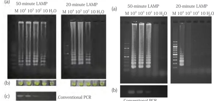
Detection limits was examined by both 20-minute and 50-minute LAMP. The LAMP products were detected by both agarose gel electrophoresis and naked eye inspection. DNA amplification was seen in 50-minute LAMP with template DNA equivalent to 10 cells. The detection limit of 20-minute LAMP was 102 cells/tube. Primer sets A and B showed the same sensitivity, and the representative results obtained using primer set A are shown in Figure 4. To examine the influence of DNA contamination on the detection limit, extracted DNAs from 102 cells of each of 20 bacterial species other than R. mucilaginosa were mixed with serially diluted R. mucilaginosa template, and LAMP was performed with primer set
Fig. 4. Detection limit of LAMP. Extracted DNAs were prepared from serial dilutions of R. mucilaginosa DY-18 cells and used as templates. Target DNA samples from 10 cells and 102 cells
showed detectable amplification on agarose gel by 50-minute LAMP and 20-minute LAMP, respectively (a). Naked eye inspection showed identical detection limits to electrophoretic detection (b). LAMP with primer sets A and B showed identical detection limits; representative results for primer set A are shown. Conventional PCR showed a detection limit identical to 50-minute LAMP (c).
Fig. 5. Influence of crude template on LAMP. Intact R. mucilaginosa cells suspended in PBS were used as the template and the detection limits for both 50-minute and 20-minute LAMP were examined (a). Although the amplicon was faint, target DNA was amplified detectably from 10 cells and 103 cells in 50-minute and 20-minute LAMP, respectively. Conventional PCR was performed simultaneously, and the results are shown in panel (b). Conventional PCR
(c)
(a)
(b)
Conventional PCR(a)
(b)
50-minute LAMP M 104 103 102 10 H 2O 50-minute LAMP M 104 103 102 10 H 2O 20-minute LAMP M 104 103 102 10 H 2O 20-minute LAMP M 104 103 102 10 H 2O 44 - 52A. Consequently, LAMP showed identical detection limits to the single template case (data not shown). The detection limits by naked eye inspection were equivalent to electrophoretic detection in both 20- and 50-minute LAMP. Conventional PCR showed the same detection limit as 50-minute LAMP (10 cells/ tube). 4. Intact cells as template To simplify the LAMP procedure, the DNA extraction step was omitted and intact cells were used as the template. The influence on the detection limit was examined using tenfold serial dilutions of R. mucilaginosa cells in 20-minute and 50-minute LAMP. The detection limit of 50-minute LAMP was 10 cells/ tube. A faint amplicon was seen in the reaction with 10 cells. The 20-minute LAMP showed a detection limit of 103 cells/tube, and a faint amplicon was seen in the reaction with 103 cells. The conventional PCR method showed identical sensitivity to 50-minute LAMP. LAMP with primer sets A and B showed the same detection limit, and representative results with primer set A are shown in Figure 5. Discussion Serious systemic infection caused by R. mucilaginosa has become a significant clinical concern in immunocompromised hosts1). In this study, two LAMP primer sets were designed for R. mucilaginosa. The 16S rDNA, a multicopy gene with species-specific sequences, was selected as the target. As the 16S rDNA nucleotide sequences were very similar among Rothia species, the primers were designed to include R. mucilaginosa-specific sequences. Primer sets A and B contained specific nucleotide sequences on 5/117 and 2/118 sites in the total sequences of four basic LAMP primers (FIP, BIP, F3, and B3), respectively. On the other hand, strain-specific sequences were found between R. mucilaginosa DY-18 and ATCC 25296, and therefore mixed primers were included in the primer sets. LAMP methods have been reported to be applicable to detect single nucleotide polymorphisms17,18). However, nonspecific
amplification was seen in LAMP with primer set B for R. aeria and R. dentocariosa. LAMP primers containing only a few specific sites may not consistently show high specificity. The amplicon of LAMP with primer set B was seen only in three Rothia species. Although they may be applicable as common primers for Rothia species, better primer design would be possible by searching for details of specific sites common among Rothia species. Primer set A showed higher specificity than primer set B. The LAMP amplicon was seen only for R. mucilaginosa but not for R. aeria, R. dentocariosa, or other reference strains. Primer set A contained more R. mucilaginosa-specific nucleotide sites than primer set B, which may have been responsible for the observed specificity. Selective medium19) and conventional PCR methods16) have been reported for detection of R. mucilaginosa. In this study, conventional RCR was performed simultaneously with LAMP. Although the detection limit of 20-minute LAMP was inferior to that of conventional PCR, 50-minute LAMP showed an equivalent detection limit to PCR in accordance with previous reports10,13). In addition, as naked eye inspection was feasible, LAMP may have advantages in both rapidity and simplicity compared to the PCR method. The direct use of intact cells as template decreased the detection limit of 20-minute LAMP by one digit. Although a faint amplicon was seen in the case of 10 cells, 50-minute LAMP showed the same detection limit for both extracted DNA and intact cells as the template. Conventional PCR using intact cells showed the equivalent detection limit to 50-minute LAMP. Considering the result of reducing the amount of amplicon, around 10% of bacterial cells were estimated to be damaged or lysed during manipulation and incubation at 64°C. Although the direct use of clinical specimens in LAMP would be difficult, LAMP is probably applicable as a method for identification of R. mucilaginosa from cultivated colonies without a DNA extraction step.
Culture- and PCR-based methods may be better in facilities with sophisticated laboratories.
JJ S E D P Vol. 10 No. 1: , 2018 On the other hand, the simplicity and rapidity of the LAMP assay would be advantageous, allowing examination without special equipment. Endodontic focal infection theory is a matter in controversy. However, many reports have suggested relations with systemic disease20-22). Oral mucositis and dental root canals are likely sources of R. mucilaginosa infection, and therefore bacterial examination of oral specimens would be required. The LAMP assay would be applicable for detection of R. mucilaginosa, and may be especially useful in facilities without sophisticated laboratories, such as dental offices. Conclusion Application of LAMP enabled the development of a rapid and simple detection assay for R. mucilaginosa. LAMP may provide an alternative detection assay for clinical specimens. Conflicts of interest The authors have no conflicts of interest to declare. Acknowledgments This study was supported in part by a Grant-in-Aid for Scientific Research (Grant number: 15K11404) from the Japan Society for the Promotion of Science. References 1) Abidi MZ, Ledeboer N, Banerjee A, Hari P: Morbidity and mortality attributable to Rothia bacteremia in neutropenic and nonneutropenic patients, Diagn Microbiol Infect Dis, 85: 116-120, 2016 2) Bruminhent J, Tokarczyk MJ, Jungkind D, Desimone JA Jr: Rothia mucilaginosa prosthetic device infections: a case of prosthetic valve endocarditis, J Clin Microbiol, 51: 1629– 1632, 2013 3) Lee AB, Harker-Murray P, Ferrieri P, Schleiss MR, Tolar J: Bacterial meningitis from Rothia mucilaginosa in patients with malignancy or undergoing hematopoietic stem cell transplantation, Pediatr Blood Cancer, 50: 673–676, 2008 4) Maraki S, Papadakis IS: Rothia mucilaginosa pneumonia: a
literature review, Infect Dis (Lond), 47: 125-129, 2015 5) Oie S, Mochizuki K, Ishida K, Nakayama A, Ohkusu K:
Case of late-onset bleb associated endophthalmitis caused by Rothia mucilaginosa, J Infect Chemother, 22: 645-647, 2016
6) Ramanan P, Barreto JN, Osmon DR, Tosh PK: Rothia bacteremia: a 10-year experience at Mayo Clinic, Rochester, Minnesota, J Clin Microbiol, 52: 3184-3189, 2014 7) Yamane K, Ogawa K, Yoshida M, Hayashi H, Nakamura T, Yamanaka T, Tamaki T, Hojoh H, Leung KP, Fukushima H: Identification and characterization of clinically isolated biofilm-forming Gram-positive rods from teeth associated with persistent apical periodontitis, J Endo, 35: 347-352, 2009
8) Notomi T, Okayama H, Masubuchi H, Yonekawa T, Watanabe K, Amino N, Hase T: Loop-mediated isothermal amplification of DNA, Nucleic Acids Res, 28: E63, 2000 9) Iwamoto T, Sonobe T, Hayashi K: Loop-mediated
isothermal amplification for direct detection of Mycobacterium tuberculosis complex, M. avium, and M. intracellulare in sputum samples, J Clin Microbiol, 41: 2616–2622, 2003
10) Maeda H, Kokeguchi S, Fujimoto C, Tanimoto I, Yoshizumi W, Nishimura F, Takashiba S: Detection of periodontal pathogen Porphyromonas gingivalis by loop-mediated isothermal amplification method, FEMS Immunol Med Microbiol, 43: 233–239, 2005 11) Miyagawa J, Maeda H, Murauchi T, Kokeguchi S, Yamabe K, Tanimoto I, Nishimura F, Fukui K, Takashiba S: Rapid and simple detection of eight major periodontal pathogens by the loop-mediated isothermal amplification method, FEMS Immunol Med Microbiol, 53: 314-321, 2008 12) Francois P, Tangomo M, Hibbs J, Bonetti EJ, Boehme CC, Notomi T, Perkins MD, Schrenzel J: Robustness of a loop-mediated isothermal amplification reaction for diagnostic applications, FEMS Immunol Med Microbiol, 62: 41-48, 2011 13) Koide Y, Maeda H, Yamabe K, Naruishi K, Yamamoto T, Kokeguchi S, Takashiba S: Rapid detection of mecA and spa by the loop-mediated isothermal amplification (LAMP) method, Lett Appl Microbiol, 50: 386-392, 2010 14) Maeda H, Fujimoto C, Haruki Y, Maeda T, Kokeguchi S, Petelin M, Arai H, Tanimoto I, Nishimura F, Takashiba S: Quantitative real-time PCR using TaqMan and SYBR Green for Actinobacillus actinomycetemcomitans, Porphyromonas gingivalis, Prevotella intermedia, tetQ gene and total bacteria, FEMS Immunol Med Microbiol, 39: 81-86, 2003
15) Nagamine K, Hase T, Notomi T: Accelerated reaction by loop-mediated isothermal amplification using loop primers, Mol Cell Probes, 16: 223-229, 2002
16) Tsuzukibashi O, Uchibori S, Shinozaki-Kuwahara N, Saito M, Kobayashi T, Fukumoto M: New primer design for identification of oral rothia, including R. aeria, using multiplex PCR, Int J Oral-Med Sci, 12: 85-89, 2013 17) Iwasaki M, Yonekawa T, Otsuka K, Suzuki W, Nagamine K, Hase T, Tatsumi K, Horigome T, Notomi T, Kanda H: Validation of the loop-mediated isothermal amplification method for single nucleotide polymorphism genotyping with whole blood, Genome Lett, 2: 119-126, 2003 18) Yongkiettrakul S, Kampeera J, Chareanchim W,
Rattanajak R, Pornthanakasem W, Kiatpathomchai W, Kongkasuriyachai D: Simple detection of single nucleotide polymorphism in Plasmodium falciparum by SNP-LAMP assay combined with lateral flow dipstick, Parasitol Int, 66: 964-971, 2017 19) Kobayashi T, Uchibori S, Tsuzukibashi O, Goto H, Aida M: A selective medium for Rothia mucilaginosa and its distribution in oral cavities, J Microbiol Methods, 91: 364-365, 2012
20) Blount CA, Leser C: Multisystem complications following endodontic therapy, J Oral Maxillofac Surg, 70: 527-530,
2012
21) Khalighinejad N, Aminoshariae MR, Aminoshariae A, Kulild JC, Mickel A, Fouad AF: Association between systemic diseases and apical periodontitis, J Endod, 42: 1427-1434, 2016
22) Virtanen E, Nurmi T, Söder P, Airila-Månsson S, Söder B, Meurman JH: Apical periodontitis associates with cardiovascular diseases: a cross-sectional study from Sweden, BMC Oral Health, 17: 107, 2017