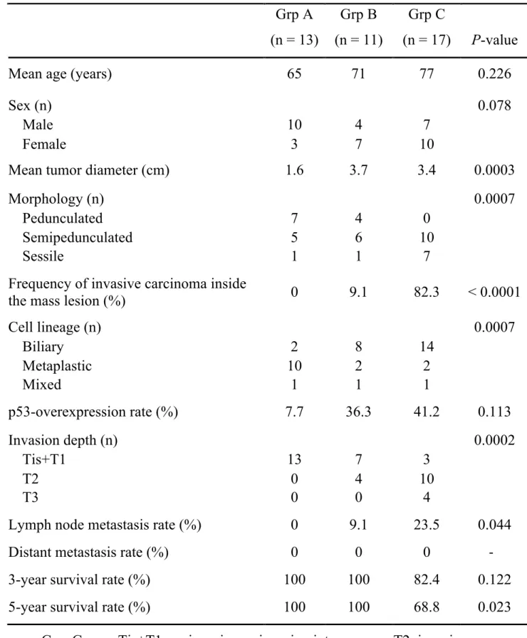
• 検索結果がありません。

Clinicopathological study of mass-forming gallbladder cancer focusing on the grade of cellular dysplasia Nobukazu SHIMA

N/A
N/A
Protected

Academic year: 2021

シェア "Clinicopathological study of mass-forming gallbladder cancer focusing on the grade of cellular dysplasia Nobukazu SHIMA"

Copied!
22
0
0

読み込み中.... (全文を見る)

全文

(1)

1

Clinicopathological study of mass-forming gallbladder cancer focusing on the grade of cellular dysplasia

Nobukazu SHIMA

1)

, Nobuyuki OHIKE

2)

, Reika SUZUKI

2)

, Tomoko NOROSE

2)

, Tomohide ISOBE

2)

, Akira SHIOKAWA

2)

, Nobuyuki TAKEYAMA

3)

, Junichi TANAKA

4)

, Masahiko MURAKAMI

5)

, Masafumi TAKIMOTO

1)

1)

Department of Pathology, Showa University School of Medicine, 1-5-8, Hatanodai, Shinagawa-Ku, 142-8555, Tokyo, Japan.

2)

Department of Pathology, Showa University Fujigaoka Hospital

3)

Department of Radiology, Showa University Fujigaoka Hospital

4)

Department of Gastroenterological and General Surgery, Showa University Fujigaoka Hospital

5)

Department of Surgery, Division of General and Gastroenterological Surgery, Showa University School of Medicine.

Corresponding Author: Nobukazu Shima

Affiliation: Department of Pathology Clinico-diagnostic Pathology,

Showa University School of Medicine

(2)

2

Address: 1-5-8, Hatanodai, Shinagawa-Ku, 142-8555, Tokyo, Japan Tel.: 03-3784-8119

Fax: 03-3784-2959

E-mail: [email protected]

Running title: Clinicopathological study of gallbladder cancer

(3)

3

Abstract: The relationship between the clinicopathological features and the grade of cellular dysplasia of the neoplastic glands in mass-forming gallbladder cancer was investigated. In this retrospective study, 41 mass-forming (≧1 cm) gallbladder cancer specimens from 83 resected cases were examined. Tumors were classified into three groups: Group A had intraluminal masses consisting of neoplastic glands with only low-grade dysplasia; Group B had mixed low- and high-grade dysplasia, and Group C had only high-grade dysplasia. Of the 41 tumors, 13 were classified as Group A, 11 as Group B, and 17 as Group C. For Group A, B, and C, respectively, the mean tumor diameter was 1.6, 3.7 and 3.4 cm;

macroscopic type (pedunculated/semi-pedunculated/sessile) was 7/5/1, 4/6/1 and 0/10/7; frequency of an invasive component inside the mass was 0%, 9% and 82%; and cell lineage (biliary/metaplastic/mixed) was 2/1/10, 8/1/2 and 14/1/2. In addition, invasion depth (Tis+T1/T2/T3) was 13/0/0, 7/4/0 and 3/10/4; lymph node metastases were present in 0%, 9% and 24%

of patients; 3-year survival rate was 100%, 100% and 82%; and 5-year survival rate was 100%, 100% and 69%, for A, B and C, respectively.

Significant intergroup differences were seen for positive lymph node

metastasis rate and 5-year survival rate. The present study indicates that the

clinicopathological features of mass-forming gallbladder cancer are

(4)

4

different depending on the grade of cellular dysplasia of the mass lesion.

The tumors in Groups A and B were of lower malignancy than those in Group C and the prognosis of patients in the former groups was excellent.

Group A and B tumors may be intracholecystic papillary-tubular neoplasms, a recently proposed new disease concept.

Key words: mass-forming gallbladder cancer, cellular dysplasia,

clinicopathological study, intracholecystic papillary-tubular neoplasm

(ICPN)

(5)

5 Introduction

Opportunities for the detection of elevated lesions in the lumen of the

gallbladder are increasing with the widespread use of, and advances in,

clinical imaging. These elevated lesions form a diverse group that includes

not only cholesterol polyps, hyperplastic polyps, and adenomyomatosis, but

also adenomas and adenocarcinomas. Adenocarcinomas are further

subdivided into low-grade tumors, which may be confused with adenomas

and hyperplastic polyps, and obviously high-grade tumors

1,2,3)

. The present

study focused on the characteristics of the neoplastic cells in intraluminal

elevated lesions in patients diagnosed with mass-forming gallbladder

cancer, as well as the influence of the cellular dysplastic grade on the

clinicopathological features, including staging and prognosis.

(6)

6 Subjects and Methods

From 83 patients who underwent surgical resection at our institutions (Showa University Hospital and Showa University Fujigaoka Hospital) and were histologically diagnosed with primary adenocarcinoma of the gallbladder between 1998 and 2015, 46 patients with macro-sized (approximately ≧ 1 cm) polypoid or papillary mass lesions in the gallbladder lumen were initially selected. This retrospective study was approved by the institutional review board and was conducted in accordance with the Health Insurance Portability Accountability Act. The institutional review board waived the requirement for informed patient consent.

Clinicopathological study

Tumors were classified into the following three groups, based on the

dysplastic grade of the tumor cells that composed the elevated lesions

protruding into the lumen of the gallbladder, and their clinicopathological

features were compared. Group A tumors were intraluminal masses

consisting solely of low-grade dysplastic glands (Fig. 1a), Group B were

intraluminal masses with a mixture of low-grade and high-grade dysplastic

glands (arbitrarily with each component accounting for at least 25% of the

(7)

7

whole tumor), and Group C were intraluminal masses consisting extensively of high-grade dysplastic glands (usually showing complex papillo-tubular structures and cribriform structures; Fig. 1b). Low-grade dysplastic glands were characterized by the regular arrangement of uniform and relatively simple glands with mild nuclear enlargement, while high-grade dysplastic glands were characterized by the irregular arrangement of complex glands with prominent nuclear enlargement and more significant hyperchromasia. The mean age, sex ratio, macroscopic type, mean tumor diameter, presence of an invasive carcinoma component inside the mass lesion, presence of tumors exhibiting p53-overexpression, invasion depth, rate of lymph node metastasis, rate of distant metastasis, and postoperative prognosis were investigated to make comparisons among these three groups. The duration of postoperative follow-up ranged from 45 to 4137 days, and 5 patients who died of other causes (such as myocardial infarction and pneumonia) were excluded from the analysis of prognosis.

Statistical analysis was carried out using the Kruskal-Wallis test, with the log-rank test for Kaplan-Meier curves; P < 0.05 was regarded as significant.

Investigation of cell lineage

(8)

8

The cell lineage of the neoplastic glands composing the mass lesion was investigated histomorphologically and immunohistochemically for all cases.

(a) Histomorphological observation:

Tumor cells were categorized as biliary type, intestinal (INT) type, or

gastric (GAS) type through observation of hematoxylin and eosin-stained

specimens under light microscopy, and each tumor was classified as either

biliary, metaplastic (GAS or INT), or mixed (biliary + GAS and/or

INT)

1,2,4)

. Briefly, the histological characteristics of these different cell

lineages were as follows: biliary type was characterized by cuboidal or

low-columnar epithelial cells with oval nuclei and scant mucinous

cytoplasm resembling proper biliary tract epithelium. INT type consisted of

tall columnar epithelium with spindle-shaped, pseudostratified nuclei and

strongly-stained cytoplasm resembling colorectal adenoma. The presence

of goblet cells and Paneth cells was also considered indicative of INT

lineage. GAS type consisted of tall columnar epithelium with mucinous

cytoplasm resembling gastric foveolar epithelium or small tubular units

similar to gastric pyloric glands. If more than one of these characteristics

were present simultaneously, the most prominent cell type was used, taking

into account the predominant features and degree of cellular dysplasia.

(9)

9 (b) Immunohistochemical analysis:

For each patient, thin sections (3 µm) were prepared for immunostaining using one or two formalin-fixed, paraffin-embedded tissue sections including the maximum area of the tumor. Staining was performed using the avidin-biotin complex detection method with a BenchMark automated immunostainer (Ventana Medical Systems, Inc., Tucson, AZ, USA). The primary antibodies used were MUC1 (Ma695, Leica Biosystems Newcastle Ltd, Newcastle, UK; 100-fold dilution), MUC2 (Ccp58, Leica; 200-fold dilution), MUC5AC (CLH2, Leica; 200-fold dilution), MUC6 (CLH5, Leica; 50-fold dilution), and CDX-2 (AMT28, Leica; 50-fold dilution).

MUC1 was used as a marker for biliary type, MUC2 and CDX-2 for INT type, and MUC5AC and MUC6 for GAS type

1,2,4-6)

. The proportion of cells staining positive for each marker as a percentage of the total number of neoplastic cells was calculated. Immunostaining for p53 (DO-7, Leica;

800-fold dilution) was also carried out following the same staining

procedure

1,2,3)

.

(10)

10 Results

Of the 41 patients, 13 were classified with tumors belonging to Group A, 11 to Group B, and 17 to Group C.

Clinicopathological study (Table 1)

The mean age was 65 years in Group A, 71 years in Group B, and 77

years in Group C, and the male:female ratio was 10:3 in Group A, 4:7 in

Group B, and 7:10 in Group C. Groups B and C tended to be older and to

include more women than Group A, but these differences were not

significant. The mean tumor diameter was 1.6 cm in Group A, but 3.7 cm

in Group B and 3.4 cm in Group C, with the latter groups being

significantly larger than Group A. When the tumors were classified as

pedunculated, semipedunculated, or sessile on the basis of their

macroscopic morphology, Group A included 7 pedunculated, 5

semipedunculated, and 1 sessile tumor, Group B included 4 pedunculated,

6 semipedunculated, and 1 sessile tumor, and Group C included only 10

semipedunculated, and 7 sessile tumors, with Group C containing

significantly more sessile tumors than the other groups. The frequency of

the presence of invasive carcinoma inside the mass was 0% in Group A and

9% in Group B, while it was significantly higher, at 82%, in Group C.

(11)

11

Invasion depth was classified as no invasion (Tis), invasion into the mucosa (T1), invasion into the muscularis propria (T2), or invasion into the subserosa (T3), and expressed as Tis+T1/T2/T3. The number of tumors of each invasion depth was 13/0/0 in Group A, 7/4/0 in Group B, and 3/10/4 in Group C, and these differences were significant. The rate of lymph node metastasis was 0% in Group A, 9.1% in Group B, and 23.5% in Group C, and these differences were also significant. The 3-year survival rate was 100% in Groups A and B, but only 82.4% in Group C. The 5-year survival was 100% in Groups A and B, but significantly lower in Group C at 68.8%.

The significance of this difference was also confirmed by the log-rank test (P = 0.02).

Investigation of cell lineage and immunohistochemical studies

When cell lineages were histomorphologically classified as biliary,

metaplastic, or mixed, 2 of the tumors in Group A were classified as biliary,

1 as metaplastic, and 10 as mixed; in Group B, 8 were classified as biliary,

1 as metaplastic, and 2 as mixed; and in Group C, 14 were classified as

biliary, 1 as metaplastic, and 2 as mixed. Group A contained significantly

more mixed-type tumors, while Groups B and C contained significantly

more biliary-type tumors.

(12)

12

In Group A, 33.6% of tumors were immunohistochemically positive for MUC1, 0.8% for MUC2, 26% for MUC5AC, 73.8% for MUC6, and 32.5%

for CDX2. Similarly, the percentages in Group B were 20%, 7%, 23%,

48%, and 36%, and those in Group C were 37%, 2.6%, 15.7%, 27%, and

29.8%, respectively. Group A contained a conspicuously high proportion of

MUC6-positive tumor cells. The frequency of tumors exhibiting

p53-overexpression was 7.7% in Group A, but significantly higher at

36.3% in Group B and 41.2% in Group C.

(13)

13 Discussion

This study showed that the clinicopathological features of mass-forming gallbladder cancer were different depending on the grade of cellular dysplasia of the neoplastic glands within the intraluminal elevated lesions.

In Group A, with mass lesions consisting of low-grade dysplastic glands,

there was a high proportion of men, more lesions were pedunculated

masses of mean diameter <2 cm, they did not contain an invasive

carcinoma component, the rate of p53-overexpression was low, tumors

invaded no deeper than the mucosa, there were no lymph node metastases,

and the 5-year survival rate was 100%. The cell type was most commonly

mixed, consisting of biliary+GAS, and a high proportion stained positive

for MUC6. In contrast, in Group C with mass lesions consisting of

high-grade dysplastic glands, there was a high proportion of women, more

tumors were semipedunculated or sessile masses with mean diameters

greater than 3 cm, most elevated lesions already contained an invasive

carcinoma component, the p53-overexpression rate exceeded 40%, many of

the lesions had invaded the muscularis or deeper, lymph node metastases

were present in almost a quarter of patients, and the 5-year survival rate

was 68%. The cell type was most commonly biliary. Group B was

intermediate between Groups A and C in most respects, but the 5-year

(14)

14

survival rate was 100%, the same positive outcome as in Group A. Group C had tumors with a deeper invasion and poorer prognosis than Group A or B.

The difference between these groups may be due to the difference in stage.

In the future, legitimate evaluation of these tumors should be performed to accumulate a larger number of cases for analysis.

Histological classification focusing on the grade of cellular dysplasia has

not been well established for gallbladder carcinoma. Recently, however, a

new disease concept of intracholecystic papillary-tubular neoplasm (ICPN)

has been proposed

1,2,4,7-12)

. ICPNs are defined as intraluminal mass-forming

preinvasive neoplasms occurring within the gallbladder which are highly

analogous to pancreatic or biliary intraductal papillary and tubular

neoplasms, as evidenced by their papillary and/or tubular growth, variable

cell lineage, and spectrum of dysplastic change (adenoma-carcinoma

sequence)

9,13)

. ICPNs are biologically indolent; noninvasive examples show

an excellent prognosis, whereas those with invasion exhibit a malignant,

but nevertheless significantly better prognosis, compared to ordinary

invasive carcinomas unaccompanied by ICPN. The majority of tumors in

Groups A and B in the present study would appear to be classified as

ICPNs, while the majority of tumors in Group C would be excluded from

ICPN and should be classified as ordinary high-grade “invasive papillary

(15)

15

adenocarcinoma” or “tubular adenocarcinoma” because many of them contained an invasive carcinoma component within the intraluminal mass lesion.

The present results may be useful for several issues in the diagnosis and treatment of mass-forming gallbladder cancer. For instance, low-grade adenocarcinoma, as shown in tumors in Group A, might be regarded as hyperplasia or adenoma depending on the pathologist

13)

. However, the present study shows that a different diagnosis would not be a crucial problem because such tumors usually show an almost “benign” course.

Mass-forming gallbladder cancer may be associated with intraepithelial

spread along a Rokitansky-Aschoff sinus (RAS), which often is confused

with non-neoplastic RAS or invasive tubular adenocarcinoma. The present

study indicates that in tumors of Group A which are rarely associated with

an invasive component, it is appropriate to firstly consider non-neoplastic

RAS or intraepithelial spread, even if glandular ducts are present in the

deep gallbladder wall. Conversely, in tumors in Group C, the possibility of

invasive tubular adenocarcinoma should be aggressively pursued. Finally,

as for treatment strategies for mass-forming gallbladder cancer, particularly

surgical procedures, simple cholecystectomy seems to be sufficient for

tumors in Group A. In Groups B or C with lymph node metastasis, however,

(16)

16

a surgical procedure including lymph node dissection must be considered.

Therefore, if preoperative classification of the tumor into one of the Groups A–C is feasible, it would be a highly worthwhile process.

Conclusion

The present study indicated that the clinicopathological features of mass-forming gallbladder cancer were different depending on the cellular dysplastic grade of the mass lesion. Tumors with an intraluminal mass consisting of low-grade dysplastic glands were of lower malignancy and the prognosis of patients with such tumors was excellent. These results may support the significance of the disease concept of ICPN.

Conflict of interest statement

None of the authors have any conflicts of interest or financial ties to disclose.

Acknowledgments

We would like to thank Ms. Tomoko Nagai for technical assistance with

the immunohistochemical studies.

(17)

17 References

1) Albores-Saavedra J, Chablé-Montero F, González-Romo MA, et al.

Adenomas of the gallbladder. Morphologic features, expression of gastric and intestinal mucins, and incidence of high-grade dysplasia/carcinoma in situ and invasive carcinoma. Hum Pathol.

2012;43:1506-1513.

2) Adsay V, Jang KT, Roa JC, et al. Intracholecystic papillary-tubular neoplasms (ICPN) of the gallbladder (neoplastic polyps, adenomas, and papillary neoplasms that are ≥1.0 cm): clinicopathologic and immunohistochemical analysis of 123 cases. Am J Surg Pathol.

2012;36:1279-1301.

3) You Y, Bui K, Bui M, et al. Histopathological and immunophenotypical features of intestinal-type adenocarcinoma of the gallbladder and its precursors. Cancer Control. 2014;21:247-250.

4) Bennett S, Marginean EC, Paquin-Gobeil M, et al. Clinical and pathological features of intraductal papillary neoplasm of the biliary tract and gallbladder. HPB (Oxford). 2015;17:811-818.

5) Sasaki M, Yamato T, Nakanuma Y, et al. Expression of MUC2,

MUC5AC and MUC6 apomucins in carcinoma, dysplasia and

non-dysplastic epithelia of the gallbladder. Pathol Int. 1999;49:38-44.

(18)

18

6) Sato R, Ando T, Tateno H, et al. Intracystic papillary neoplasm with an associated mucinous adenocarcinoma arising in Rokitansky-Aschoff sinus of the gallbladder. Surg Case Rep.

2016;2:62.

7) Washington K, Gottfried MR. Expression of p53 in adenocarcinoma of the gallbladder and bile ducts. Liver. 1996;16:99-104.

8) Sai JK, Suyama M, Nobukawa B, et al. Severe dysplasia of the gallbladder associated with occult pancreatobiliary reflux. J Gastroenterol. 2005;40:756-760.

9) Hughes NR, Bhathal PS. Adenocarcinoma of gallbladder: an immunohistochemical profile and comparison with cholangiocarcinoma. J Clin Pathol. 2013;66:212-217.

10) Argon A, Barbet FY, Nart D. The relationship between intracholecystic papillary-tubular neoplasms and invasive carcinoma of the gallbladder. Int J Surg Pathol. 2016;24:504-511.

11) Hashimoto S, Horaguchi J, Fujita N, et al. Intracholecystic papillary-tubular neoplasm of the gallbladder presenting with jaundice.

Intern Med. 2014;53:2313-2317.

12) Isozaki M, Ohike N, Tajiri T, et al. Clinicopathological study of

intracholecystic papillary-tubular neoplasms (ICPNs) of the

(19)

19

gallbladder. Showa Univ J Med Sci. 2014;26:17-26.

13) Kai K. Organ-specific concept and controversy for premalignant

lesions and carcinogenesis of gallbladder cancer. Hepatobiliary Surg

Nutr. 2016;5:85-87.

(20)

20

Table 1. Clinicopathological features of mass-forming gallbladder cancer

Grp, Group; Tis+T1, no invasion or invasion into mucosa; T2, invasion into muscularis propria; T3, invasion into subserosa.

Grp A (n = 13)

Grp B (n = 11)

Grp C

(n = 17) P-value

Mean age (years) 65 71 77 0.226

Sex (n) 0.078

Male 10 4 7

Female 3 7 10

Mean tumor diameter (cm) 1.6 3.7 3.4 0.0003

Morphology (n) 0.0007

Pedunculated 7 4 0

Semipedunculated 5 6 10

Sessile 1 1 7

Frequency of invasive carcinoma inside

the mass lesion (%) 0 9.1 82.3 < 0.0001

Cell lineage (n) 0.0007

Biliary 2 8 14

Metaplastic 10 2 2

Mixed 1 1 1

p53-overexpression rate (%) 7.7 36.3 41.2 0.113

Invasion depth (n) 0.0002

Tis+T1 13 7 3

T2 0 4 10

T3 0 0 4

Lymph node metastasis rate (%) 0 9.1 23.5 0.044

Distant metastasis rate (%) 0 0 0 -

3-year survival rate (%) 100 100 82.4 0.122

5-year survival rate (%) 100 100 68.8 0.023

(21)

21 Figure legends

Figure 1. Microscopic features of mass-forming gallbladder cancer. (a-1) Intraluminal mass consists extensively of low-grade dysplastic glands.

Hematoxylin and eosin (H&E) stain, x50 magnification. (a-2) Intraluminal

mass consists extensively of low-grade dysplastic glands. H&E stain, x200

magnification. (b-1) Intraluminal mass consists extensively of high-grade

dysplastic glands. H&E stain, x50 magnification. (b-2) Intraluminal mass

consists extensively of high-grade dysplastic glands. H&E stain, x200

magnification.

(22)

a-1

a-2

b-1

b-2

Table 1. Clinicopathological features of mass-forming gallbladder cancer

参照

関連したドキュメント

doi: 10.4021/gr336e.. We present the HES case with hepatic involvement. The patient is 70-year-old male. He complained fever and back pain. Blood examination showed marked

まず,PREG 及び PROG の重水素標識体をアルカリ条 件下での交換反応により合成し,それぞれを IS として Fig.. 7) .コント

熱力学計算によれば、この地下水中において安定なのは FeSe 2 (cr)で、Se 濃度はこの固相の 溶解度である 10 -9 ~10 -8 mol dm

As far as local conditions at infinity are concerned, it is shown that at energy zero the Dirac equation without mass term has no non-trivial L 2 -solutions at infinity for

At Geneva, he protested that those who had criticized the theory of collectives for excluding some sequences were now criticizing it because it did not exclude enough sequences

We study the basic preferential attachment process, which generates a sequence of random trees, each obtained from the previous one by introducing a new vertex and joining it to

In this article we study a free boundary problem modeling the tumor growth with drug application, the mathematical model which neglect the drug application was proposed by A..

Answering a question of de la Harpe and Bridson in the Kourovka Notebook, we build the explicit embeddings of the additive group of rational numbers Q in a finitely generated group