Introduction
There have been no published records of adult digeneans (Trematoda) from freshwater fishes of Wakayama Prefecture (Kinki), and Tokushima and Kochi Prefectures (Shikoku), Japan (Shi- mazu, 1999, 2003b). Freshwater fishes collected in several rivers in these prefectures were exam- ined for adult digeneans from 1998 to 2000. This paper reports the results of these examinations and discusses the final fish hosts, geographical distributions, and life cycles, where known, for each of the digeneans, with respect to Japan.
Materials and Methods
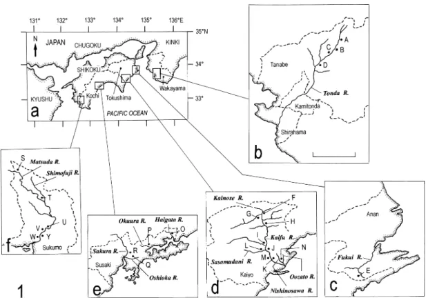
Freshwater fishes were collected in rivers in Wakayama Prefecture (Kinki), and Tokushima and Kochi Prefectures (Shikoku), Japan, for ex- amination for adult digeneans. Figure 1 and
Table 1 show the prefectures, rivers, sampling sites, and dates.
Unidentified fishes of the genus Rhinogobius were classified into five types, CB, CO, DA, LD, and OR, according to Kawanabe and Mizuno (1989).
Fish specimens were examined fresh for adult digeneans. Digenean worms found were treated as follows: either (1) flattened under pressure of cover slip, fixed with AFA, and stained with Hei- denhain’s iron hematoxylin; or (2) flattened, fixed with 70% ethanol, and stained with Grenacher’s alum carmine; or (3) fixed in hot 10% neutralized formalin and stained with acetocarmine. These stained specimens were mounted in Canada bal- sam.
Some related institutional specimens were bor- rowed from Meguro Parasitological Museum, Tokyo (MPM); the National Museum of Nature and Science, Tokyo (formerly the National Sci- ence Museum, Tokyo, NSMT); and Dr. Misako Urabe of the University of Shiga Prefecture, Hikone, Shiga Prefecture. Drawings were made with the aid of a drawing tube. Measurements
Digeneans (Trematoda) Found in Freshwater Fishes of Wakayama, Tokushima, and Kochi Prefectures, Japan
Takeshi Shimazu*
Nagano Prefectural College, 8–49–7 Miwa, Nagano, 380–8525 Japan
Abstract Freshwater fishes (25 identified and 5 unidentified species) were caught in rivers in Wakayama Prefecture (Kinki), and Tokushima and Kochi Prefectures (Shikoku), Japan, in 1998, 1999, and 2000 and examined fresh for adult digeneans (Trematoda). Digeneans of nine species were found, mainly in the digestive tract. Eight of them were identified as previously known species, and one remained unidentified. A single specimen of the latter, Allocreadiumsp., was found in Zacco temminckii(Temminck and Schlegel) (Cyprinidae) (Japanese name kawamutsu) from the Kaifu River in Kaiyo, Tokushima Prefecture. It probably represents an undescribed species. The digeneans are described and figured. Their final fish hosts, geographical distributions, and life cycles, where known in Japan, are discussed.
Key words : Trematoda, digeneans, freshwater fishes, Wakayama Prefecture, Tokushima Prefec- ture, Kochi Prefecture, Japan.
* Present address: 10486-2 Hotaka-Ariake, Azumino, Nagano, 399–8301 Japan
E-mail: [email protected]
Fig. 1. Maps showing the prefectures, rivers, and sampling sites. Scale bar: 10 km.
Table 1. The prefectures, rivers, sampling sites, and dates. The former three correspond to those in Fig. 1.
Prefecture
Sampling site Date
River
Wakawama Prefectue, Kinki (Fig. 1a, b)
Tonda River Nakahechi, Tanabe: A, Hyozei; B, Fukusada; 2–4 August 1999 C, Ookawa; D, Kurisugawa
Tokushima Prefecture, Shikoku (Fig. 1a, c, d)
Fukui River (a tributary) Fukui, Anan: E, Kono 13 September 1998
Kainose River Ogawa, Kaiyo: F, Kainose 12 September 1998
Kaifu River Ogawa, Kaiyo: G, Uke; H, Higashikuwabara 11–16 September 1998
Aikawa, Kaiyo: I, Nakano 11–16 September 1998
Kaiyo: J, Ooi; K, Yoshino 11–16 September 1998
Kaifu River (a small tributary) Kaiyo: M, Yoshida 12 September 1998
Sasamudani River Aikawa, Kaiyo: L, Sasamudai 12 September 1998
Nishinosawa River Kaiyo: N, Shihohara 13 and 15 September 1998
Oozato River Kaiyo: N, Shihohara 12 September 1998
Kochi Prefecture, Shikoku (Fig. 1a, e, f)
Haigata River Susaki: O, Uranouchihaigata 30 July 2000
Okuura River Susaki: P, Uranouchihigashibun 30 July 2000
Oshioka River Susaki: Q, Oshioka 30 July 2000
Sakura River Susaki: R, Koda 29 July 2000
Matsuda River Hashikami, Sukumo: S, Idei 5–7 August 2000
Sukumo: U, Nakatsuno; V, Ninomiya; W, Chuo; Y, Wada 5–7 August 2000
Shimofuji River Hashikami, Sukumo: T, Sakamoto 5 August 2000
(length by width) are given in millimeters unless otherwise stated. The digenean specimens found have been deposited in the National Museum of Nature and Science, Tokyo, under the name ab- breviation NSMT-Pl.
Results
Table 2 summarizes the freshwater fishes ex- amined and the adult digeneans found in the pres- ent examinations. The following are the digenean species found.
Class Trematoda Subclass Digenea Family Derogenidae Genarchopsis goppoOzaki, 1925
(Fig. 2)
Genarchopsis goppo Ozaki, 1925: 101–103, figs. 1–3;
Takahashi, 1929: 1928–1929, pl. 3, fig. 12; Yamaguti, 1934: 500–501, fig. 128; Yamaguti, 1938: 133, in part;
Yamaguti, 1942: 388–389; Urabe, 2001: 1407, fig. 3E;
Shimazu and Urabe, 2005: 2–3, figs. 1–3.
Progonus goppo: Srivastava, 1933: 55.
Genarchopsis anguillaeYamaguti, 1938: 132–133, fig. 81;
Yamaguti, 1942: 388; Shimazu, 1995: 11, fig. 6.
Genarchopsis gigi Yamaguti, 1939: 227, pl. 29, fig. 6;
Shimazu, 1995: 9, fig. 5.
Genarches anguillae: Skryabin and Gushanskaya, 1955:
680, fig. 199.
Genarches gigi: Skryabin and Gushanskaya, 1955: 680, 685, fig. 200.
Genarches goppo: Skryabin and Gushanskaya, 1955:
685–686, 689, fig. 201.
Genarchapsis[sic] goppo: Shimazu, 1995: 6–9, figs. 1–5.
Specimens studied. One mature and 1 imma- ture and 16 mature specimens (NSMT-Pl 5515 and 5516) found in the stomach of Rhinogobius giurinus(Rutter) (Gobiidae) from the Kaifu (sam- pling site, K) and Oshioka (Q) rivers, respectively;
1 and 7 mature specimens (NSMT-Pl 5517 and 5518–5520) found in the stomach of Odontobutis obscura (Temminck and Schlegel) (Odontobuti- dae) from the Fukui (E) and Matsuda (U, V, Y) rivers, respectively; 23 and 15 mature specimens (NSMT-Pl 5521 and 5522) found in the stomach of Gymnogobius petschiliensis (Rendahl) (Gobi-
idae) from the Okuura (P) and Oshioka (Q) rivers, respectively; 1 mature specimen (NSMT- Pl 5531) found in the stomach of Rhinogobius flumineus(Mizuno) from the Sakura River (R); 2 immature and 4 mature specimens (NSMT-Pl 5523) found in the stomach of Rhinogobius sp.
CB (Gobiidae) from the Oshioka River (Q); and 6 mature specimens (NSMT-Pl 5524) found in the stomach of Tridentiger brevispinis Katsuya- ma, Arai, and Nakamura (Gobiidae) from the Oshioka River (Q).
Description. Ten gravid specimens with nu- merous uterine eggs measured. Body 1.36–2.64 by 0.51–0.77; forebody 0.72–1.44 long, occupy- ing 50–56% of total body length. Oral sucker 0.12–0.21 by 0.15–0.24. Pharynx 0.05–0.09 in diameter. Esophagus short, with small esophageal pouch. Intestines fusing to form cyclocoel anteri- or to vitellaria. Ventral sucker large, about equa- torial, 0.28–0.43 by 0.28–0.44; sucker width ratio 1 : 1.60–2.00. Testes 0.16–0.22 by 0.17–0.22.
Ovary immediately anterior to vitellaria, smaller than testes, 0.12–0.15 by 0.09–0.16. Uterus much folded in forebody and hindbody. Eggs numer- ous, elongate-oblong, slightly reniform, fully em- bryonated when laid, 48–70 by 24–35mm, with a long anopercular filament. Vitellaria consisting of 2 elliptical symmetrical or diagonal masses measuring 0.12–0.16 by 0.09–0.12, near posteri- or end of body.
The uterus was more folded, especially in the forebody, and uterine eggs were more numerous in larger specimens.
Discussion. These specimens agree in mor- phology and measurements with Genarchopsis goppo as redescribed by Shimazu (1995). Their eggs (48–70 by 24–35mm) are larger than those (46–50 by 25–26mm) in Ozaki’s (1925) original description and those (40–45 by 18–19mm (col- lapsed)) in one specimen (MPM Coll. No.
30028) in Ozaki’s Collection (Shimazu, 1995).
However, eggs are 51–84 by 21–42mm (com- bined) in other previous descriptions for G.
goppo from Japan (Yamaguti, 1934, 1942; Shi- mazu, 1995; Urabe, 2001; Shimazu and Urabe, 2005).
Table2.Fishes examined and digeneans found. Fish examinedDigenean found Number of Body size2) Species name (Japanese name)River (sampling site)1)fish of fish Species name (sampling site3))Prevalence5) examinedexamined Adrianichthyidae Orygias latipes (medaka)Oozato R. (N)1130–4) Amblycipitidae Liobagrus reinii(akaza)Kaifu R. (H, I, K)2465–90– Anguillidae Anguilla japonica(unagi)Kaifu R. (J)3380–410Phyllodistomum anguilae(J)1/3 Sakura R. (R)1210– Cobitidae Cobitis biwae(shimadojo)Tonda R. (D)1105– Kaifu R. (G, H, I, K)2870–120– Okuura R. (P)1240–90– Misgurnus anguillicaudatus(dojo)Okuura R. (P)455–75– Cottidae Cottus kazika(ayukake)Kaifu R. (K)2150–185– Haigata R. (O)1160– Cyprinidae Carassius auratus buergeriOshioka R. (Q)1155– (ookimbuna)Sakura R. (R)1735–133– Ca. a. langsdorfii(gimbuna)Kaifu R. (I)660–120– Matsuda R. (U)590–100– Cyprinus carpio carpio(koi)Matsuda R. (V)2110–200– Moroco jouyi(takahaya)Kaifu R. (H, I)2865–85– Sasamudani R. (L)190– Sakura R. (R)1436–90– Pseudogobio esocinus(kamatsuka)Kaifu R. (K)595–185– Tribolodon hakonensis(ugui)Tonda R. (B, D)3150–195Asymphylodora macrostoma(D)1/3 Kaifu R. (G, H, I, K)3575–225– Sasamudani R. (L)595–120– Sakura R. (R)3125–250– Matsuda R. (S, U)935–175As. macrostoma(S)2/9 Zacco platypus(oikawa)Tonda R. (C)2105–110Neoplagioporus zacconis(C)2/2 Kaifu R. (G, H, I, K)4580–135N. zacconis(H, I, K)10/45 Oshioka R. (Q)390–100– Sakura R. (R)970–115–
Table2.Continued Fish examinedDigenean found Number of Body size2) Species name (Japanese name)River (sampling site)1) fish of fish Species name (sampling site3) )Prevalence5) examinedexamined Zacco platypus(oikawa)Matsuda R. (S, U, V)1280–110N. zacconis(V)1/12 Z. temminckii(kawamutsu)Tonda R. (B, C, D)1780–145N. zacconis(B, C, D)7/17 Kaifu R. (G, H, I, K)4845–150Allocreadiumsp. (K)1/48 Oshioka R. (Q)665–75– Sakura R. (R)1570–14N. zacconis(R)4/15 Matsuda R. (S, U, V)2370–160– Eleotridae Eleotris oxycephala(kawaanago)Haigata R. (O)2110–145– Matsuda R. (V)3165–190– Gobiidae Gymnogobius petschiliensisHaigata R. (O)190– (sumiukigori)Okuura R. (P)945–80Genarchopsis goppo(P)6/9 Oshioka R. (Q)945–55G. goppo(Q)5/9 Sakura R. (R)145– Matsuda R. (W)170– Rhinogobius flumineusTonda R. (D)430–37– (kawayoshinobori)Kaifu R. (H)2035–50Dimerosaccus oncorhynchi(H)3/20 Sakura R. (R)730–45G. goppo(R)1/7 D. oncorhynchi(R)5/7 R. giurinus(gokurakuhaze)Kaifu R. (K)2745–60G. goppo(K)1/27 Haigata R. (O)440–60– Oshioka R. (Q)335–50G. goppo(Q)2/3 Sakura R. (R)140– Rhinogobiussp. CBTonda R. (D)2735–65D. oncorhynchi(D)5/27 (shimayoshinobori)Kaifu R. (K)1035–50– Oshioka R. (Q)540–45G. goppo(Q)1/5 D. oncorhynchi(Q)1/5 Sakura R. (R)1930–60– Matsuda R, (S, W, Y)1135–65D. oncorhynchi(S)1/11 Rhinogobiussp. CO Tonda R. (B, C)260–77D. oncorhynchi(B, C)2/2 (ruriyoshinobori)Oshioka R. (Q)535–50– Matsuda R. (W)355–70D. oncorhynchi(W)1/3 Shimofuji R. (T)940–90– Rhinogobiussp. DAKaifu R. (H)170D. oncorhynchi(H)1/1 (kuroyoshinobori)Haigata R. (O)150–
Table2.Continued Fish examinedDigenean found Number of Body size2) Species name (Japanese name)River (sampling site)1)fish of fish Species name (sampling site3))Prevalence5) examinedexamined Rhinogobiussp. LD Kaifu R. (H, I)465–90D. oncorhynchi(H)1/4 (ooyoshinobori)Oshioka R. (Q)2035–65– Sakura R. (R)235–45– Matsuda R. (S)175– Rhinogobiussp. OR (toyoshinobori)Kaifu R. (H, K)1335–55D. oncorhynchi(H, K)4/13 Sicyopterus japonicus(bozuhaze)Tonda R. (C)1100– Kaifu R. (H, K)1040–85– Sakura R. (R)570–90– Matsuda R. (Y)295–115– Tridentiger brevispinisTonda R. (C, D)655–85D. oncorhynchi(C)1/6 (numachichibu)Kaifu R. (H, K)4335–100Coitocaecum plagiorchis(K)6/43 Haigata R. (O)230–80– Oshioka R. (Q)830–40G. goppo(Q)2/8 Sakura R. (R)660–100– Matsuda R. (W)545–65– T. obscurus(chichibu)Sakura R. (R)175– Odontobutidae Odontobutis obscura(donko)Fukui R. (E)965–115G. goppo(E)1/9 Sakura R. (R)1100– Matsuda R. (S, U, V, Y)1965–160G. goppo(U, V, Y)5/19 Plecoglossidae Plecoglossus altivelis altivelis Tonda R. (A, B, C, D)40120–175N. ayu(B, C)2/40 (ayu)Kaifu R. (G, H, I, K)21105–195– Sakura R. (R)2100–115– Matsuda R. (U, V)20135–180N. ayu(U, V)4/20 Salmonidae Oncorhynchus masou ishikawaeKainose R. (F)2170–200D. oncorhynchi(F)292 (amago)Sasamudani R. (L)990–170D. oncorhynchi(L)8/9 Siluridae Silurus asotus(namazu)Kaifu R. (M)1125Pseudexorchis major(M)1/1 Nishinosawa R. (N)3230–410Ps. major(N)3/3 1)Correspond to those in Fig. 1 and Table 1. 2)Standard body length (mm). 3)The sampling site where the digenean species was obtained. 4)Not infected. 5)Number of individuals of a particular fish species infected with a particular digenean species/number of individuals of the fish species examined for the digenean species from the river.
It seems evident from the above description that, as worms grow further after attaining sexual maturity in the final host, the uterus becomes much more folded, especially in the forebody, and uterine eggs increase in number.
Fish species previously recorded as the final host of Genarchopsis goppoincluding G. anguil- laeand G. gigiin Japan are: Odontobutis obscura (syn. Mogurnda obscura (Temminck and Schlegel)) (Odontobutidae), Gymnogobius casta- neus(O’Shaughnessy) (syn. Chaenogobius laevis (Steindachner)) (Japanese name juzukakehaze), Gy. isaza (Tanaka) (syn. Ch. isaza Tanaka) (isaza), Gy. urotaenia(Hilgendorf) (syn. Ch. uro- taenia (Hilgendorf), Ch. annularis urotaenia)
(ukigori), Rhinogobius flumineus, Rhinogobius sp. OR, Rhinogobiussp. (or possibly spp.) (syn.
R. brunneus (Temminck and Schlegel), Gobius similis(Gill)) (yoshinobori), Tridentiger brevispi- nis(Gobiidae), Pelteobagrus nudiceps(Sauvage) (Bagridae) (gigi), Cottus polluxGünther (kajika), Co. reiniiHilgendorf (Cottidae) (utsusemikajika), Anguilla japonica (Anguillidae), Silurus asotus (Siluridae), Lepomis macrochirusRafinesque (bu- rugiru), and Micropterus salmoides Lacepède (Centrarchidae) (ookuchibasu) in Ibaraki, Nagano, Fukui, Shiga, Kyoto, Nara, Okayama, and Hi- roshima Prefectures (Ozaki, 1925; Yamaguti, 1934, 1938, 1942; Takahashi, 1929; Shimazu, 1995, 2007; Nakamura et al., 2000; Urabe, 2001;
Fig. 2. Genarchopsis goppo. Adult specimen found in Odontobutis obscurafrom the Matsuda River (U), entire body, ventral view. Anopercular filament of eggs omitted. Scale bar: 0.5 mm.
Figs. 3–5. Allocreadiumsp. 3, adult specimen found in Zacco temminckiifrom the Kaifu River (K), entire body, ventral view; 4, terminal genitalia, ventral view; 5, Ovarian complex, dorsal view. cvd, common vitelline duct; cp, cirrus pouch; ed, ejaculatory duct (cirrus); ga, genital atrium; gp, genital pore; lc, Laurer’s canal; m, metraterm; mg, Mehlis’ gland; o, ovary; od, oviduct; ot, ootype; pc, prostatic cells; pp, pars prostatica; sr, seminal receptacle; sv, seminal vesicle; u, uterus. Scale bars: 1 mm in Fig. 3; 0.5 mm in Figs. 4 and 5.
Shimazu and Urabe, 2005). In addition, the fish (Japanese name “gori”) is known as the final host (Shimazu, 1995, 2000). Rhinogobius giurinus and Gymnogobius petschiliensis, at least, are new host records for G. goppo. New locality records are the Fukui River in Anan and the Kaifu River in Kaiyo, Tokushima Prefecture; and the Okuura, Oshioka, and Sakura rivers in Susaki and the Matsuda River in Sukumo, Kochi Prefecture.
Urabe (2001) experimentally elucidated the life cycle of Genarchopsis goppo in Nara, Nara Prefecture: a natural first intermediate host was a pleurocerid snail, Semisulcospira libertina (Gould) (Japanese name kawanina), in which a cystophorous cercaria similar to Cercaria yoshi- dae Cort and Nichols, 1920 was produced in a redia; experimental second intermediate hosts were copepods, Mesocyclops leuckarti (Claus) (asagaokemmijinko), Thermocyclops hyalinus (Rehberg) (Japanese name not yet given), and Eucyclops serrulatus (Fischer) (nokogirikemmi- jinko), in which an unencysted metacercaria grew; and experimental and natural final hosts were Rhinogobius sp. OR and Odontobutis ob- scura. Urabe (2001) discussed the importance of each of the host fish species known at that time as the final host in the life cycle of G. goppo.
Family Allocreadiidae Allocreadiumsp.
(Figs. 3–5)
Specimen studied. One mature specimen (NSMT-Pl 5525) found in the intestine of Zacco temminckii(Temminck and Schlegel) (Cyprinidae) from the Kaifu River (sampling site, K).
Description. Body elongate, 6.40 by 2.19;
forebody 1.28 long, occupying 20% of body length. Tegument smooth. Eyespot pigment not seen. Oral sucker globular, subterminal, 0.40 by 0.41. Prepharynx almost absent. Pharynx globu- lar, 0.20 in diameter. Esophagus slightly undulat- ing, bifurcating dorsally to ventral sucker, sur- rounded anteriorly by numerous small gland cells. Intestinal ceca terminating some distance
from posterior end of body. Ventral sucker globu- lar, located at about junction of anterior and sec- ond fifths of body, 0.94 by 0.98; sucker width ratio 1 : 2.35. Testes slightly oblique, separated, in middle fifth of hindbody; anterior testis triangu- lar, apparently atrophied, 0.38 by 0.55; posterior testis elliptical, healthy, 0.69 by 0.44. Cirrus pouch claviform, large, 1.16 by 0.31, dextral to ventral sucker, extending posteriorly slightly be- yond middle level of ventral sucker. Seminal vesicle large, occupying posterior two-thirds of cirrus pouch, constricted to form small anterior portion and large posterior portion, making a small loop posteriorly. Pars prostatica oblong, 0.02 by 0.01; prostatic cells small. Ejaculatory duct long, winding, everted into genital atrium.
Genital atrium small. Genital pore median, im- mediately in front of ventral sucker. Ovary glob- ular, median, slightly posterior to ventral sucker, 0.57 in diameter. Seminal receptacle retort- shaped, submedian, between ovary and anterior testis, 0.63 by 0.14. Laurer’s canal short, running forward, sinistral to ventral sucker. Ootype post- ovarian. Uterus coiled inter-cecally between poste- rior tests and ventral sucker, extending into inter- testicular region of body; metraterm weakly de- veloped, short. Eggs numerous, operculate, oval, 80–86 by 51–56mm, not embryonated when laid.
Vitelline follicles large, distributed from bifurcal level to posterior end of body, separated anterior- ly, almost confluent in post-testicular region of body. Excretory vesicle I-shaped, ending anteri- orly some distance from posterior testis; excreto- ry pore postero-dorsal.
Discussion. This specimen is characterized by a small oral sucker, a long esophagus bifurcat- ing dorsally to the ventral sucker, a large ventral sucker, a large sucker width ratio (1 : 2.35), and a large cirrus pouch extending posteriorly slightly beyond the middle level of the ventral sucker. In these characteristics, the specimen differs from all of the species of the genus Allocreadium Looss, 1900 previously known from Japan: A.
gotoi (Hasegawa and Ozaki, 1926) Shimazu, 1988, A. hasuOzaki, 1926, A. japonicumOzaki, 1926, A. tosaiShimazu, 1988, A. brevivitellatum
Shimazu, 1992, A. tribolodontis Shimazu and Hashimoto, 1999, A. shinanoenseShimazu, 2003, A. aburahayaShimazu, 2003, Allocreadiumspp.
1–3 of Shimazu, 1999 (namely, Allocreadiumsp.
of Kataoka and Momma, 1934 and Allocreadium spp. of Shimazu, 1988), and Allocreadiumsp. of Shimazu, 2005 (Hasegawa and Ozaki, 1926;
Ozaki, 1926; Kataoka and Momma, 1934; Ya- maguti, 1934; Shimazu, 1988a, 1992a, 1999, 2003a, 2005; Shimazu and Hashimoto, 1999).
From India there have previously been described two species with the sucker width ratio of 1 : more than 2.0: A. kamalaiGupta, 1956, from Chela bacaila (Hamilton) (Cyprinidae); and A. mehrai Gupta, 1956, from Rhynochobdella aculeata (Bloch) (Mastacembelidae) (Gupta, 1956). The present specimen is different from these species in other morphological features, host fish species, and geographical distribution. It is likely to represent an undescribed species of the genus. However, it remains unidentified until additional specimens are available for definite identification.
Family Gorgoderidae
Phyllodistomum anguilaeLong and Wai, 1958
(Fig. 6)
Phyllodistomum (Phyllodistomum) anguilae Long and Wai, 1958: 351–352, 365–366, fig. 3.
Phyllodistomum anguilae: Shimazu, 2005: 142–143, figs.
7–9; Shimazu, 2007: 11–12, figs. 16–19.
Specimen studied. One mature specimen (NSMT-Pl 5526) found in the urinary bladder of Anguilla japonica Temminck and Schlegel (An- guillidae) from the Kaifu River (sampling site, J).
Description. Body 3.84 by 1.89; forebody 1.15 long, occupying 30% of body length. Oral sucker 0.30 by 0.32. Esophagus thick-walled, winding, bifurcating at about junction of anterior and middle thirds of forebody; intestinal ceca slightly undulating, ending some distance from posterior extremity of body. Ventral sucker at about junction of anterior and middle thirds of body, 0.40 by 0.42; sucker width ratio 1 : 1.31.
Testes lobed irregularly, oblique, separated, inter-
cecal, in middle third of hindbody; anterior testis 0.25 by 0.31, posterior testis 0.30 by 0.38. Com- mon sperm duct anterior to ventral sucker, short.
Seminal vesicle pyriform, median, dorsal to me- traterm, 0.23 by 0.13. Ejaculatory duct long, dis- tally surrounded by gland cells, anterior to me- traterm, slightly everted through genital pore.
Genital atrium large, shallow. Genital pore large, median, slightly postbifurcal. Ovary cordate, dextro-submedian, intercecal, slightly anterior to anterior (or left) testis, 0.21 by 0.20 in diameter.
Ovarian complex median, posterior to ventral sucker. Uterus much folded in hindbody, inter- and post-cecal; metraterm well developed, anteri- or to ventral sucker; uterine seminal receptacle seen. Uterine eggs numerous, slightly curved, large embryonated eggs 68–77 by 46–56mm, miracidia not seen in uterus. Vitellaria in form of 2 compact masses, elliptical, submedian, separat- ed, inter-cecal, 0.10–0.23 by 0.17–0.25. Excreto- ry vesicle I-shaped, extending anteriorly to level of ovary; excretory pore postero-terminal.
Discussion. This specimen is similar in mor- phology and measurements to those described as Phyllodistomum anguilae by Shimazu (2005, 2007) from Anguilla japonica caught in Lake Ogawara at Kamikita, Aomori Prefecture, and Lake Suwa at Suwa, Nagano Prefecture, respec- tively. Close examination of the present specimen and Shimazu’s (2005, 2007) ones (NSMT-Pl 5247 and 5322–5325) suggested that “weakly-em- bryonated eggs” and “fully-embryonated eggs” in Shimazu (2007) should be read “large embry- onated eggs” and “miracidia in uterus,” respec- tively. Most of the miracidia were found enclosed tightly by their thin, torn respective eggshells.
The Kaifu River in Kaiyo, Tokushima Prefec- ture, is a new locality record for P. anguilae. The life cycle of P. anguilaeis not known.
Family Opecoelidae
Coitocaecum plagiorchisOzaki, 1926
(Fig. 7)
?CercariaNo. 16 of Nakagawa, 1915: 117, fig. 16.
?Cercaria distyloidesFaust, 1924: 295.
Coitocoecum plagiorchisOzaki, 1926: 125–128, no fig- ure; Yoshida and Urabe, 2005: 239, fig. 1.
Coitocaecum plagiorchis: Ozaki, 1929; 77–78, 80–82, figs. 1–3; Yamaguti, 1934: 359–360, fig. 56; Yamaguti, 1939: 218–219; Yamaguti, 1942: 351–352; Shimazu, 1988b: 6–7, figs. 1–4; Shimazu, 2000: 18–19, figs.
1–4.
Ozakia plagiorchis: Wi´sniewski, 1934: 36–38.
Specimens studied. Seventeen mature speci- mens (11 flattened and 6 hot formalin-fixed, NSMT-Pl 5527) found in the intestine of Tri- dentiger brevispinis (Gobiidae) from the Kaifu River (sampling site, K).
Description. Based on 11 flattened, gravid specimens, with measurements of 6 hot formalin- fixed specimens in parentheses. Body 1.68–2.56 (1.44–1.55) by 0.72–1.14 (0.46–0.56); forebody
0.69–0.99 (0.59–0.64), occupying 35–43 (38–
44)% of total body length. Oral sucker 0.17–0.25 (0.14–0.19) by 0.19–0.28 (0.16–0.19). Pharynx 0.11–0.15 (0.09–0.10) by 0.11–0.19 (0.10–0.12).
Esophagus short, 0.06–0.19 long. Cyclocoel pres- ent, near posterior end of body. Ventral sucker usually larger than testes but rarely as large as or slightly smaller than them, 0.26–0.37 (0.22–0.31) by 0.32–0.44 (0.24–0.34); sucker width ratio 1 : 1.55–2.00 (1 : 1.63–2.00). Testes diagonal to nearly tandem, contiguous, usually in front of cy- clocoel but rarely anterior testis in front of cyclo- coel and posterior testis behind it; anterior testis 0.23–0.44 (0.17–0.27) by 0.32–0.49 (0.21–0.31), posterior testis 0.27–0.45 (0.19–0.28) by 0.35–
0.50 (0.24–0.28). Cirrus pouch thick-walled, muscular, sinistro-submedian, in front of left in-
Fig. 6. Phyllodistomum anguilae. Adult specimen found in Anguilla japonicafrom the Kaifu River (J), entire body, ventral view. Scale bar: 1 mm.
Fig. 7. Coitocaecum plagiorchis. Adult specimen found in Tridentiger brevispinisfrom the Kaifu River (K), en- tire body, ventral view. Scale bar: 1 mm.
testine, 0.20–0.40 by 0.06–0.09, enclosing tubu- lar internal seminal vesicle 0.04–0.09 long, oval pars prostatica 0.05–0.07 long, and thick-walled, straight ejaculatory duct 0.03–0.05 long. Exter- nal seminal vesicle voluminous, convoluted be- tween intestinal bifurcation and ventral sucker or extending farther between ventral sucker and left intestine, but not beyond ventral sucker. Prostatic cells present mostly around anterior part of exter- nal seminal vesicle. Ovary usually globular but rarely triangular, 0.19–0.29 (0.11–0.23) by 0.20–
0.37 (0.12–0.16), dextro-submedian, antero-dex- tral or dextral to anterior (left) testis. Uterus pre- ovarian and pretesticular, rarely extending poste- riorly on left side of anterior testis to middle level of anterior testis. Eggs numerous, 54–64 by 35–41mm, not embryonated. Excretory vesicle I- shaped, extending to anterior testis; excretory pore postero-terminal.
Discussion. These specimens are similar in general morphology to, but slightly larger in measurements than, those previously described as Coitocaecum plagiorchis by Ozaki (1926, 1929), Yamaguti (1934, 1939, 1942), Shimazu (1988b, 2000), and Yoshida and Urabe (2005).
Ozaki (1929) states that the type locality of this species is Saijo (now Higashihiroshima), Hi- roshima Prefecture, Japan. Although Ozaki (1926, 1929, fig. 1) described the testes as small- er than the ventral sucker, the testes were usually smaller than the ventral sucker but rarely as large as or slightly larger than the ventral sucker in the present specimens, as seen in those previously described by Yamaguti (1934, 1939, 1942), Shi- mazu (1988b, 2000), and Yoshida and Urabe (2005). My reexamination of Yoshida and Urabe’s specimens (5 gravid specimens, NSMT-Pl 5437 and 5441; and 11 gravid specimens in Urabe’s personal collection) has confirmed their descrip- tion. In one of the last specimens, the ovary is exceptionally anterosinistral to the anterior testis.
In Japan, the adult of Coitocaecum plagiorchis has previously been recorded from Odontobutis obscura (Odontobutidae), Gymnogobius urotae- nia (syn. Chaenogobius annularis urotaenia), Gy. isaza (syn. Ch. isaza), “small goro” (?Gy.
isaza), Rhinogobius flumineus, Rhinogobius sp.
(syn. Gobius similis) (Gobiidae), Coreoperca kawamebari (Temminck and Schlegel) (syn.
Bryttosus kawamebari(Temminck and Schlegel)) (Percichthyidae) (Japanese name kawamebaru), Cottus reinii (Cottidae), Misgurnus anguillicau- datus (Cobitidae), and Pelteobagrus nudiceps (Bagridae) in Shiga, Kyoto, Hyogo, Hiroshima, Fukuoka, and Ooita Prefectures (Ozaki, 1926, 1929; Yamaguti 1934, 1939, 1942; Shimazu, 1988b, 2000; Yoshida and Urabe, 2005). The fish (Japanese name “gori”) (other data not given) is also known as the final host (Shimazu, 1995, 2000). Tridentiger brevispinis is a new host record for C. plagiorchis. The Kaifu River in Kaiyo, Tokushima Prefecture, is a new locality record.
Yoshida and Urabe (2005) experimentally elu- cidated the life cycle of Coitocoecum plagiorchis in the Futatsu River at Mitsuhashi, Fukuoka Pre- fecture, and the Chikugo River at Hita, Ooita Prefecture, Kyushu, and in the laboratory: natural first intermediate hosts were pleurocerid snails, Semisulcospira reiniana (Brot) (Japanese name chirimenkawanina), Se. libertina, and a hybrid between them, in which a cotylomicrocercous cercaria with a 2-point stylet was produced in an elongate sporocyst; an atyid shrimp, Neocaridina denticulata (de Haan) (Japanese name mi- naminumaebi) acted as a natural and an experi- mental second intermediate hosts, in which an encysted metacercaria grew; and natural final hosts were Coreoperca kawamebari, Odontobutis obscura, Rhinogobius flumineus, and Rhino- gobiussp. Yoshida (1917) detected CercariaD, a cotylomicrocercous cercaria with a 2-point stylet produced in an elongate sporocyst (not “redia”), in Melania (syn. of Semisulcospira, species not specified) from Tomioka, now in Anan, Tokushi- ma Prefecture. Yoshida’s CercariaD is similar to Yoshida and Urabe’s cercaria. Shimazu (2003b) and Yoshida and Urabe (2005) mention other previous records of the metacercaria of C. pla- giorchisfrom Japan and China.
Yoshida and Urabe (2005) identified their cer- caria as Cercaria distyloides Faust, 1924. It was
CercariaNo. 16 of Nakagawa (1915) that Faust (1924) originally named Ce. distyloides. This cercaria, “Microcerke Cercarien,” was found in an elongate “redia” (most presumably sporocyst) in the “liver” of a freshwater snail (Japanese name “kawanina B”) (?Semisulcospirasp.) from Nanga-sho, Shinchiku-cho, Taiwan (Nakagawa, 1915). Nakagawa (1915) says nothing about a stylet in the cercaria, but the figure (fig. 16) sug- gests the presence of a 1-point stylet in the posi- tion of the mouth of the oral sucker. The cercaria is different from Yoshida and Urabe’s cercaria in having a larger body and larger organs and in the site of infection. Yoshida and Urabe’s cercaria cannot be referred at present to Ce. distyloides until further studies indicate that Nakagawa’s and Yoshida and Urabe’s cercariae are identical in morphology and that C. plagiorchis actually oc- curs at the same locality in Taiwan.
Nicoll (1915) erected the genus Coitocoecum to contain a new species, gymnophallus. Subse- quently, Ozaki (1926) added two new species to the genus, plagiorchis and orthorchis. However, later and without explanation, Ozaki (1929) used the spelling Coitocaecum(substituting an “a” for the original “o”) and attributed it to the author- ship and date of the original spelling, namely, CoitocaecumNicoll, 1915, for the generic name when describing further new taxa and erecting a new family Coitocaecidae. Since then this spelling, Coitocaecum Nicoll, 1915, has usually been used for the genus (see Yamaguti, 1971), although the original spelling Coitocoecum has occasionally been used, most recently by Yoshida and Urabe (2005). Since the International Com- mission on Zoological Nomenclature (1999) pro- vides the rulings (ICZN Articles 33.2.1, 33.2.3, 33.2.3.1, 33.3, 33.3.1, and 33.5) that justify the emendation to and maintenance of the combina- tion Coitocaecum Nicoll, 1915 as correct, I as- sert that this spelling of the generic name should be adopted to avoid future confusion.
Dimerosaccus oncorhynchi(Eguchi, 1931)
(Figs. 8 and 9)
Allocreadium oncorhynchiEguchi, 1931: 21–22; Eguchi, 1932: 24–28, 1 pl., figs. 1–6.
Plagioporus oncorhynchi: Peters, 1957: 140.
Dimerosaccus oncorhynchi: Shimazu, 1980: 164, 166, figs. 1–7; Shimazu, 1988b: 10–11, figs. 5–7; Shimazu, 2000: 25–26, figs. 11–13; Shimazu and Urabe, 2005:
4–5, figs. 4–7.
Plagioporus honshuensisMoravec and Nagasawa, 1998:
283–284, fig. 1.
Specimens studied. Thirteen and 48 mature specimens (NSMT-Pl 5528 and 5529) found in the intestine of Oncorhynchus masou ishikawae Jordan and McGregor in Jordan and Hubbs (Salmonidae) from the Kainose (sampling site, F) and Sasamudani (L) rivers, respectively; 1 im- mature and 2 mature and 11 mature specimens (NSMT-Pl 5530 and 5531) found in the intestine of Rhinogobius flumineus (Gobiidae) from the Kaifu (H) and Sakura (R) rivers, respectively; 2 immature and 11 mature, 1 mature, and 1 mature specimens (NSMT-Pl 5532, 5533, and 5534) found in the intestine of Rhinogobius sp. CB from the Tonda (D), Oshioka (Q), and Matsuda (S) rivers, respectively; 2 mature and 1 immature specimens (NSMT-Pl 5535–5536 and 5537) found in the intestine of Rhinogobius sp. CO from the Tonda (B, C) and Matsuda (W) rivers, respectively; 3 immature and 1 mature specimens (NSMT-Pl 5538) found in the intestine of Rhino- gobiussp. DA from the Kaifu River (H); 2 ma- ture specimens (NSMT-Pl 5539) found in the in- testine of Rhinogobius sp. LD from the Kaifu River (H); 9 immature specimens (NSMT-Pl 5540–5541) found in the intestine of Rhinogob- iussp. OR from the Kaifu River (H, K); and 1 mature specimen (NSMT-Pl 5542) found in the intestine of Tridentiger brevispinis (Gobiidae) from the Tonda River (C).
Description. Measurements taken on 5 large, gravid specimens from Oncorhynchus masou ishikawae, with those taken on 5 large, gravid specimens from gobiids (6 species of Rhinogob- ius and Tridentiger brevispinis) in parentheses.
Body 2.64–3.12 by 0.91–1.12 (1.24–2.36 by 0.51–0.85); forebody 0.88–1.12 (0.56–0.96), oc- cupying 33–37% (37–45%) of total body length.
Oral sucker 0.17–0.22 by 0.23–0.27 (0.13–0.20 by 0.15–0.25). Pharynx 0.17–0.19 by 0.19–0.22 (0.11–0.15 by 0.09–0.15). Esophagus 0.17–0.21 (0.11–0.27) long, bifurcating between pharynx and ventral sucker. Ventral sucker 0.32–0.37 by 0.36–0.41 (0.25–0.31 by 0.28–0.37); sucker width ratio 1 : 1.45–1.70 (1 : 1.10–1.87). Testes trans- versely elongated or elliptical, median; anterior testis 0.18–0.25 by 0.34–0.44 (0.06–0.17 by 0.20–0.31), posterior testis 0.25–0.31 by 0.34–
0.44 (0.09–0.20 by 0.17–0.29). Cirrus pouch large, divided into anterior and posterior por- tions; anterior portion thick-walled, muscular, small, 0.11–0.15 by 0.08–0.11 (0.07–0.15 by 0.05–
0.08), enclosing small distalmost part of seminal
vesicle, small pars prostatica surrounded by a small number of prostatic cells, and short ejacu- latory duct; posterior portion thin-walled, large, 0.58–0.72 by 0.19–0.25 (0.22–0.50 by 0.11–
0.15), anterior to posterior margin of ventral sucker, enclosing greater part of undulating tubu- lar seminal vesicle and a large number of prostat- ic cells. Genital pore sinistro-submedian, located at pharyngeal level or slightly posterior to it.
Ovary transversely elongated or elliptical, sub- median, 0.14–0.20 by 0.26–0.34 (0.07–0.18 by 0.16–0.26). Uterus usually pretesticular (Fig. 8) but rarely extending posteriorly to level of poste- rior testis (Fig. 9). Eggs numerous, not embry- onated, 48–57 by 29–33mm (49–59 by 29–40
Figs. 8 and 9. Dimerosaccus oncorhynchi. 8, adult specimen found in Oncorhynchus masou ishikawaefrom the Kainose River (F), entire body, ventral view; 9, adult specimen found in Rhinogobiussp. CB from the Tonda River (D), entire body, ventral view. Scale bar: 1 mm.
Fig. 10. Neoplagioporus ayu.Adult specimen found in Plecoglossus altivelis altivelisfrom the Tonda River (C), entire body, ventral view. Scale bar: 1 mm.
mm). Vitelline follicles anteriorly separate and distributed usually to postbifurcal level (Fig. 8) but rarely to middle level of esophagus in the specimens from On. masou ishikawae, but on the contrary, usually to middle level of esophagus (Fig. 9) but rarely to postbifurcal level in those from the gobiids; posteriorly confluent and ex- tending to posterior end of body. Excretory vesi- cle I-shaped, usually reaching anteriorly to ovary;
excretory pore postero-terminal.
Discussion. The specimens obtained from Oncorhynchus masou ishikawae differ from those obtained from the gobiids in that the body is larger; that the ventral sucker is located slight- ly more posterior; and that the anterior distribu- tion of the vitelline follicles is limited more pos- teriorly, namely, usually postbifurcal instead of prebifurcal. They closely resemble one another in other morphological features. It is uncertain that the above differences are sufficient to separate species. All the present specimens are assigned at present to Dimerosaccus oncorhynchi as de- scribed by Eguchi (1931, 1932), Shimazu (1980, 1988b, 2000), and Shimazu and Urabe (2005).
Fish species previously recorded as the final host of Dimerosaccus oncorhynchi are: On- corhynchus masou ishikawae, On. masou masou (Brevoort) (Japanese name sakuramasu or yamame), Salvelinus leucomaenis leucomaenis (Pallas) (amemasu), S. leucomaenis pluvius (Hilgendorf) (Salmonidae) (nikkoiwana), Cottus nozawae Snyder (hanakajika), Co. pollux Günther (Cottidae) (kajika), Rhinogobius flu- mineus(Gobiidae), and Liobagrus reinii(Ambly- cipitidae) from Hokkaido, Iwate, Nagano, Toya- ma, Gifu, and Nara Prefectures (Eguchi, 1931, 1932; Shimazu, 1980, 1988b, 2000; Moravec and Nagasawa, 1998; Nakamura et al., 2000; Shi- mazu and Urabe, 2005). Yoshida and Urabe (2005) recorded D. oncorhynchifrom the Chiku- go River at Hita, Ooita Prefecture, without stat- ing the host fish. Rhinogobiussp. CB, CO, DA, LD, and OR and Tridentiger brevispinis are new host records. New locality records are the Tonda River in Tanabe, Wakayama Prefecture; the Kaifu, Kainose, and Sasamudani rivers in Kaiyo,
Tokushima Prefecture; and the Oshioka and Sakura rivers in Susaki and the Matsuda River in Sukumo, Kochi Prefecture.
Some well-grown adults of Dimerosaccus on- corhynchi have been found in Liobagrus reinii from the Sho River in Ohta, Toyama Prefecture (Moravec and Nagasawa, 1998; Shimazu, 2000), and the Takami River in Higashiyoshino, Nara Prefecture (Shimazu and Urabe, 2005). It re- mains to be explained why L. reinii was not in- fected with D. oncorhynchi in the Kaifu River (sampling sites, H, I, K) though Rhinogobius flu- mineus and Rhinogobius spp. DA, LD, and OR were infected in the same river (H, K) (Table 2).
The life cycle of D. oncorhynchiis not known.
Neoplagioporus ayu(Takahashi, 1928)
(Fig. 10)
Podocotyle ayuTakahashi, 1928: 51–55, figs. 1–3; Taka- hashi, 1929: 1927–1928, fig. 8; Yamaguti, 1934: 295, fig. 22.
Neoplagioporus ayu: Shimazu, 1990: 390–391, figs. 6–9.
Specimens studied. Two and 6 mature speci- mens (NSMT-Pl 5543–5544 and 5545–5546) found in the intestine and pyloric ceca of Plecoglossus altivelis altivelis (Temminck and Schlegel) (Plecoglossidae) from the Tonda (sam- pling sites, B, C) and Matsuda (U, V) rivers, respectively.
Description. Three intact, gravid specimens measured. Body elongated, 3.04–3.92 by 0.96–
1.15; forebody 0.88–1.12 long, occupying 25–
28% of total body length. Oral sucker 0.17–0.23 by 0.20–0.23. Pharynx 0.14–0.17 by 0.13–0.17.
Esophagus 0.22–0.25 long. Ventral sucker 0.27–
0.39 by 0.30–0.39; sucker width ratio 1 : 1.37–
1.93. Testes in middle third of hindbody; anterior testis 0.44–0.48 by 0.47–0.51, posterior testis 0.61–0.63 by 0.46–0.49. Cirrus pouch 0.47–0.52 by 0.12–0.19, extending posteriorly to middle level of ventral sucker. Seminal vesicle distinctly bipartite. Genital pore sinistro-submedian, at esophageal level. Ovary deeply trilobed, dextro- anterior to anterior testis, 0.19–0.28 by 0.34–
0.47. Seminal receptacle medial to ovary, 0.19–
0.44 by 0.07–0.19. Eggs numerous, 64–72 by 45–48mm. Vitelline follicles extending anteriorly to level of posterior margin of ventral sucker and posteriorly to near posterior end of body. Excre- tory vesicle I-shaped, reaching anteriorly to ovary or not; excretory pore postero-subterminal.
Discussion. These specimens agree in mor- phology and measurements with the descriptions of Neoplagioporus ayu found in Plecoglossus altivelis altivelis from Kyoto and Okayama Pre- fectures (Takahashi, 1929; Yamaguti, 1934; Shi- mazu, 1990). Yoshida and Urabe (2005) recorded N. ayu from the Chikugo River at Hita, Ooita Prefecture, without mentioning the fish host. The Tonda River in Tanabe, Wakayama Prefecture, and the Matsuda River in Sukumo, Kochi Prefec- ture, are new locality records. The life cycle of N.
ayuis not known.
Neoplagioporus zacconis(Yamaguti, 1934)
(Fig. 11)
Caudotestis zacconis Yamaguti, 1934: 292–294, fig. 21;
Yamaguti, 1938: 20, plate-fig. 1; Yamaguti, 1942: 332–
333.
Plagioporus(Caudotestis) zacconis: Yamaguti, 1954: 76.
Plagioporus(Plagioporus) zacconis: Skryabin and Koval’, 1958: 533–534, fig. 180.
Neoplagioporus zacconis: Shimazu, 1990: 387–388, figs.
1–5; Shimazu and Urabe, 2005: 7–8, figs. 11–14.
Specimens studied. Five mature, 1 immature and 16 mature, and 1 mature specimens (NSMT- Pl 5547, 5548–5550, and 5551) found in the in- testine of Zacco platypus (Temminck and Schlegel) (Cyprinidae) from the Tonda (sampling site, C), Kaifu (H, I, K), and Matsuda (V) rivers, respectively; and 14 mature and 1 immature and 12 mature specimens (NSMT-Pl 5552–5554 and
Fig. 11. Neoplagioporus zacconis.Adult specimen found in Zacco platypusfrom the Tonda River (C), entire body, ventral view. Scale bar: 1 mm.
Fig. 12. Asymphylodora macrostoma.Adult specimen found in Tribolodon hakonensisfrom the Tonda River (D), entire body, ventral view. Uterine eggs omitted. Scale bar: 0.5 mm.
5555) found in the intestine of Z. temminckii from the Tonda (B, C, D) and Sakura (R) rivers, respectively.
Description. Ten large, gravid specimens measured. Body oval, 1.87–2.96 by 0.91–1.29;
forebody 0.72–1.36 long, occupying 38–48% of total body length. Oral sucker 0.19–0.27 by 0.23–0.28. Pharynx 0.10–0.17 by 0.10–0.15.
Esophagus 0.09–0.24 long, bifurcating about halfway between two suckers; intestinal ceca ex- tending to about middle level of to posterior bor- der of posterior testis. Ventral sucker 0.37–0.49 by 0.32–0.48; sucker width ratio 1 : 1.54–1.75.
Testes globular to transversely elongated, entire or indented, median, tandem or slightly oblique, contiguous, in posterior half of hindbody; anteri- or testis 0.19–0.31 by 0.25–0.50, posterior testis 0.19–0.37 by 0.25–0.50. Cirrus pouch claviform, lying diagonally in front of ventral sucker, 0.36–0.63 by 0.06–0.22. Genital pore sinistro- submedian, at pharyngeal level. Ovary deeply trilobed, with lobes rarely further lobulated, dex- tro-anterior to or side by side with anterior testis, 0.12–0.25 by 0.25–0.37. Seminal receptacle dex- tro-submedian, 0.08–0.25 by 0.08–0.14. Uterus coiled between anterior testis and ventral sucker, extending into extracecal fields; metraterm well developed. Eggs numerous, not embryonated, 61–75 by 40–46mm. Vitelline follicles distrib- uted along intestinal ceca, anteriorly extending to pharyngeal level, separate there, posteriorly en- tering post-testicular region, confluent there, ab- sent from peripheral lateral fields of body. Excre- tory vesicle I-shaped, reaching to anterior testis;
excretory pore postero-subterminal.
Discussion. These specimens are closely similar to Neoplagioporus zacconisas redescribed by Shimazu (1990) and Shimazu and Urabe (2005).
Fish species previously recorded as the final host of N. zacconisin Japan are: Zacco platypus, Z. temminckii, Pungtungia herzi Herzenstein (Cyprinidae) (Japanese name mugitsuku), On- corhynchus masou masou(Salmonidae), and Li- obagrus reinii (Amblycipitidae) from Ibaraki, Saitama, Nagano, Kyoto, Nara, Hyogo, and Hi-
roshima Prefectures (Yamaguti, 1934, 1938, 1942; Shimazu, 1990; Nakamura et al., 2000;
Shimazu and Urabe, 2005). Yoshida and Urabe (2005) recorded N. zacconis from the Chikugo River at Hita, Ooita Prefecture, without mention- ing the fish host. The Tonda River in Tanabe, Wakayama Prefecture; Kaifu River in Kaiyo, Tokushima Prefecture; and Sakura River in Susa- ki and Matsuda River in Sukumo, Kochi Prefec- ture, are new locality records. The life cycle of N.
zacconisis not known.
Family Lissorchiidae
Asymphylodora macrostomaOzaki, 1925
(Fig. 12)
CercariaH of Kobayashi, 1918: 70–73, 1 pl., fig. 16.
CercariaeumA of Kobayashi, 1922: 266–267.
Cercariaeum innominatum: Faust, 1924: 295.
Asymphylodora macrostoma Ozaki, 1925: 104–106, fig.
4; Yamaguti, 1934: 393; Shimazu, 1992b: 8–10, figs.
6–11; Shimazu and Urabe, 2005: 11–12, figs. 18–20 Parasymphylodora macrostoma: Szidat, 1943: 44–45,
table 1, fig. 12.
Cercaria innominatum[sic]: Ito, Mochizuki, and Noguchi, 1959: 918; Ito, 1960: 67–68, fig. 13.
Orientotrema macrostoma: Tang, 1962: 169, 182.
Specimens studied. Six and 14 mature speci- mens (NSMT-Pl 5556 and 5557) found in the in- testine of Tribolodon hakonensis (Günther) (Cyprinidae) from the Tonda (sampling site, D) and Matsuda (S) rivers, respectively.
Description. Measurements taken on each of 5 gravid specimens from the Tonda and Matsuda rivers. Body 0.89–1.35 by 0.41–0.58; forebody 0.28–0.51, occupying 31–40% of total body length. Oral sucker 0.12–0.16 by 0.14–0.20. Phar- ynx 0.08–0.10 by 0.07–0.10. Esophagus 0.06–
0.07 long. Ventral sucker 0.15–0.19 by 0.15–
0.25; sucker width ratio 1 : 1.00–1.48. Testes 0.22–0.31 by 0.17–0.27. Cirrus pouch 0.17–0.27 by 0.07–0.10. Seminal vesicle 0.08–0.17 by 0.06–0.09. Cirrus 0.06–0.09 long. Ovary 0.11–
0.19 by 0.12–0.22. Uterus much folded in all available space of hindbody even in post-testicu- lar region; its proximal parts folded dorsally and slightly anteriorly to ventral sucker, acting as a
uterine seminal receptacle. Metraterm 0.07–0.13 by 0.05–0.06. Eggs 22–25 by 11–14mm.
Discussion. These specimens agree in mor- phology and measurements with Asymphylodora macrostoma as redescribed by Shimazu (1992b) and Shimazu and Urabe (2005).
Fish species previously recorded as the final host of Asymphylodora macrostomain Japan are:
Odontobutis obscura (syn. Mogurnda obscura) (Odontobutidae), Hemibarbus barbus (Tem- minck and Schlegel) (Japanese name nigoi), Mo- roco steindachneri (Sauvage) (aburahaya), Op- sariichthys uncirostris (Temminck and Schlegel) (hasu), Tribolodon hakonensis, the fish (Japanese name “bote”), the fish (Japanese name “ukika- matsuka(?)”) (Cyprinidae), Gymnogobius isaza (syn. Chaenogobius isaza), and Tridentiger bre- vispinis (Gobiidae) from Ibaraki, Saitama, Nagano, Toyama, Fukui, Shiga, Kyoto, Nara, and Hiroshima Prefectures (Ozaki, 1925; Yamaguti, 1934; Shimazu, 1992b, 2003b, 2007; Nakamura et al., 2000; Shimazu and Urabe, 2005). In addi- tion, the fish (Japanese name “gori”) (other data not given) and Op. uncirostris from the Yodo River (locality not specified) have been recorded (Yamaguti, 1934; Shimazu, 1992b). The Tonda River in Tanabe, Wakayama Prefecture; and Ma- tsuda River in Sukumo, Kochi Prefecture, are new locality records.
Shimazu (2007) elucidated the life cycle of Asymphylodora macrostoma in the laboratory and field in Nagano Prefecture: natural first inter- mediate hosts were pleurocerid snails, Semisul- cospira libertina and Se. dolorosa (Gould) (Japanese name kitanokawanina), in which a tailless cercaria (Cercaria innominata) was pro- duced in a redia; natural and experimental second intermediate hosts were cyprinids, in which an encysted metacercaria grew; Tribolodon hako- nensis served as a natural and an experimental final host.
Family Heterophyidae
Pseudexorchis major(Hasegawa, 1935)
(Fig. 13)
Exorchis majorHasegawa, 1935a: 1193–1197, 1 pl., figs.
1–2; Hasegawa, 1935b: 1546, 1 pl.
Pseudexorchis major: Yamaguti, 1938: 66, 68; Shimazu, 2007: 24–25, figs. 31–34.
Specimens studied. Eight mature and many immature and mature specimens (NSMT-Pl 5558 and 5559) found in the intestine of Silurus asotus Linnaeus (Siluridae) from a small tributary of the Kaifu River (M) and the Nishinosawa River (N), respectively.
Description. Ten large, gravid specimens measured. Body 0.26–0.31 by 0.22–0.28; fore- body 0.11–0.13 long, occupying 41–44% of total body length. Oral sucker 0.07–0.09 by 0.10–
0.11. Pharynx 0.02–0.03 in diameter. Esophagus short; intestinal ceca ending at about middle level of hindbody. Ventral sucker slightly anterior to middle level of body, 0.03–0.04 by 0.04; sucker width ratio 1 : 0.35–0.45. Ventrogenital sac small, shallow, enclosing antero-ventral half of ventral sucker. Ventral invagination present between two suckers, usually inverted but rarely everted.
Testes 0.05–0.08 by 0.04–0.06. Seminal vesicle bipartite, 0.07–0.12 by 0.04–0.07. Pars prostatica antero-internal to seminal vesicle. Hermaphrodit- ic duct fairly long, opening into ventrogenital sac through genital pore on anterior wall of ventro- genital sac. Ovary 3-lobed, 0.06–0.10 by 0.05–
0.06. Seminal receptacle 0.04–0.09 by 0.03–0.06.
Eggs 29–38 by 14–21mm. Vitelline follicles large, 7 each making compact cluster at level of and dorsally to ovary on either side of body. Ex- cretory vesicle Y-shaped, with arms extending anteriorly to level of ventral sucker but not beyond it; excretory pore postero-dorsal or -terminal.
Discussion. These specimens agree well in morphology and measurements with Pseudex- orchis major as redescribed by Shimazu (2007).
The Kaifu and Nishinosawa rivers in Kaiyo, Tokushima Prefecture, are new locality records.
The life cycle of Pseudexorchis majoris well known (Ito, 1956, 1964; Komiya, 1965; Shi- mazu, 1999, 2003b). Pleurocerid snails, Semisul- cospira spp., serve as the first intermediate host, in which a parapleurolophocercous cercaria is produced in a redia. Fishes of various species act as the second intermediate host, in which a
metacercaria encysts. Silurus asotus is the final host. Ito (1956) found the cercaria in Se. japoni- ca(Reeve) (Japanese name misujikawanina, syn.
of Se. libertina) from Hatta-gun (correctly Hata- gun) (locality not specified), Kochi Prefecture.
Sukumo (sampling sites, S-Y) is included in Hata-gun, but Si. asotuswas not examined there in the present study.
Acknowledgments
I am grateful to Dr. Yasuhiko Jo (formerly the Director of Tokushima Prefectural Fisheries Ex- perimental Station, Hiwasa) and Mr. Ken’ichi Hashimoto (Kochi) for collecting and identifying the fishes examined in Tokushima and Kochi Pre- fectures; Dr. Toshiaki Kuramochi (the National Museum of Nature and Science, Tokyo), Mr. Jun Araki (MPM, Tokyo), and Dr. Misako Urabe (the University of Shiga Prefecture, Hikone) for the
loan of the specimens; Kochi Prefectural Fish- eries Experimental Station (Sukumo) for making facilities available for examining fishes; the Fish- ermen’s Unions of the Kaifu River (Kaiyo) and the Matsuda River (Sukumo) for permitting me to collect fishes in the Kaifu and Matsuda rivers.
Thanks are also due to Dr. Lester Cannon (Bris- bane, Australia) for reviewing the manuscript.
References
Eguchi, S., 1931. [On a new species of the trematode genus Allocreadiumparasitic in Oncorhynchus macro- stomus.] Nihon Kiseichugakkai Kiji, (3): 20–22. (In Japanese.)
Eguchi, S., 1932. Studies on some parasites of On- corhynchus in Japan. I. A new trematode from On- corhynchus macrostomus or “amago.” Osaka Koto Igaku Semmongakko Zasshi, 1: 24–29, 1 pl.
Faust, E. C., 1924. Notes on larval flukes from China. II.
Studies on some larval flukes from the central and south coast provinces of China. American Journal of Fig. 13. Pseudexorchis major.Adult specimen found in Silurus asotusfrom the Nishinosawa River (N), entire
body, ventral view. vi, ventral invagination; mvs, mouth of ventrogenital sac. Scale bar: 0.1 mm.