Received May 31,2016;Accepted August 25,2016
CABG f or cor onar y ar t er y l es i ons of Kawas aki di s eas e at a Japanes e hos pi t al:Summar y of a 30- year exper i ence
Satoshi Marutani,Tohoru Shinohara,Tsukasa Takemura Department of Pediatrics Kindai University Faculty of Medicine
Abstract In a recent national survey concerning
Kawasaki disease,cardiac sequellae were repor- ted in 2.6% of patients. Stenotic coronary lesions significantly affected the long-term prog- nosis.To assess the long-term outcome follow- ing coronary artery bypass grafting(CABG)for coronary artery lesions of Kawasaki disease,the medical records of 16 patients who underwent CABG at our hospital between 1985 and 2015 were analyzed retrospectively.The bypass graft remained patent in 11 patients during a mean postoperative follow-up period of 10.5 years.
One patient died, another developed graft stenosis,and three were lost to follow-up.
Although the postoperative course of patients with internal thoracic artery(ITA)grafts was generally uneventful,competition between graft blood flow and flow in the original coronary may be problematic.We believe that a more rigorous selection of CABG candidates will improve the long-term prognosis of patients with coronary artery lesions of Kawasaki disease.
Key words:Kawasaki disease, coronary artery,bypass grafting
Introduction
First described by Kawasaki et al.in 1967, Kawasaki disease is characterized by systemic vascultis affecting medium-sized blood vessels throughout the body.웋In a national survey in Japan during 2013 and 2014,워more than 30,000 persons were affected by Kawasaki disease dur- ing the survey period,among whom 2.6% had cardiac sequellae.These coronary artery lesions impair myocardial blood flow,and eventually are likely to calcify,further compromising the myocardial blood supply.웍
Medical treatments for coronary artery lesions affecting the myocardial blood flow generally include balloon dilation,stent placement,and rotary ablation procedures for calcified lesions. Surgical options for children include coronary artery bypass grafting(CABG),as initially repor- ted in 1976.웎In our hospital,we have extensive surgical experience with adults,having aggres- sively performed CABG for coronary artery lesions of Kawasaki disease in collaboration
with cardiovascular surgeons.
Objective
To assess the long-term outcome following CABG for coronary artery lesions of Kawasaki disease.
Subjects and Methods Subjects
Sixteen patients(12 men and 4 women)who had been diagnosed with coronary artery lesions of Kawasaki disease with anticipated or sympto- matic myocardial ischemia underwent CABG in the Department of Cardiovascular Surgery at Kinki University Hospital between 1985 and 2015.
Methods
The clinical course,prognosis,and graft paten- cy of these patients were evaluated retrospective- ly based on medical records.Graft patency was assessed by contrast-enhanced catheter angiocar-
Acta Med Kindai Univ Vol.41,No.2 53‑56,2016
53
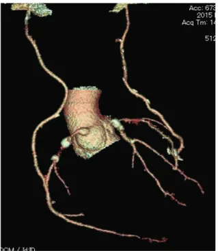
diography or multi-detector computed tomogra- phy(MDCT,Figures 1 and 2).
In the assessment by MDCT,volume rendering images were created from the image data obtained to evaluate the status of the vascular graft and coronary artery. If the vascular mor- phology of the graft,junction,and its periphery were visualized,the graft and coronary artery were assessed as patent,with sufficient blood flow (Fig.1). Since many vascular clips are usually attached to the graft,it was difficult to visualize and assess the surrounding vascular morphology.In view of the hemodynamics,the graft and coronary artery were assessed as patent if the blood flow between the central side of the
graft and the graft-artery junction was visualized.
If the blood flow was poor,and could not be visualized on volume rendering images,the graft- artery junction was evaluated using CT data (Fig.2).
Results
Table 1 presents data describing the 16 patients with a total of 23 grafts. In the early period of this series,the great saphenous vein (GSV)was used in 2 patients(2 grafts),1 of whom died suddenly 15 years postoperatively, possibly of myocardial infarction caused by graft obstruction.Since the other patient was lost to follow-up at 16 years postoperatively,his remain- ing course was unclear. From 1987,internal thoracic artery(ITA)grafts became the mainstay in CABG,with graft patency confirmed in all patients on follow-up.Both the ITA and GSV were used in 2 patients,in 1 of whom both grafts were confirmed to be patent 26 years postoper- atively. For the other patient,who changed hospitals,graft patency is unclear.In only 1 of the 16 patients,blood flow through the bypass graft was evaluated as poor at 9 years postoper- atively. Nonetheless,the graft was patent,and remained so for at least 15 years postoperatively (Figure 2).
In 13 patients with a total of 19 grafts who had sufficient follow-up,the survival rate was 92.3%.
The patency of 18 grafts(17 ITA and 1 GSV) was maintained for a maximum of 29 years,with an overall patency rate of 94.7%.Patency rates of ITA and GSV grafts were 100 and 50%,respec- tively.
All patients have been resumed their normal activities, with some limitations due to anticoagulant therapy and heart failure that there was since before CABG.
Discussion
Before the widespread use of CABG using the ITA,22 and 87%of patients with coronary artery lesions of Kawasaki disease were reported to die between the first and third episodes of myocar- dial infarction respecrively.웏 Considering the need for the long-term prevention of myocardial ischemia caused by coronary artery lesions of Kawasaki disease,ITA-CABG became widely used in children as well as in adults.
The results of the present study suggest that Fig.2 Evaluation of CABG by volume-rendering imaging
with MDCD (patient 16,poor blood flow). Fig.1 Evaluation of CABG by volume-rendering imaging
with MDCD (patient 10).
S.Marutani et al.
54
when the ITA is used for CABG for coronary artery disease due to Kawasaki disease,long- term graft patency is likely to be achievable,and cardiac events can be prevented.Kitamura et al. reported in 1988 that in children undergoing CABG using ITA grafts,growth of the grafts in proportion with somatic growth could be expect- ed.원When CABG is needed at a young age,as in coronary artery disease of Kawasaki disease, ITA grafts,therefore,are preferable.Our experi- ence led to a similar conclusion.
The use of GSV grafts is controversial as they show a low rate of long-term graft patency.웑웦웒We used GSV grafts in the 1980s,in part because of the paucity of published reports.However,since the report of Kitamura et al,웎웦웏we have relied mainly on ITA grafts.If more than one bypass graft is required,a GSV graft may be included.
A GSV graft in 1 of our patients with sufficient follow-up remained patent for an extended period.
The pathologic lesions peculiar to Kawasaki disease appear to be involved when stenosis occurs in ITA grafts that were expected to remain patent for a long time.Coronary artery lesions of Kawasaki disease are highly likely to calcify over time,which differs from the course of lesions in typical coronary artery disease. Such changes can interfere with evaluation of the degree of stenosis.We suspect that if CABG is
performed without sufficient evaluation of the stenotic lesions requiring intervention,residual blood flow through the original coronary artery with an uncertain degree of stenosis can compete with blood flow through the graft-artery junc- tion.Such competition could reduce blood flow to the graft artery,leading to narrowing of the graft. We consider a similar mechanism to be partly responsible for the narrowing of grafts in the patients in this study.However,ITA grafts in functionally no-flow situations can became recanalized several years later.웓Careful follow- up will be continued of our group of patients.
Conclusion
The use of ITA grafts in CABG for coronary artery lesions of Kawasaki disease contributes to a favorable long-term prognosis.
References
1.Kawasaki T et al.(1974)A new infantile acute febrile mucocutaneous lymph node syndrome(MCLS) prevailing in Japan.Pediatrics 54:271‑276
2.Yashiro M et al.(2014)22th Kawasaki disease national survey results.Jap J of Ped 77:271‑290[in Japanese]
3.Kato H et al.(1996)Long-term consequences of Kawasaki Disease.Circulation 94:1379‑1385 4.Kitamura S et al.(1985)Severe Kawasaki heart
Table 1 Sixteen patients with Kawasaki disease undergoing CABG in our hospital
Patient no.
Current age,
years Sex Age at CABG,
years
Years from disease onset to CABG
Postoperative period (years)
Recipient coronary
arteries Graft Postoperative course and graft
1 41 M 11 9 - #7 GSV Death(15 years after CABG) 2 41 M 13 12 28 #3 GSV Further participation declined 3 35 M 7 7 28 #7 ITA Patent
4 36 F 8 6 28 #9 ITA Unknown(follow-up refused) 5 38 M 9 8 29 #7#13 ITA GSV Patent
6 31 M 7 2 24 #3#7 ITA Patent
7 48 M 37 37 11 #8#4PD ITA GSV Unknown
(followed at another hospital) 8 16 M 5 5 11 LAD ITA Patent
9 37 M 26 23 11 #3 ITA Patent 10 12 M 12 12 0 #8#3 ITA Patent 11 15 M 8 8 7 #11#7 ITA Patent 12 12 F 7 2 5 #7 ITA Patent 13 40 F 39 33 1 #3 ITA Patent 14 12 F 12 10 0 #7#3 ITA Patent 15 18 M 18 12 0 #3 ITA Patent 16 43 M 28 25 15 #2#7 ITA Poor blood flow
CABG for coronary artery lesions of Kawasaki disease at a Japanese hospital:Summary of a 30-year experience
55
disease treated with an internal mammary artery graft in pediatric patients. A first successful report. J Thorac Cardiovasc Surg 89:860‑866
5.Kato H et al.(1986) Myocardial infarction in Kawasaki disease:clinical analyses in 195 cases. J Pediatr 108:923‑927
6.Kitamura S et al.(1988)Excellent patency and growth potential of internal mammary artery grafts in pediatric coronary artery bypass surgery.New evidence for a“live”conduit.Circulation 78:1129‑1139
7.Kitamura S et al.(1994)Long-term outcome of myocardial revascularization in patients with Kawasa- ki coronary artery disease:a multicenter cooperative study.J Thorac Cardiovasc Surg 107:663‑674 8.Kitamura S et al.(2009)Twenty-five-year outcome of
pediatric coronary artery bypass surgery for Kawasaki disease.Circulation 120:60‑68
9.Akasaka T et al.(1998)Flow dynamics of angiogra- phically no-flow patent internal mammary artery grafts. J Am Coll Cardiol 31:1049‑1056
S.Marutani et al.
56