Inhibitory Effect on Melanin Formation by Marine Bacterial Glycosaminoglycan
Katsuichiro OKAZAKI and Koichi OKUTANIAbstract
The polysaccharide produced by a marine bacterial strain designated P-21295 was found to be a high molecular weight glycosaminoglycan composed of galactose, galacturonic acid, glucosamine, galactosamine, alanine, and py-ruvate, together with N- and O-acetyl groups. This glycosaminoglycan suppressed the melanin formation in mouse B16F10 cells. The effect was dose dependent without any observed impact on the cell proliferation, indicating that this glycosaminiglycan will be a useful candidate of skin whitening cosmetics. Phenotypic and genetic features of the strain P-21295 suggest that this strain belongs to the Gram-negative Cobetia marina in the class
Gammaproteo-bacteria containing the genus Pseudomonas and Vibrio. However, some important phenotypic features of the strain
P-21295 differed with those of the type strain C. marina DMS4741. Key words: glycosaminoglycan; Cobetia sp. P21295; melanin; whitening.
Introduction
Naturally occurring glycosaminoglycans are widely distri-buted in higher organisms and of broad biochemical interests for their functional importance. Many microorganisms are also known to produce glycosaminoglycans such as sugar moieties of lipopolysaccharides and cell wall polysaccharides. How-ever, there are only a few reports on production of extracel-lular glycosaminoglycans by microorganisms.(1-5) During the
course of the screening of bioactive polysaccharide produced by marine microorganisms, the highly viscous glycosamino-glycan-producing bacterium was found from seawater sample in the Seto Inland Sea. This polysaccharide showed inhibitory effect on the melanin formation in mouse melanoma B16F10 cells. In the present paper, we describe the isolation, purifica-tion, and inhibitory effect on melanin formation of this poly-mer as well as characterization of the bacterium.
Materials and Methods
Bacterial Strain
The bacterial strain P-21295 used in the present experiment was a stock culture originally isolated from seawater in the Seto Inland Sea, Japan. Pure cultures were maintained on the agar medium contained 30 g of sucrose, 5 g of peptone and 1
g of yeast extract, and 15 g of agar per liter of seawater. Phenotypic Analysis
Cells for assays were grown for 24 hr at 25℃ on MB2216 agar (Becton Dickinson, MD, USA). Cells were examined by light microscopy (Phase-contrast) with Olympus BX50F4 (Olympus, Tokyo) to determine their shape and cell size. The biochemical properties such as catalase, oxidase, and oxida-tion-fermentation on glucose (O/F) was tested as described in Cowan and Steel s Manual.(6)
Gram stain was determined by using Favor Nissui G (Nissui Pharm, Tokyo). Phenotypic characteristics were also examined using a AP120NE Kit (bioMerieux, Lyon, France) according to the manufacturer
protocol.
Genetic Analysis
The 16S rDNA gene was amplified and sequenced as fol-lows. Chromosomal DNA of the strain was extracted with a InstaGene Matrix (BioRad, CA, USA) according to the manufacturer protocol. The 16S rDNA was amplified as tem-plate of the strain P-21295 DNA by polymerase chain reaction (PCR) using PrimeSTAR HS DNA Polymerase (TAKARA BIO, Shiga). PCR amplification was done using conserved primer 9F (5 -GAGTTTGATCCTGGCTCAG-3 ) and 1510R
*
(5 -GGCTACCTTGTTACGA-3 ) corresponded to 16S-rDNA positions 9 and 1501 of Escherichia coli, respectively. Nu-cleotides of the amplified fragmant were sequenced with ABI PRISM 3100 genetic analyzer using BigDye Terminator v3.1 Cycle Sequencing Kit (Applied Biosystems, CA, USA). Ho-mology search was conducted using BLAST program, and the nucleotide sequence of strain P-21295 was aligned with that of Cobetia marina DMS4741 using CLUSTAL W program of DDBJ (DNA Data Bank of Japan) online search and analysis. Isolation and Purification of the Polysaccharide
The bacterial medium for polysaccharide production con-tained 30 g of sucrose, 5 g of peptone and 1 g of yeast extract per liter of seawater. Each 200 ml of the medium in a 500 ml Erlenmyer flask was inoculated with 10 ml of seed culture which had been incubated for 24 hr at 25℃ in statically in the same components of the medium. After incubation statically for 5-7 days at 25 ℃, the viscous cultures were stirred with the addition of water and then bacterial cells were removed by centrifugation followed by filtration through GF-75 glass fiber filter (Advantec, Tokyo) using Radiolite #500 (Showa Chemical, Tokyo) as a filter bed. The polysaccharide material was precipitated from the clear supernatant solution by the addition of 2 volumes of ethanol. The precipitate obtained was dissolved in water and reprecipitated with the addition of 5% cetyltrimethylammonium bromide (Cetavlon). The precipitated Cetavlon-polysaccharide complex was collected by filtration and redissolved in 4 M NaCl solution. The poly-saccharide component was recovered by precipitation with 2 volumes of ethanol, followed by filtration. This material was dissolved in water and dialyzed against deionized water, followed by freeze-drying to afford the sodium salt of the polysaccharide. Purity of the polysaccharide was checked by electrophoretic analysis.
Component Analysis of the Polysaccharide
Electrophoresis was performed on cellulose acetate strips (Sartorius 1120, 57 × 145 mm), for 20 min at 150V in 0.2 M calcium acetate buffer (pH 7.5) or for 25 min at 150V in 0.05M sodium borate buffer (pH 9.4). Poly- and monosac-charide bands were visualized with 0.1% alcian blue in 10% acetic acid and alkaline silver nitrate reagent, respectively. Paper chromatography (PC) was carried out by the descend-ing method on Whatman No.1 paper with ethyl acetate-acetic acid-formic acid-water (18:3:1:4) as the solvent. Sugars and amino compounds were detected either with alkaline silver
ni-trate, p-anisidine hydrochloride, or ninhydrin reagents. Sugars were also analyzed with a Hitachi 655 HPLC equipped with a refractive index (RI) detector on a Wakopak WBT 130E co-lumn (Wako Pure Chemicals, 7.8×300 mm) using water as a mobile phase at 60℃ at a flow rate of 0.5 ml/min.
Amino acids and amino sugars were also determined with a Hitachi L-8500 or JEOL JCL-500/V amino acid analyzer. Pyruvate was determined by ion chromatography (IC) and as well as by 1
H-NMR spectroscopy. For IC, the hydrolysates of the polysaccharide with 0.05 M trifluoroacetic acid (TFA) for 12 hr at 100℃ or 0.05 N H2SO4 for 1 hr at 100℃ were
ana-lyzed on a Shimpak IC-101H column (Shimadzu, 7.9 × 300 mm) at 50 ℃ with 5 mM perchloric acid as a mobile phase using a Shimadzu HIC-6A ion chromatograph equipped with a UV detector (at 210 nm).
The average molecular size of the polysaccharide was de-termined by gel permeation chromatography (GPC) with a Hitachi 655 HPLC equipped with an RI detector on an Asa-hipak GFA 7M column (Asahi Chemicals, 7.6 × 500 mm). For minimizing the association effect of the polysaccharide solution, 0.1 M NaCl solution was used as a mobile phase at 30℃ and at a flow rate of 0.4 ml/min. The calibration of the molecular size was made with pullulans of various molecular sizes (Shodex Standard Kit P-82, Showa Denko). Protein concentrations were determined by the method of Lowry et
al. (7)
For the analyses of neutral sugars, the polysaccharide was hydrolyzed with 2 M TFA for 12 hr at 100℃ in a sealed glass tube. After the acid was removed by several evaporations under reduced pressure with water, the hydrolysates were ana-lyzed by PC, electrophoresis, and HPLC. For the analyses of amino acids and amino sugars, the polysaccharide was hydro-lyzed with 4 N HCl for 12 hr at 100℃ , followed by concen-tration to dryness in vacuo. The residue was analyzed by PC, electrophoresis, and amino acid analyzer. Uronic acids were identified by electrophoresis after hydrolysis of the polysac-charide with 2 M TFA for 12 hr at 100℃ . Uronic acids were also determined by the carbazole-sulfuric acid method. The
1H-NMR spectrum was obtained with a Varian Unity INOVA
500 spectrometer (1H 500MHz) at 85℃ . Cell Culture
Mouse melanoma B16F10 cells were grown in Dulbecco s minimum essential medium (DMEM) supplemented with 10% fetal bovin serum (FBS) in 5% CO2 incubator. Cells
were suspended in the growth medium at 104
seeded on 12 well plate. After incubation for 48 hr, the culture fluid in the well was changed to DMEM supplemented with 10% FBS containing various concentrations of the test sam-ple. After 3-day incubation at 37 ℃ in 5% CO2, the number
of viable cells was determined by the WST-1 method (Dojin, Kumamoto). The effect on cell proliferation of the compound against B16F10 cells was determined with the viability of cells, as monitored by absorbance at 450 nm. To determine melanin content, cells were washed, dissolved in 1N NaOH solution containing 10% dimethylsulfoxide. The absorbance at 475 nm was measured, and the melanin content was measured using synthetic melanin (Sigma,Mo, USA) as the authentic standard. Each experiment carried out in triplicate.
Results and Discussion
Bacterial Taxonomy
The strain P-21295 exhibited 99.4% similarity of 16S rDNA gene sequence with the sequence of Cobetia marina DMS4741(8) (Accession number AJ306890), type strain of C.
marina (Fig. 1). As shown in Table 1, the phenotypic
proper-ties of the strain P-21295 also correspond to those of reported for Cobetia marina.(9)
However, the type strain of Cobetia
marina (DMS4741) was rod-shaped cell and was motile,
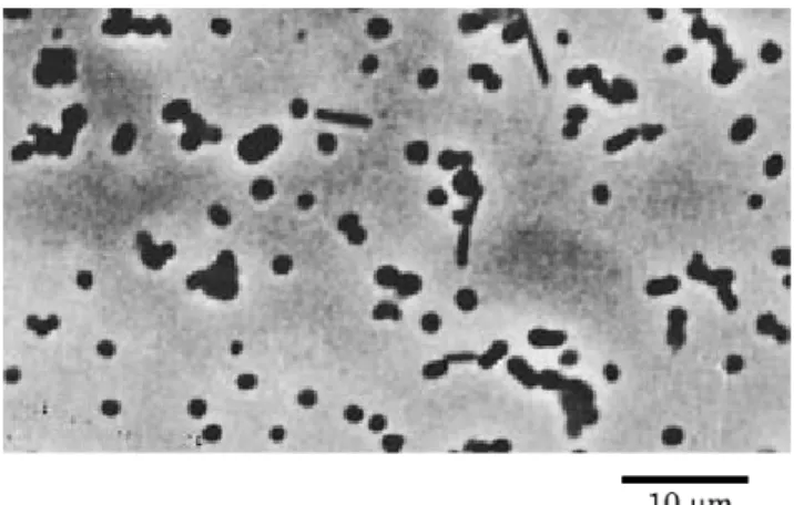
while the strain P-21295 was a non-motile and showed
pleo-morphic growth (Fig. 2). We also found some phenotypic characteristics that distinguished the type strain and the strain P-21295. For example, the type strain did not grow at 45 ℃ and utilized L-serine, while the strain P-21295 grew at 45 ℃
and did not utilize L-serine. Detail analyses for the genetic and
phenotypic characteristics for distinguishing taxonomically the strain P-21295 and the type strain of Cobetia, together with other reported strains of the Cobetia are in progress. In addition, the genus Cobetia belongs to the family
Haromona-daceae among the class Gammaproteobacteria containing the
families Pseudomonadaceae and Vibrionaceae by the data-base from DDBJ.
Fig. 1. Comparison of the nucleotide sequences of 16S ribosomal DNA from the strain P-21295(P)and Cobetea marina DMS4741(DMS).
The numbers of the first nucleotide in upper sequence are shown on the left. A hyphen(−)in sequence of Cobetia
ma-rina DMS4741 indicates identical nucleotides. The underlines indicate sequences for design PCR primers, as described in
the text. Nucleotide Y in the position 462 of the strain P-21295 is C or T.
containing polysaccharides produced by microorganisms. Some of them are known to contain lysine, glycine, glutamic acid, alanine, and N-(2-hydroxyethyl)-D-alanine.(4 , 5 , 13, 14)
Among them a glycosaminoglycan had approximately identi-cal components with that in the present experiment, but was the production by different bacterial species.(5)
Further stud-ies of the structure of the glycosaminoglycan produced by the strain P-21295 are in progress.
Preparation and Purification of the Polysaccharide The purified polysaccharide was homogeneous in elec-trophoresis and did not contain proteins. Owing to the high viscosity of the polysaccharide solution, only a broad peak was observed on GPC analyses, estimating to be approximate molecular size of 1.0-1.5×10⁶
. Component Analyses
The acid hydrolysates of the polysaccharide gave galact-uronic acid, galactose, glucosamine, galactosamine, pyruvic acid, and alanine. The presence of galacturonic acid was also confirmed by the isolation of uronic acid from the acid hydro-lysate using Amberlite IR 410 (formate form) and PC, fol-lowed by carboxyl-reduction to galactose. Glucosamine, ga-lactosamine, and alanine were determined in the approximate molar ratios of 1:2:1 by HPLC and amino acid analyzer. The
1
H NMR spectrum of the polysaccharide showed the presence of a signal at δ= 2.2 attributable to O–acetyl groups (Fig. 3). In addition, 1
H NMR spectra revealed the presence of methyl signals of N-acetyl moieties around at δ =2.0, indicating the presence of N-acetylamino sugar residues.(10-12)
There are only a few reports on extracellular amino
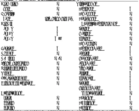
acid-Table1.Phenotypic properties of the strain P-21295
Gram stain − Utilization of
Spore − D-glucose +
Mobility − D-mannose −
Shape rod(pleomorphic) L-arabinose −
Growth N-acetyl-D-glucosamine −
at 10℃ + lactose −
at 37℃ + maltose −
at 45℃ +W sucrose −
D-mannitol −
Catalase + D-gluconic acid +
Oxidase − acetate +
O/F test −/− n-capric acid −
Indole production − adipic acid −
Nitrate reduction − dl-malate −
Urease − citrate +
B-Galactosidasse − succinate +
Arginine dihydrolase − lactate +
propionic acid +
Hydrolysis of Phenylacetate −
starch − L-alanine +
gelatin − L-histidine −
esculin − L-serine −
O/F, Oxidation/Fermentation; +, prsitive reaction; −, negative reaction; W, week reaction.
References
Inhibitory Effect on Melanin Formation
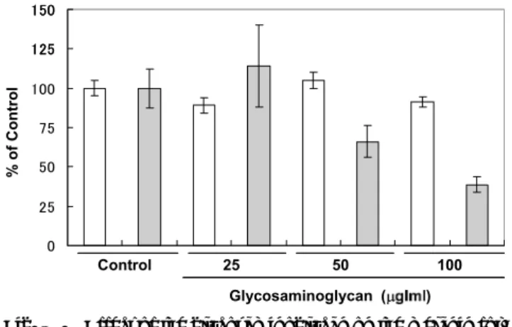
As shown in Fig. 4, this polysaccharide inhibited the mela-nin formation in mouse B16F10 cells at a concentration of 50 and 100 μg/ml in a concentration dependent fashion. No inhibitory effect on the cell proliferation was observed at a
concentration up to 100 μg/ml. When a glycosaminogly-can known to have inhibitory effect on melanin formation in mouse B16 cells was examined in the same assay system using as the positive reference,(15)
it was also found to have inhibitory effect on melanin formation in mouse B16F10 cells at similar concentrations.
Melanin biosynthesis is regulated by melanogenic enzymes such as tyrosinase, tyrosinase-related protein 1 (TRP-1) and dopachrome tautomerase (DCT or TRP-2).(16)
Synthesis of melanin starts from the conversion of the amino acid L
-tyro-sine to dopaquinone by tyrosinase. The control of melano-genesis is an important strategy in treatment of abnormal skin pigmentation for cosmetic purposes. Modulators of melano-genesis can act directly on tyrosinase, tyrosinase gene expres-sion, or the mechanisms responsible for the transfer of mela-nosomes from melanocytes to keratinocytes.(17)Therefore, the
novel glycosaminoglycan produced by strain P-21295 can be useful for whitening agent. However, further experiments are required to find inhibitory mechanism of melanin biosynthesis by the glycosaminoglycan.
⑴ Bonde, GJ., Carlsen, FE., and Jensen, DE.: Production of hyaluronic Acid by Pseudomonas aeruginosa. Act.
Phar-macol. Toxicol., 13, 205-212 (1957).
⑵ Pierce, WA., White. J., and White, ACG.: Hyaluronic acid formation by Streptococcus pyogenes. Proc. Soc.
Exp. Biol. Med., 83, 50-54 (1954).
⑶ Izumi, K.: Mucopolysaccharides produced by a strain of Clostridium perfringens. J. Bacteriol., 83, 956-959 (1962).
⑷ Shamsuddin, AA., Matsuda, M., Nomura, M. and Oku-tani, K.: Structure of a glutamic acid-containing gly-cosaminoglycan from a marine Pseudomonas species.
Fisheries. Sci., 64, 469-473 (1998).
⑸ Tandavanitj, S., Ishida, S., and Okutani, K.: Isolation and characterization of an extracellular mucopolysaccharide produced by a marine strain of Pseudomonas. Nippon
Suisan Gakkaishi, 55, 2015-2019 (1989).
⑹ Barrow G.I. and Feltham, R.K.A.: Cowan and Steel s Manual for the Identification of Medical Bacteria, 3rd ed., Cambridge University Press, Cambridge (1993). ⑺ Lowry, O.H., Rosebrough, N.J., Farr, A.L., and Randall,
R.J.: Protein measurement with the Folin phenol reagent.
J. Biol. Chem., 103, 265-275 (1951).
Fig. 4. Effect of the glycosaminoglycan on the melanin for-mation and the cell proliferation in B-16F10 cells. The cell number (open column)and melanin con-tent(gray column)were measured as described in the text.
⑻ Arahal,D.R., Castillo, A. M.,Ludwig,W., Schleifer,K. H. and Ventosa,A.: Proposal of Cobetia marina gen. nov., comb. nov., within the family Halomonadaceae to include the species Halomonas marina. System. Appl.
Microbiol., 25, 207-211 (2002).
⑼ Brenner, D.J., Krieg, N.R., Staley, J.T. and Garrity, G.M.: Bergey s manual of Systematic Bacteriology. Vol. 2, The Proteobactera, Part B. The Gammapeoteobacteria. 2005. Springer.
⑽ Knirel, Y.A., Dashunin, V.V., Shashkov, A.S., Kochetkov, N.K., Dmitriev, B.A., and Hofman, I.L.: Somatic anti-gens of Shigella structure of the O-specific polysaccha-ride chain of the Shigella dysenteriae type 7 lipopolysac-charide. Carbohydr. Res., 179, 51-60 (1988).
⑾ Helander, P.B., Kenne, L., Lindberg, B., Peterson, K., and Unger, R.: Structural studies of two capsular poly-saccharides elaborated by different Strains of
Haemophi-lis influenzae type e. Carbohydr. Res., 88, 77-84 (1981).
⑿ Katzenellenbogen, E., Ekiel, I., and Romanowska, E.: The Structure of the O-specific polysaccharide chain from Citrobacter O23 lipopolysaccharide. Cabohydr.
Res., 179, 349-357 (1988).
O-specific chains of Proteus mirabilis 1959, Eur, J. Bio-chem.,62, 391-399 (1976).
⒁ Vinogradov, E.A., Kaca, W., Shashkov, A., Pietrasik, D.K., Rozalski, A., Knirel, Y.A., and Kochetkov, N.K.: The structure of Proteus mirabilis O3 O-specific polysac-charide containing N-(2-hydroxyethyl)-D-alanine. Eur. J.
Biochem., 188, 645-651 (1990).
⒂ Okazaki, K. and Okutani, K.: Glycosaminoglycan-devel-opment of whitening cosmetics by marine microbial utili-zation. Fragrance J., No.8, 61-64 (2004). (in Japanese) ⒃ Ohguchi, K., Akao, Y. and Nozawa, Y.: Stimulation of melanogenesis by the citrus flavonoid naringenin in mouse B-16 melanoma cells. Biosci. Biotechnol.
Bio-chem., 70, 1499-1501 (2006).
⒄ Kim,Y.J., No, J.K., Lee, J.K., Kim, M.S. and Chung, H.Y.: Antimelanogenic activity of 3,4-dihydroxyacetophenone: inhibition of tyrosinase and MITF. Biosci. Biotechnol.
Biochem., 70, 532-534 (2006).
海洋細菌P-21295株の生産する多糖は,N- や O-アセチル基を持つガラクトース,ガラクツロン酸,グルコサミン,ガ ラクトサミン,アラニンとピルビン酸から構成される高分子グリコサミノグリカンであることが見出された.このグリ コサミノグリカンはマウスメラノーマB-16F10細胞のメラニン形成を抑制した.その効果は濃度依存的であり,細胞の 増殖にも影響がなかったことから,このグリコサミノグリカンは美白化粧品開発に有用であると考えられた.P-21295 株の生理学的および遺伝的性質から,本菌株は、シュードモナス属やビブリオ属が含まれているガンマプロテオバクテ リアクラス内のグラム陰性のCobetia marinaに属することが示唆された.しかしながら,P-21295株のいくつかの重要な 生理学的性質は、基準株であるDMS4741株と異なっていた.