H
emophilia is one of the most common inherited bleeding disorders due to the deficiency in clot-ting factor VIII (hemophilia A) or IX (hemophilia B). The estimated frequency of hemophilia is reported 1 in 5000-10000 birth [1]. Patients with hemophilia have been reported to have an increased risk of hepatitis virus infection and hepatocellular carcinoma (HCC) due to frequent blood transfusions [2]. Repeated transfu-sions of coagulation factors VIII or IX are known to induce the inhibitor of each coagulation factor [3]; approximately 5% of patients with hemophilia A are reported to have the inhibitor to coagulation factor VIII [4,5].Several reports have been published focusing on hepatectomy for HCC in patients with hemophilia, regardless of the high incidence of HCC and hepatitis
[6-11]. In these previous reports, no patients under-went laparoscopic resection, and only one had the inhibitor to coagulation factor VIII [11]. Herein, we report our experience of laparoscopic hepatectomy for a patient with hemophilia A with high titer acquired fac-tor VIII inhibifac-tor.
Case
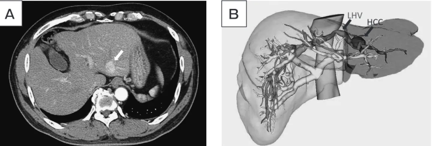
A 60-year-old man with hemophilia A and transfu-sion-acquired hepatitis C was diagnosed with HCC in segment II. He was referred to our department of hepa-tobiliary and pancreatic surgery for liver resection. The tumor was 2.1 cm in diameter and located close to the left hepatic vein; next, laparoscopic left lateral seg-mentectomy was planned (Fig.1A,B). In addition, the patient experienced cholecystitis twice; laparoscopic
CopyrightⒸ 2021 by Okayama University Medical School.
http ://escholarship.lib.okayama-u.ac.jp/amo/
Case Report
Laparoscopic Hepatectomy for the Patient with Hemophilia
A with High Titer Factor VIII Inhibitor
Tatsuo Matsuda
a,b, Yuzo Umeda
a*, Kazuhiro Yoshida
a, Tadakazu Matsuda
b,
Masatoshi Uno
c, Masaya Abe
d, Noboru Asada
d, Yoshinobu Maeda
d,
Takahito Yagi
a, and Toshiyoshi Fujiwara
aDepartments of aGastroenterological Surgery, dHematology and Oncology, Okayama University Graduate School of Medicine,
Dentistry and Pharmaceutical Sciences, Okayama 700-8558, Japan,
bDepartment of Surgery, Matsuda Hospital, Kurashiki, Okayama 710-0056, Japan, cDepartment of Internal Medicine, Kaneda Hospital, Maniwa, Okayama 719-3193, Japan
We present the first case of laparoscopic left lateral segmentectomy for hepatocellular carcinoma (HCC) in a patient with hemophilia A, acquired hepatitis C, and high-titer factor VIII inhibitor, which was confirmed by preoperative diagnosis. He underwent laparoscopic left lateral segmentectomy with the administration of recombinant activated factor VII. Surgery could be performed with reduced intraoperative hemorrhage. He experienced postoperative intra-abdominal wall hemorrhage, which was successfully managed with red cell concentrates transfusion and administration of recombinant activated factor VII. Laparoscopic hepatectomy can be applied for hemophilia patients with high titer inhibitors.
Key words: hemophilia A, hepatectomy, inhibitor, laparoscopy, hepatocellular carcinoma
Received June 17, 2020 ; accepted October 19, 2020.
*Corresponding author. Phone : +81-86-235-7257; Fax : +81-86-221-8775
cholecystectomy was planned simultaneously. We con-sulted the hematology department about perioperative management of his hemophilia; preoperative blood test showed extremely prolonged activated partial thrombo-plastin time (APTT) (106.8 sec). He had been treated with 2000 IU of full-length recombinant human coagu-lation factor VIII (ADYNOVATE®, Takeda, Tokyo) twice a week for his hemophilia A. Due to The pro-longed APTT, inhibitor formation for coagulation fac-tor VIII was suspected, and high-titer inhibifac-tor of coagulation factor VIII was detected (10 IU/ml). Patients with hemophilia without inhibitors or low titer inhibitors can be treated with high doses of FVIII or FIX for perioperative management; however, this treat-ment is ineffective for hemophilia patients with high titer inhibitors [12,13]. Afterward, we decided to use recombinant activated factor VII (rFVIIa, NovoSeven®, Novo Nordisk, Bagsvaerd, Denmark) [11,13,14] for his perioperative management.
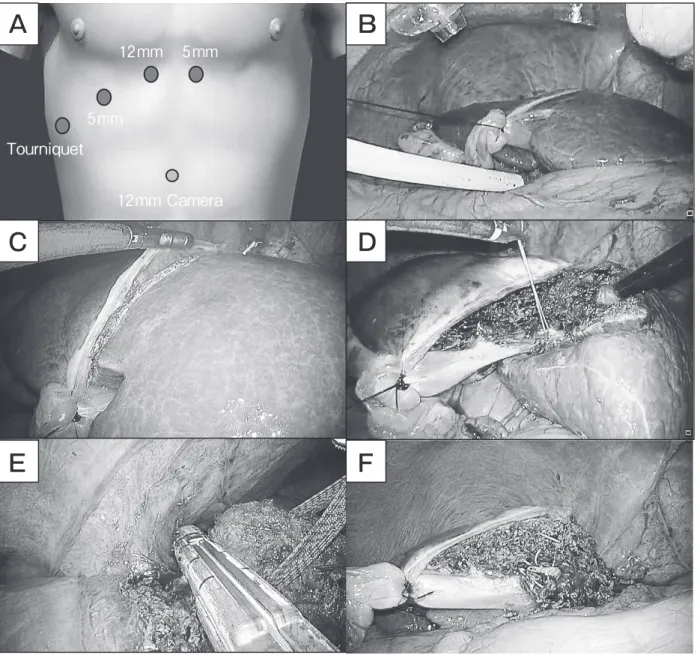
Surgical procedure. A camera port was inserted through a 12-mm port in the umbilicus, using the open technique; three additional ports (two 5-mm ports and a 12-mm port) were inserted under pneumoperito-neum (10 mmHg) (Fig.2A). Tourniquet method was performed, using a 20 Fr silicone tube during transec-tion (Fig.2B). Liver parenchymal transectransec-tion with a vessel-sealing system (LigaSure®, Medtronic, Dublin, Iceland) and endoscopic Cavitron ultrasonic surgical aspirator (CUSA, Olympus, Tokyo, Japan) were per-formed after the laparoscopic ultrasound examination,
along the lateral border of the falciform-round ligament (Fig.2C). Pedicles of segments II and segment III were identified and divided along the transection plane (Fig.2D). At the end of the resection, the left hepatic vein was exposed and dissected with a vascular stapler (Fig.2E,2F).
The specimen was placed in the specimen bag. After laparoscopic cholecystectomy, these specimens were delivered through a Pfannenstiel incision. Two drains were inserted through the 5-mm port site and a 20 Fr silicone tube inserted hole. After hemostasis was secured, the ports were removed and the wound (including the port site) was closed, except the drain inserted wound. Total intraoperative blood loss was 50 ml and total surgery time was 6 h (3.5 h for hepatec-tomy, 2.5 h for cholecystectomy).
Pre- and perioperative management of hemophilia.
Based on previous reports, 100 μg/kg of rFVIIa per dose bolus were administered just before surgery, and bolus injection (100 μg/kg rFVIIa) were administered every 2 h during surgery [14,15].
Postoperative course. Administration of rFVIIa at a dose of 100 μg/kg every 2 h was planned to con-tinue for 48 h postoperatively. Twenty four h after sur-gery, he was transferred to a general ward from the intensive care unit, without any sign of postoperative bleeding; 40 h postoperatively, subcutaneous hemor-rhage around the port site (20 Fr silicone tube insertion point) and significant decrease in hemoglobin level of 10.6 g/dl to 6.8 g/dl were observed. At the time of
A
B
LHV HCCFig. 1 (A) Computed tomography (CT) images before liver resection. Dynamic CT scan showing HCC of 2.1 cm in diameter, located at segment II. (B) 3D imaging by the Synapse Vincent system (Fuji Film); green transparent area, regions for planned resection; pink area, the portal vein; blue area, the portal vein, and the inferior vena cava; red area, HCC in segment II.
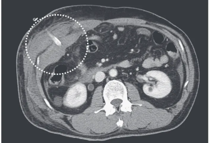
bleeding, rFVIIa was still injected every two hours. Enhanced computed tomography image showed the right lateral abdominal wall hematoma around the drain tube (placed through the port site) (Fig.3). There was no intra- or extra-abdominal hemorrhage; only intra-abdominal wall hemorrhage was observed. A total transfusion of 840 ml of red cell concentrates was per-formed. We decided to continue injecting rFVIIa every 2 h, until hemostasis was achieved; 2 days after the
intra-abdominal wall bleeding, we confirmed the hemostasis and removed both drain tubes placed though the port site. Thick bite sutures were made at the port sites to prevent rebleeding from the abdominal wall. Dosage interval of rFVIIa infusion was gradually extended 6 days postoperatively (Fig.4). We stopped the infusion of rFVIIa 17 days postoperatively and switched to the injection of emicizumab-kxwh (Hemlibra®, Genentech, Inc., San Francisco, CA, USA)
5mm 12mm 5mm 12mm Camera Tourniquet
A
B
C
D
E
F
Fig. 2 (A) Trocar placement for laparoscopic left lateral segmentectomy. (B) Tourniquet method with a 20 Fr silicone tube. (C) Planned resection line. (D) Pedicle to segment III was identified and divided. (E) Left hepatic vein dissection with a vascular stapler. (F) Laparoscopic left lateral segmentectomy is completed.
as a regular therapy for hemophilia. Emicizumab-kxwh is a bispecific humanized monoclonal antibody that restores the function of missing activated FVIII by bridging activated FIX and FX. It was approved by the Japanese Ministry of Health, Labor, and Welfare in 2018, for hemophilia A inhibitor patients [16]. He was discharged 25 days postoperatively, without any addi-tional postoperative bleeding or other complications. Hemophilia A has been successfully managed by inject-ing emicizumab-kxwh; HCC recurrence was not observed 1 year postoperatively.
Discussion
Although hemophilia is of the most common inher-ited bleeding disorders, most surgeons do not have
experience with hemophilia A patients. We believe that our experience is worth reporting for other surgeons. Guidelines for the management of the patient with hemophilia have been published, and postoperative management has been described [13]. However, pre-operative management of liver resection with elevated risks for bleeding is largely unknown. Several reports have been published about liver resection for patients with hemophilia; only one patient with high-titer inhibitor of coagulation factor VIII has been reported (Table 1) [6-11]. Considering the high risk of hepatitis virus infection and HCC [2], this number of previous reports seems to be extremely small. Surgeons might be hesitant to perform liver resection and select other types of treatment, such as radiofrequency ablation (RFA) or transcatheter arterial chemoembolization instead. However, liver resection has the greatest therapeutic effect on resectable HCC. Hasegawa et al. reported the therapeutic effect of surgical resection (SR) in the Japanese nationwide survey of patients with HCC treated by SR, percutaneous ethanol injection (PEI), or RFA. The HCC patients who had no more than 3 tumors and liver damage of class A or B (like our case) were analyzed. The hazard ratio for death was signifi-cantly lower in the SR group than in the RFA (SR vs. RFA: 0.84, p=0.006) and PEI groups (SR vs. PEI: 0.75, p=0.0001) [17]. In all previously reported cases and our case no severe postoperative complications were noted (Clavien-Dindo Classification III-V) [18]. Thus, liver resection can be safely performed in patients with hemophilia, with appropriate perioperative manage-ment of coagulation factors.
In our case, the inhibitor development had not been detected before preoperative examination for hepatec-tomy. The patient had undergone joint replacement three times (bilateral total knee and right hip) within 2 years, at another hospital. He was perioperatively treated with recombinant factor VIII. This large amount of factor VIII transfusion might result in inhibitor for-mation. As per the guidelines, preoperative assessment should include inhibitor screening assay [13].
Patients with hemophilia with low titer inhibitors (<5 BU/ml) can be treated with high doses of FVIII or FIX for perioperative management [3,12,14]. This treatment is mostly ineffective for hemophilia patients with high titer inhibitors (>5 BU/ml) (like in our case) and likely to induce additional inhibitors. Our case had a high titer inhibitor (10 BU/ml) and bypassing agents
Fig. 3 Dynamic CT scan showing intra-abdominal wall bleeding around the drain site.
6 7 8 9 10 11 50 100 150 200 250 -2 1 2 3 4 5 6 8 10 13 15 PT APTT * * * * * PT (Sec) APPT(Sec) Bleeding2h 3h 4h 6h 8h 12h
Post operative day
Surgery
interval of rFVIIa infusion
Fig. 4 Summary of post-operative course. Asterisks indicate PT (sec) is unmeasurable due to over coagulation.
should be used for perioperative management [12]. Two bypassing agents are currently used: recombinant acti-vated factor VII (rFVIIa) (NovoSeven®, Novo Nordisk) and activated prothrombin complex concentrate (FEIBA, Shire). In our case, we selected rFVIIa as the bypassing agent. The response to these two bypassing therapy is different among patients with hemophil-ia; thus, preparing both agents is preferable for high bleeding risk surgery [12]. FEIBA was used in the pre-vious case report about hepatectomy for the hemophilia patient with high titer factor VIII inhibitor [11]. Because the patient had always been very responsive to FEIBA during his hemophilia bleeds. The patient was infused 50 U/kg of FEIBA twice daily, from 2 h before surgery and continued until discharge.
Development of laparoscopic hepatectomy is rela-tively slow compared to other abdominal procedures due to the complexity of the biliary and vascular struc-tures and high risk of bleeding. Advancements in lapa-roscopic surgery have turned lapalapa-roscopic left lateral segmentectomy into a standard procedure [19]. The advantages of laparoscopic surgery are smaller incision and decreased operative blood loss compared to open surgery. We selected the laparoscopic hepatectomy for these advantages. This is the first report on laparo-scopic hepatectomy for a patient with hemophilia. We did not experience hemostatic difficulty during surgery
with rFVIIa infusion (total intraoperative blood loss: 50 ml). Laparoscopic hepatectomy with appropriate perioperative management (including administration of the coagulation factor) can be a safe procedure, even for patient with hemophilia. In our case, we experienced postoperative hemorrhage around the drain site. We closed the wound (including the port site) except for the drain inserted into the wound at the time of surgery. The drain tube size was smaller than the port site. Postoperative hemorrhage could have been prevented if the port site was stitched and reefed.
Laparoscopic hepatectomy could be performed in this patient, irrespective of the high FVIII inhibitor titer. The patient experienced postoperative intra- abdominal wall hemorrhage which was conservatively managed with rFVIIa administration. The patient is alive 12 months postoperatively, with no evidence of HCC recurrence. We believe that laparoscopic hepatec-tomy can be even applied for hemophilia patients with high titer inhibitors.
References
1. Mannucci PM and Tuddenham EG: The hemophilias--from royal genes to gene therapy. N Engl J Med (2001) 344: 1773-1779. 2. Darby SC, Ewart DW, Giangrande PL, Spooner RJ, Rizza CR,
Dusheiko GM, Lee CA, Ludlam CA and Prestonet FE: Mortality from liver cancer and liver disease in haemophilic men and boys in
Table 1 Previously reported articles of hemophilia patients with HCC who undergo hepatectomy
Authors Year Number of cases Inhibitor Surgical procedures Perioperative replacementof Clotting factor Shen. et al. (7) 1994 Hemophilia A: 1 No Partial hepatectomy: 1 Factor VIII product Uchino. et al. (8) 2013 Hemophilia A: 1 No Partial hepatectomy: 1 unknown
Narushima. et al. (9) 2004 Hemophilia A: 2 No Partial hepatectomy: 1SubSegmentectomy 8: 1 Factor VIII product
Inokawa. et al. (6) 2014 Haemophilia A: 5Haemophilia B: 1 No
Left lateral sectionectomy: 1 Right posterior sectionectomy: 1 Left hepatectoy: 1
Sub segmentectomy 5: 1 Sub segmentectomy 8: 1 Partical hepatectomy: 1
Factor VIII product for Hemophilia A Factor IX product for Hemophilia B
Kobayashi. et al. (10) 2019 Haemophilia A: 7 No
Right hepatectoy: 1 Sub segmentectomy 1: 1 Sub segmentectomy 6: 1 Sub segmentectomy 8: 3 Partical hepatectomy: 1
Factor VIII product Jones. et al. (11) 2009 Hemophilia A: 1 Yes Left lateral sectionectomy: 1 FVIII inhibitorbypassing activity
UK given blood products contaminated with hepatitis C. UK Haemophilia Centre Directorsʼ Organisation. Lancet (1997) 350:
1425-1431.
3. Eckhardt CL, van der Bom JG, van der Naald M, Peters M, Kamphuisen PW and Fijnvandraat K: Surgery and inhibitor devel-opment in hemophilia A: a systematic review. Journal of thrombo-sis and haemostathrombo-sis. J Thromb Haemost (2011): 1948-1958. 4. Peyvandi F, Garagiola I and Young G: The past and future of
hae-mophilia: diagnosis, treatments, and its complications. Lancet (2016): 187-197.
5. McMillan CW, Shapiro SS, Whitehurst D, Hoyer LW, Rao AV and Lazerson J: The natural history of factor VIII: C inhibitors in patients with hemophilia A: a national cooperative study. II. Observations on the initial development of factor VIII: C inhibitors. Blood (1988): 344-348.
6. Inokawa Y, Sugimoto H, Kanda M, Yamada S, Fujii T, Nomoto S, Takeda S, Suzuki N, Matsushita T and Kodera Y: Hepatectomy for hepatocellular carcinoma in patients with hemophilia. J Hepatobiliary Pancreat Sci (2014): 824-828.
7. Shen SC, Harada A, Kurokawa T, Nakao A, Nonami T, Isobe K, Imoto M, Takamatsu J and Takagi H: Partial hepatectomy for hepatocellular carcinoma in a patient with hemophilia: a case report. Hepatogastroenterology (1994): 283-286.
8. Uchino K, Tateishi R, Nakagawa H, Shindoh J, Sugawara Y, Akahane M, Shibahara J, Yoshida H and Koike K: Uninodular combined hepatocellular and cholangiocarcinoma with multiple non-neoplastic hypervascular lesions appearing in the liver of a patient with HIV and HCV coinfection. J Clin Virol (2013): 173-177.
9. Narushima Y, Ishiyama S, Kawashima K, Shimamura H, Yamaki T and Yamauchi H: Operated hepatocellular carcinoma in two HIV- and HCV-positive hemophilic patients. J Hepatobiliary Pancreat Surg (2004): 207-210.
10. Kobayashi K, Kokudo T, Yamaguchi T, Shirata C, Uldry E, Akamatsu N, Arita J, Kaneko J, Yasumoto A, Demartines N, Hasegawa K and Halkic N: Hepatectomy in patients with inherited
blood coagulation disorders can be safely performed with adequate coagulation factor replacement. Haemophilia (2019): 463-467. 11. Jones AE, Roy A, Armstrong T, Rees M and Welsh FK:
Successful liver surgery in a haemophilia patient with high titre factor VIII inhibitor. Haemophilia (2009): 1332-1333.
12. Giangrande PLF, Hermans C, O'Mahony B, de Kleijn P, Bedford M, Batorova A, Blatný J and Jansone K: European principles of inhib-itor management in patients with haemophilia. Orphanet J Rare Dis (2018): 66.
13. Srivastava A, Brewer AK, Mauser-Bunschoten EP, Key NS, Kitchen S, Llinas A, Ludlam CA, Mahlangu JN, Mulder K, Poon MC and Street A: Guidelines for the management of hemophilia. Haemophilia (2013): e1-47.
14. Rodriguez-Merchan EC, Rocino A, Ewenstein B, Bartha L, Batorova A, Goudemand J and Gringeri A: Consensus perspec-tives on surgery in haemophilia patients with inhibitors: summary statement. Haemophilia (2004): 50-52.
15. Saba HI, Morelli GA, Azam RR, Klein CJ and Letson GD: Efficacy of NovoSeven during surgery on a haemophiliac with previous his-tory of inhibitors. Haemophilia (2003): 131-136.
16. Scott LJ and Kim ES: Emicizumab-kxwh: First Global Approval. Drugs (2018): 269-274.
17. Hasegawa K, Kokudo N, Makuuchi M, Izumi N, Ichida T, Kudo M, Ku Y, Sakamoto M, Nakashima O, Matsui O and Matsuyama Y: Comparison of resection and ablation for hepatocellular carci-noma: a cohort study based on a Japanese nationwide survey. J Hepatol (2013): 724-729.
18. Clavien PA, Barkun J, de Oliveira ML, Vauthey JN, Dindo D, Schulick RD, de Santibañes E, Pekolj J, Slankamenac K, Bassi C, Graf R, Vonlanthen R, Padbury R, Cameron JL and Makuuchi M: The Clavien-Dindo classification of surgical complications: five- year experience. Ann Surg (2009): 187-196.
19. Yang T-H, Chen J-L, Lin Y-J, Chao, Y-J, Shan Y-S, Hsu H-P, Su Z-M, Chou C-C and Yen Y-T: Laparoscopic surgery for large left lateral liver tumors: safety and oncologic outcomes. Surg Endosc (2018): 4314-4320.