Yonago Acta medica 2005;48:63–74
Cell Death Induction of Thymidine Kinase Gene Transfer
Followed by Ganciclovir Treatment in Oral Squamous Cell
Carcinoma Cell Lines
Itaru Kuratate*†, Mitsuhiko Osaki* and Kazuo Ryoke†
*Division of Organ Pathology, Department of Microbiology and Pathology, †Division of Oral and Maxillofacial Biopathological Surgery, Department of Medicine of Sensory and Motor Organs, School of Medicine, Tottori University Faculty of Medicine, Yonago 683-8503 Japan
The tumoricidal effect of herpes simplex virus thymidine kinase (HSV-tk) gene trans-fer followed by ganciclovir (GCV) treatment has been demonstrated in relation to the bystander effect. We examined the mode of cell death after GCV treatment in three human oral squamous cell carcinoma (SCC) cell lines that had been infected with ad-enovirus possessing HSV-tk gene. Oral SCC cell lines displayed high susceptibility to HSV-tk/GCV treatment despite low transduction. Evidence suggests that apoptosis was not found by internucleosomal DNA fragmentation, cell cycle kinetics or gene expres-sions indicative of apoptosis. However, we observed different levels of expression of annexin V-positive/propidium iodide-negative cells in all the cell lines and occasional TUNEL-positive cells in one cell line. To address these controversial findings, we further confirmed the morphological phenotypes of cell death with semi-thin and ultra-thin sections, which revealed that the cells undergoing death were consistent with necrosis, i.e. swelling of cytoplasm and intracellular organs with membrane disruption. Further-more, no apoptotic bodies were detected within cytoplasm of apparently intact cells. A nonapoptotic mechanism may play a central role in the HSV-tk/GCV-induced cell death in oral SCC cell lines.
Key words: apoptosis; ganciclovir; necrosis; thymidine kinase
Squamous cell carcinoma (SCC) is the most fre-quent malignancy in the oral cavity. The prognosis of advanced oral SCC has remained poor over the last few decades despite progress in aggressive ap-proaches using combination therapy. Novel strate-gies should be explored in the oral cancer field. One of the potential improvements in cur-rent therapeutic strategies is herpes simplex virus thymidine kinase (HSV-tk) gene transfer fol-lowed by ganciclovir (GCV) treatment based on
Abbreviations: Ad, adenovirus; FITC, fluorescein isothiocyanate; GCV, ganciclovir; HSV-tk, herpes simplex virus thymidine kinase; IC50, median inhibitory concentration; MOI, multiplicity of infection; PBS, phosphate-buffered saline; pfu, plaque forming unit; PI, propidium iodide; SCC, squamous cell carcinoma; TUNEL, terminal deoxy-nucleotidyl transferase-mediated dUTP-digoxigenin nick-end labeling
the enzyme/prodrug system concept (Borrelli et al., 1988). HSV-tk-transduced cells selectively convert prodrug GCV into monophosphate GCV. Triphosphate GCV is the final activated form with an ability to kill cells by interfering in DNA syn-thesis through chain termination and single-strand breaks (Elion, 1982). Another promising aspect of this strategy is the existence of the so-called “by-stander effect” (Freeman et al., 1993; Colombo et al., 1995), which mediates cytotoxicity to the
adja-I. Kuratate et al.
cent untransduced cells. There have been two hy-pothesis proposed to explain the bystander effect: (i) transfer of cytotoxic molecules via gap junctions because phosphorylated GCV is unable to diffuse freely across the cytoplasmic membrane (Bi et al., 1993) and (ii) phagocytosis of apoptotic bodies con-taining GCV-metabolites from HSV-tk-transduced cells by untransduced intact cells (Freeman et al., 1993; Colombo et al., 1995).
HSV-tk/GCV treatment has been successfully applied in both human and animal tumor cell lines of different origins (Moolten et al., 1990; Glaser et al., 2001; Lanuti et al., 1999; Li et al., 1999; Boucher et al., 1998). There have been, however, few studies on oral SCC cell lines (O’Malley et al., 1996; Fukui et al., 2001). While the validity of this approach has been confirmed, the molecular mech-anism of HSV-tk/GCV-induced cell death remains to be elucidated. The most commonly described cell death mechanism was related to the induction of apoptosis, not only for the execution pathway but also the bystander effect (Colombo et al., 1995; Samejima and Maruelo, 1995). On the other hand, a few studies have suggested nonapoptotic induc-tion, i.e. necrosis (Kaneko and Tsukamoto, 1995; Vile et al., 1997; Katabi et al., 2002). Recently, activation of caspase-independent apoptotic path-ways was also suggested because of the incomplete inhibition of cell death by caspase inhibitors such as z-VAD-fmk (Beltinger et al., 1999; Rivas et al., 2001). Both molecular biological analysis and morphological examinations are indispensable for apoptotic evaluation. Most studies exposing the HSV-tk/GCV-induced cell death mechanism have been performed with apoptosis-related molecular biological analysis, while a few studies using ultra-thin examinations have been performed where apoptosis was originally evaluated from the
mor-phological changes that occurred at ultra-structural levels (Kerr et al., 1972).
The purpose of the present study was to as-sess the feasibility of adenovirus-mediated HSV-tk gene transfer followed by GCV to oral SCC cell lines, in addition to verifying whether apoptotic re-sponses are associated with HSV-tk/GCV-induced cell death. In addition, we examined the ultra-structural level alterations to identify molecular biological changes.
Materials and Methods
Cell lines
Three cell lines derived from human tongue SCCs were used (Table 1). We obtained HSC-3 and HSC-4 from the Health Science Research Resourc-es Bank (Osaka, Japan), and SCCKN from Riken Gene Bank (Tsukuba, Japan). Cell line 293 derived from human embryonic kidney was used for both virus purification and titer determination. All cell lines were grown in Dulbecco’s MEM (COSMO BIO, Tokyo, Japan) complemented with 1/100 di-luted Gibco solution (29.2 mg/mL L-glutamine
10,000 unit/mL penicillin G, 10,000 µg/mL strep-tomycin) and 10% inactivated fetal bovine serum and maintained at 37˚C in an incubator under 5% CO2/95% air atmosphere.
Adenovirus vectors
Two replication-deficient recombinant adenovi-ruses containing LacZ marker gene (Ad.β -gal) as a control and luciferase vector, and adenoviruses containing HSV-tk (Ad.tk) as the therapeutic vec-tor under the control of cytomegalovirus promoter Table 1. Oral squamous cell carcinoma cell lines and p53 status
Cell line Histology* p53 status Mutation Site mutation codon
HSC-3 por mutant exon8 TAAG insersion (305 and 306)
HSC-4 well mutant exon7 248CGC to CAG
SCCKN mod mutant exon5 178CAC to -AC (frame shift)
Cell death of oral SCC by ganciclovir
were kindly provided from Professor Kenzo Sato (Division of Molecular Biology, Department of Mo-lecular and Cellular Biology, School of Medicine, Tottori University Faculty of Medicine, Yonago, Japan). The Ad.β -gal and Ad.tk were propagated in 293 cells, which include the Ad5 E1 region in chromosomal DNA. These viruses were purified by sequential centrifugation in CsCl step gradients. Virus titers were determined as plaque forming units (pfu) using a modified end-point cytopathic effect assay. The titers of stock in 5% glycerol and 95% culture medium of Ad.β -gal and Ad.tk were 2.27 × 10¹⁰ pfu/mL and 1.35 × 10¹¹ pfu/mL, respec-tively.
Adenovirus infection efficiency
Cells were seeded in 24-well culture plates at a density of 2 × 10⁴/well. After overnight
incuba-tion, stock Ad.β -gal was diluted in 100 µL of medium and added at varying multiplicities of in-fection (MOIs) of 0, 6.25, 12.5, 25, 50 or 100. The cells were exposed for 1 h with constant agitation, and then the fresh medium was added up to 500 µL. Plates were incubated for an additional 24, 48 and 72 h at 37˚C. After rinsing with phosphate-buffered saline (PBS) twice, cells were fixed with a solution of 2% formaldehyde and 0.2% glutaralde-hyde in PBS for 10 min at 4˚C. Cells were rinsed with PBS twice and incubated in a solution contain-ing the X-gal (0.2% 5-bromo-4-chloro-3-indolyl-β -D-galactopyranoside) for 4 h at 37˚C. The
num-ber of blue cells expressing transgene was counted. Adenovirus infection efficiency was expressed as a mean of the percentage of stained cells to the total cells from three randomly selected microscopic fields at ×200 magnification.
Cell viability assay
Cells were seeded in 60 mm dishes at a density of 3 × 10⁵/dish. After overnight incubation, both
stock Ad.β -gal and Ad.tk were diluted in 100 µL of medium and adjusted at MOIs of 1.25, 5, 10 and 20. Each dish was exposed for 1 h with viral diluted medium or condition medium, and then fresh me-dium was added up to 300 µL. After 16 h
incuba-tion at 37˚C, cells were trypsinized and replated in 96-well culture plates at a density of 3 × 10³/well.
After overnight incubation, the medium was re-placed with selective 100 µL medium containing different concentrations of GCV (0–1,000 µg/mL). Uninfected cells without GCV treatment were also incubated as controls. Cell viability was assessed at 96 h after GCV treatment using an MTT assay (CellTiter 96 Nonradioactive Cell Proliferation Assay, Promega, Madison, WI). According to the manufacturer’s instruction, 15 µL of the dye solu-tion was added to each well and the plates were incubated for 4 h. Subsequently, 100 µL of the solubilizing solution was added. After overnight in-cubation, the absorbance at 570 nm wavelength was measured using a microplate reader. Cell viability was expressed as the relative percentage to control. The result represents the mean ± SE of triplicates.
DNA fragmentation
Both floating and attached cells were harvested from wells at 0, 12, 24, 48 and 72 h incubation times after HSV-tk/GCV treatment. Cells were incubated for 10 min at 4˚C in 100 µL lysis buf-fer containing 10 mM Tris-HCl (pH 7.4), 10 mM EDTA and 0.5% Triton X-100. After centrifuga-tion for 5 min at 16,000 rpm, the supernatants were decanted and digested with RNase A (100 µg/mL, final) at 37˚C for 1 h and then with proteinase K (200 µg/mL, final) for 1 h at 37˚C. After these treatments, DNA was extracted by phenol, and isopropanol precipitation. These preparations were subjected to electrophoresis in 2% agarose gels and stained with 0.5 µg/mL ethidium bromide and elec-trophoretic patterns of the gel under UV light was photographed.
Cell cycle kinetics
Cells were infected with Ad.tk at MOIs of 5 for HSC-3 or 2.5 for HSC-4 and SCCKN, respectively. After 16 h incubation, the medium was exchanged with GCV medium at a concentration of 1 µg/mL. Both floating and attached cells were harvested from wells at 0, 12, 24, 48 and 72 h incubation times after HSV-tk/GCV treatment. Then, 1 × 10⁶/mL
I. Kuratate et al. Cell death of oral SCC by ganciclovir Cell death of oral SCC by ganciclovir
suspensions were prepared for flow cytometry assay. To analyze cell cycle kinetics, 2 × 10⁵ cells
were fixed in 70% ethanol and stored at 4˚C until ready for use. Cells were centrifuged and resus-pended in 1 mL PBS containing RNaseA (DNase free, 0.1 mg/mL), and stained with 50 µg/mL prop-idium iodide (PI). Samples were analyzed within 1 h after the staining procedure. Fluorescence inten-sity was determined by an EPICS XL flow cytom-eter (Coulter, FL).
Annexin V assay
To detect the early stage of apoptosis, annexin V-fluorescine isothiocyanate (FITC) and PI double staining were performed according to the manufac-turer’s instructions for the apoptosis detection kit (Trevigen, MD). Briefly, 1 × 10⁵ cells were exposed
with 100 µL of labeling reagent containinig 1 µL of annexin V-FITC conjugate and 10 µL of PI (50 µg/mL). After 15 min incubation at room tem-perature in the dark, an additional 400 µL of 1 × binding buffer was added. Samples were analyzed within 1 h after the staining procedure. Sample analysis was also performed using an EPICS XL flow cytometer.
Antibodies
The following antibodies were used for Western blotting; anti-P53 (BP53-12, diluted 1:500; Novo-castra Lab., Claremont Place, United Kingdom), anti-Bax (P-19, 1:250; Santa Cruz Biotechnology, Santa Cruz, CA), anti-Bcl-2 (100, 1:250; Santa Cruz Biotechnology, Santa Cruz, CA), anti-cas-pase-3 (CCP32, 1:250; Transduction Laboratories, Lexington, KY) and anti-β -actin (Ab-1,1:1500; SIGMA, St. Louis, MO).
Expression of apoptosis related protein
Both floating and attached cells were harvested from wells at 0, 12, 24, 48 and 72 h incubation times after HSV-tk/GCV treatment, and solubi-lized in lysis buffer with 50 mM Tris-HCl (pH 7.4), 125 mM NaCl, 0.1% NP-40, 1 mM phenyl methyl
sulfonyl fluoride, 1 ng/mL leupeptin, 10 ng/mL soy bean trypsin inhibitor, 1 ng/mL aprotinin, 10 ng/mL N-tosyl-L-phenylalanyl chloromethyl ketone
for 1 h on ice. Lysates were centrifuged at 15,000 rpm for 10 min and then supernatants were decant-ed. Protein concentrations were determined using the Bradford protein assay (Bio-Rad Lab., Rich-mond, CA). Equal amounts (25 µg) of total protein were loaded onto each lane of 10% SDS polyacryl-amide gels, electrotransferred to a polyvinylidene difluoride membrane (Millipore, Bedford, MA), and then probed with the respective antibodies. Blots were developed with peroxidase-labeled anti-mouse or anti-rabbit antibodies (1:2000; MBL, Na-goya, Japan) using enhanced chemiluminescence (ECL detection system; Amersham, Bucks, United Kingdom).
TUNEL method
Cells cultured on cover slips were transduced and incubated for 24, 48 and 72 h with and without GCV (1 µg/mL). After incubation, attached cells were washed with PBS twice. Terminal deoxynu-cleotidyl transferase-mediated dUTP-digoxigenin nick-end labeling (TUNEL) staining was per-formed according to kit protocol with minor modi-fication using an Apop Tag Plus peroxidase In Situ Apoptosis Detection Kit (Intergen, Purchase, NY). Briefly, after fixation with 95% ethanol, cells were exposed to terminal deoxynucleotidyl transferase with digoxigenin-11-dUTP and dATP in a moist chamber for 90 min at 37˚C. Antidigoxigenin antibody-peroxidase was used for detection of digoxigenin-11-dUTP labeling for 30 min at room temperature, followed by color development with 3,3'-diaminobenzidine containing H2O2 solution.
Methyl green was used for counterstaining.
Morphological examinations
Both floating and attached cells were harvested from wells at 72 h with and without GCV (1 µg/mL) in all cell lines. After centrifugation and washing with PBS twice, cell pellets were fixed in Kalnovsky solution for 4 h at 4˚C. After overnight
Cell death of oral SCC by ganciclovir Cell death of oral SCC by ganciclovir
washing with 0.1 M phosphate solution buffered at pH 7.4 at 4˚C, cell pellets were postfixed in 1% osmium tetroxide solution for 2 h. After washing with 0.1 M phosphate solution buffered at pH 7.4, cell pellets were dehydrated in a graded ethanol series and embedded in Epon 812. After polym-erization, 1 µm semi-thin sections were served for light microscopic examinations after toluidine blue staning. Then 60 nm ultra-thin sections were served for electron microscopic examination after uranyl acetate and Reynold’s lead citrate staining. Specimens were examined with an H-800 electron microscope (Hitachi, Tokyo).
Results
Adenovirus infection efficiency
Adenovirus infecction efficiencies in human oral SCC cell lines were quantified using direct cell counting with X-gal staining. We assessed the in-fection efficiency at the 48 h time point, when the β -gal expression level was sufficient. For 100% infection, an MOI 50 was needed for HSC-3 and SCCKN cell lines, and impossible for the HSC-4 cell line within an MOI 100 (Fig. 1). However, in-Fig. 1. Adenovirus infection efficiency in oral SCC cell lines. Cells were infected with Ad.β -gal at varying MOIs of 0, 6.25, 12.5, 25, 50 or 100 pfu/cell, respec-tively. At 48 h after the infection, cells were fixed and stained by X-gal. The percentages of cells expressing the transgene were counted. Infection efficiency was expressed by the percent average of blue-staining cells in three randomly selected × 200 microscopic fields. , HSC-3, , HSC-4 and , SCCKN. ��� � �� ��� ��� �� �� �� �� � �� ��� �� �� � ��� �� �
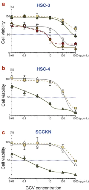
Fig. 2. Cell viabilities after HSV-tk/GCV treatment. Infected cells with either Ad.tk or Ad.β -gal were treated with GCV at concentrations ranging from 0 to 1,000 µg/mL. Uninfected cells without GCV treatment were used as control. Cell viabilities were measured at 96 h after GCV treatment using the MTT assay. Results were calculated as the percentage of viability relative to control. The result represents the mean ± SE of tripli-cates. The IC50 were determined. (a) HSC-3, (b) HSC-4
and (c) SCCKN cell lines. , GCV alone; , Ad.β -gal + GCV; , Ad.tk 1.25 MOI + GCV; , Ad.tk5 MOI + GCV; , Ad.tk 10 MOI + GCV and , Ad.tk 20 MOI + GCV. ���� ��� � �� ��� ���� ������� ��� ������������� � �� �� �� �� ��� � ��� ��� �� � ���� ��� � �� ��� ���� ������� � �� �� �� �� ��� � ��� ��� �� � ���� ��� � �� ��� ���� ������� � �� �� �� �� ��� � ��� ��� �� � ����� ����� �����
�
�
�
fection of Ad.tk at these high titers failed to be cy-totoxic to oral SCC cell lines. Then we performed an MOI escalation study to determine the sublethal
I. Kuratate et al. Cell death of oral SCC by ganciclovir
MOI value of Ad.tk infection (data not shown). The results showed MOIs of 20 and less for the HSC-3 cell line and 2.5 and less for HSC-4 and SCCKN cell lines, respectively.
Cell viability
Cell viability was determined from the colorimetric quantity of viable cells. HSV-tk gene transfer de-creased the median inhibitory concentration (IC50)
at least 2 logs lower in all cell lines (Fig. 2). Cell viability was dependent on GCV concentration. As the HSC-3 cell line only had resistance to Ad-tk infection, we could examine cell viabilities at several ranges of MOI (Fig. 2a). Though the IC50
decreased in proportion to the MOI escalation, the results were not different between MOIs of 20 and 10. This fact suggested the upper limit bounds of MOI escalation of this strategy.
DNA fragmentation
Internucleosomal DNA fragmentation was exam-ined for standard apoptotic hallmarks. They all showed smearing, whose peaks were 72 h, 72 h and 24 h for cell lines HSC-3, HSC-4 and SCCKN, re-spectively (Fig. 3).
Fig. 4. Cell cycle kinetics analysis by flow cytometry. Cells were infected with Ad.tk at MOI 5 for HSC-3, 2.5 for HSC-4 and SCCKN, and followed by 1 µg/mL GCV. Cells were harvested in a time-dependent manner. Uninfected cells without GCV treatment were used as control. The number within each panel shows the percentage of Sub-G1 fractions. C, control.
Fig. 3. DNA fragmentation after HSV-tk/GCV treat-ment. Cells were infected with Ad.tk at MOI 5 for HSC-3, 2.5 for HSC-4 and SCCKN, and followed by 1 µg/mL GCV. Cells were harvested in a time-dependent manner. Uninfected cells without GCV treatment were used as control. DNA extracts were loaded on 2% agarose gel by electrophoresis and visualized by stain-ing with 0.5 µg/mL ethidium bromide. (a) HSC-3, (b) HSC-4 and (c) SCCKN cell lines. M, marker; C, con-trol. � � � �� �� �� �� � � � �� �� �� �� � � � �� �� �� �� ���� ���� ���� ����� ����� �����
�
�
�
���Cell cycle kinetics
Figure 4 shows the results of cell cycle kinetic analysis. The cells broke down severely showing a time-dependent decrease of G1 and G2/M peaks without cell cycle arrest. Sub-G1 fraction, which means hypodiploid cells correspond to dead cells, ���� ����� ����� ����� � � �� �� �� �� ��� ���� ���� ���� ���� ����� ����� ���� ���� ���� ���� ����� ����� ���� ���� ����� ����� ����� �����
I. Kuratate et al. Cell death of oral SCC by ganciclovir Cell death of oral SCC by ganciclovir
Fig. 5. Annexin V-FITC and PI double stainning by flow cytometry. Cells were infected with Ad.tk at MOI 5 for HSC-3, 2.5 for HSC-4 and SCCKN, and followed by 1 µg/mL GCV. Cells were harvested in a time-dependent manner. Uninfected cells without GCV treatment were used as control.
a: Representative results of HSC-4. The number within each panel shows the percentage of each quadrant. C, control. b: Increases in percentage in the lower right quadrant relative to control. , HSC-3, , HSC-4 and , SCCKN. c: Increases in percentage in the upper right quadrant relative to control. , HSC-3, , HSC-4 and , SCCKN.
����
increased in a time-dependent manner in all the cell lines.
Annexin V assay
Annexin V has an affinity for the phosphatidylser-ine that is translocated from the inner leaflet to the outer leaflet of the plasma membrane in the early stage of apoptosis (Martin et al., 1995). During the late stage, the plasma membrane showed increased permeability and PI entered the cytoplasm and stained chromatin indicating necrosis. Figure 5a shows representative kinetics of HSC-4 in the cell lines. The proportion of annexin V-positive/PI-negative cells in the lower right quadrant shows the early stage of apoptosis increased in a time-depen-dent manner (Fig. 5b). The proportion of annexin V-positive/PI-positive in the upper right quadrant shows necrosis also increased in a time-dependent manner (Fig. 5c).
Expression of apoptosis related protein
Protein expression levels of P53, P21, Bax, Bcl-2 and caspase-3 activation were examined by West-ern blot analysis (Fig. 6). P53 was stably expressed in HSC-3 and HSC-4 cell lines, and undetectable at all time points in the SCCKN cell line. P21 and Bax was consistently expressed at any time points in all cell lines. On the other hand, Bcl-2 slightly but steadily increased within 24 h after treatment in HSC-3 and SCCKN cell lines. No caspase-3 cleavage or activation was observed throughout the experiment in any cell lines.
TUNEL method
In situ DNA strand breaks were examined by the TUNEL method. TUNEL -positive cells with brown-labeled nuclei were occasionally detected
I. Kuratate et al.
in only the SCCKN cell line infected with Ad.tk at MOI 2.5 followed by 1 µg/mL GCV after 24 h (Fig. 7). However, we could not evaluate correctly at 48 h and 72 h because most treated SCCKN cells had easily come off in the staining procedure. However, the untreated SCCKN cells showed no TUNEL signal.
Morphological examinations
There were no significant differences in morpho-logical findings in both semi-thin and ultra-thin sections after HSV-tk/GCV treatment among the cell lines. Semi-thin sections of untreated cells showed variations in size and shape and occasion-ally small vesicles in the cytoplasm with a few mi-toses (Fig. 8a). In contrast, a large number of treat-ed cells became swollen with or without membrane disruption, sometimes involving nuclei disruption (Fig. 8b). Cytoplasmic vesicles were markedly increased. There were few cells with chromatin ag-gregation in HSC-4 and SCCKN cell lines.
Ultra-thin sections revealed intact nuclei, intracellular organelles and their membranes in un-treated cells (Fig. 9a). Otherwise, a large number of treated cells contained enlarged and rounded nuclei in which heterochromatin was sparse. Cyto-plasmic membranes were also disrupted (Fig. 9b).
Fig. 7. TUNEL staining of treated SCCKN 24 h after GCV treatment. Cells were infected with Ad.tk at MOI 2.5, and followed by 1 µg/mL GCV. TUNEL-positive cells with brown-labeled nuclei are occasionally detect-ed in only SCCKN cell line (arrows).
Fig. 8. Semi-thin section micrographs of HSC-4 cells. (a) Untreated cells. Some cells occasionally have small vesicles, and some display mitoses. (b) Treated cells 72 h after GCV treatment. Cells were infected with Ad.tk at MOI 2.5, and followed by 1 µg/mL GCV. Cells be-come swollen and intracytoplasmic vesicles remarkably increase. Few cells show chromatin aggregation (ar-rows).
Fig. 6. Western blot analysis of P53, P21, Bax, Bcl-2 and caspase-3 of each cell lines. Cells were infected with Ad.tk at MOI 5 for HSC-3, 2.5 for HSC-4 and SCCKN, and followed by 1 µg/mL GCV. Cells were harvested in a time-dependent manner. Uninfected cells without GCV treatment were used as control. (a) HSC-3, (b) HSC-4 and (c) SCCKN cell lines. C, control.
� � �� �� �� �� � � �� �� �� �� � � �� �� �� �� ����� ���� ����� ���� ����� ���� ���
�
�
�
A few cells showing chromatin aggregation to the nuclear membranes, also displayed cytoplasmic membranes disruption. Treated cells presented structural changes of intracellular organelles in-volving enlargement of mitochondria with the dis-appearance of cristae, vesiculation and vacuoliza-tion of endoplasmic reticulum. Apoptotic bodies were not observed in any of the treated cells.
b
a
Cell death of oral SCC by ganciclovir
Fig. 9. Ultra-thin section micrograph of HSC-4 cells. (a) Untreated cells. (b) Treated cells 72 h after GCV treat-ment. Cells were infected with Ad.tk at MOI 2.5, and followed by 1 µg/mL GCV. A large number of cells contain enlarged and rounded nuclei of which hetero chromatin are sparse. Some of treated cells show chromatin aggrega-tion. But their cytoplasmic membranes are still disruptured. Inset of (b) displays enlargement of mitochondria with disappearance of cristae, vesiculation and vacuolization of endoplasmic reticula. Bars in (a) and (b) = 1 µm.
Discussion
This study clearly demonstrated that HSV-tk/GCV treatment showed diverse but obvious sensitivity to oral SCC cell lines independent of their histologi-cal status (Table 1 and Fig. 2). Oral SCC cell lines displayed high susceptibility to HSV-tk/GCV treat-ment despite low transduction, suggesting possible tumoricidal effects even in the case of inability of 100% gene delivery (Fig. 1). In the HSC-3 cell line, however, HSV-tk/GCV treatment showed the upper limit bounds at MOI 10. As reported previ-ously, a higher expression of HSV-tk gene may not be able to further enhance the tumoricidal effect, though a threshold level of HSV-tk gene expression is necessary for maximum cell killing (Chen et al., 1995).
The tumoricidal mechanism of HSV-tk/GCV treatment has focused on exploring the molecu-lar biological events associated with apoptosis (Samejima and Maruelo, 1995; Wei et al., 1998; Ri-vas et al., 2001). To date, the most reliable method for identifying apoptotic cell death is the detection of the occurrence of internucleosomal DNA strand breaks (Wyllie, 1980). Concerning past HSV-tk/GCV treatment studies, several reported the ex-istence of DNA laddering (Samejima and Maruelo, 1995; Wei et al., 1998), but several reported the
opposite (Kaneko and Tsukamoto, 1995). It is pos-sible that the DNA fragmentation assay might not detect low levels of apoptosis. Confirmation of the phenotype of dying cells is needed by reference to morphology using electron microscopy (Vile et al., 1997). In fact, some cases failed to demonstrate DNA laddering even when the presence of apop-totic cells was further confirmed using electron microscopy (Freeman et al., 1993; Craperi et al., 1999). In the present results, DNA fragmenta-tion showed unspecific DNA degradafragmenta-tion, that is, smearing (Fig. 3). Accordingly, both semi- and ultra-thin examinations revealed the phenotype of dying cells was necrotic in all cell lines.
Previous studies of flow cytometry demon-strated that HSV-tk/GCV treatment caused either S- and/or G2/M-phase cell cycle arrest before undergoing cell death (Kaneko et al., 1995; Wei et al., 1998; Craperi et al., 1999). The present re-sults showed cell death progressing with no cell cycle arrest (Fig. 4). The DNA content of sub-G1 increased in a time-dependent manner. A rapid increase was observed in the HSC-4 cell line at 72 h (42.7%), in which DNA fragmentation definitely showed smearing suggesting no occurrence of reg-ular DNA double-strand breaks. Annexin V has been used as another biological marker to detect the early stage of apoptosis before the occurrence
b
a
I. Kuratate et al. of morphological changes in single cells (Martin
et al., 1995). The present results showed that an-nexin V-positive/PI-negative cells corresponding to apoptotic cells, increased in a time-dependent manner in all cell lines (Fig. 5b), although further semi- and ultra-thin examinations revealed ne-crotic morphological features. While the external-ization of phosphatidylserine during apoptosis has been presented, intracellular events have not been adequately elucidated. Although the meaning of the annexin V-positive reaction induced by HSV-tk/GCV treatment is not contested, this phenom-enon implies the feasibility of inspection and exem-plification by phagocytic cells (Depraetere, 2000; Hanayama et al., 2002). Changes in the properties of their surface membrane would improve the com-bination with a targeting approach such as immu-notherapy.
Since the tumoricidal effect of HSV-tk/GCV treatment is conceivable via DNA damage, the loss of function of p53 can develop resistance to apop-tosis. Li et al. reported, however, the HSV-tk/GCV cytotoxic response did not depend on the expres-sion of a functional p53 (Li et al., 1999; Craperi et al., 1999). Similarly, we demonstrated an obvious tumoricidal effect without either p53 or p21 expres-sion (Figs. 4 and 6). These results were consistent in all cell lines and suggested that p53-dependent cell cycle regulation or apoptosis induction may not correlate with the tumoricidal effect of HSV-tk/ GCV treatment to oral SCC. Alternatively, HSV-tk/GCV treatment might have considerable poten-tial to oral SCC that highly expresses mutated p53 in vivo (Naglar et al., 2002) and in vitro (Sakai and Tsuchida, 1992). A previous exogenous p53 trans-duction study suggested co-expression of p53 did not enhance the cytotoxicity of HSV-tk/GCV treat-ment, although p53 was able to increase amount of apoptosis which was markedly less than the total cell death in vitro (Xie et al., 1999).
Dimerization between Bax and Bcl-2 is an important factor to direct either death promotion or death inhibition. Bax accumulation was reported after GCV exposure in glioma cell lines (Craperi et al., 1999). However, they could not rule out the possible occurrence of necrotic cell death. In
contrast, over-expression of Bcl-2 inhibited HSV-tk/GCV-induced activation of caspase and apop-tosis (Beltinger et al., 2000). However, no syn-chronous correlation could be found between the expression levels of Bcl-2 family and the sensitivity to the oral SCC cell lines.
No cleavage or activation of caspase-3 was detected in oral SCC cell lines. This suggested the existence of an alternative molecular pathway responsible for the tumoricidal effect of HSV-tk/ GCV treatment to oral SCC cell lines even though the route of cell death is unknown.
One exception in the present study was an oc-casional positive TUNEL signal in SCCKN cells (Fig. 7). However, no other evidence of apoptosis was detected with other biological assays. Caution-ary notes suggested that a positive TUNEL signal should not be considered as a specific marker of apoptosis because the assay would also suggest necrotic cell death (Charriaut-Marlangue and Ben-Ari, 1995; Grask-kraupp et al., 1995).
Apoptosis was originally described on the basis of the morphological features by electron microscopy (Kerr et al., 1972) even though some features may also be detectable by light micros-copy. Apoptotic bodies including cellular remnants should be observed either in the intercellular space or within the cytoplasm of intact cells. A large number of treated cells showed disintegration of the cell structure involving irregular scattered hetero-chromatin, swelling of cytoplasm and intracellular organelles, and cytoplasmic membrane disruption in all cell lines. Furthermore, intact cells contain-ing apoptotic bodies that deposit the fragmented components from neighboring dead cells were not found. Based on the annexin V/PI double staining at 72 h, the HSC-4 cell line should include the cells of early stage apoptosis of 19.1% (Fig. 5). How-ever, we could not find firm evidence of apoptosis in semi- and ultra-thin section examinations. Con-sequently, apoptosis may not play a central role in the tumoricidal mechanism. The hypothesis that the bystander effect is mediated by phagocytosis is unacceptable for oral SCC cells in vitro.
A previous study using Fas ligand demon-strated an artificial deficiency of caspase-8 resulted
Cell death of oral SCC by ganciclovir
in a switch of cell death from apoptosis to necrosis in Jurket cell lines (Kawahara et al., 1998). This implies that caspase activation itself was dispens-able for determining cell death. Kitanaka and Kuchino (1999) suggested the existence of caspase-independent programmed cell death with necrotic-like morphology, that can be activated either alone or in concert with the caspase-dependent apoptotic program. Differential sensitivity and the type of cell death induced by HSV-tk/GCV treatment ap-pear to be dependent upon cell-type difference, in addition to the status of the apoptosis-related gene. In summary, we demonstrated that oral SCC cell lines exhibited an obvious sensitivity to HSV-tk/GCV treatment suggesting the presence of a p53- and caspase-3-independent death signaling pathway. However, the mechanism and the molec-ular pathway responsible for HSV-tk/GCV-induced cell death are still unknown. A better understand-ing of the cell death mechanism is essential to establish an appropriate and effective modality in future studies.
Acknowledgments: All authors thank Professor Kenzo Sato (Division of Molecular Biology, Department of Molecular and Cellular Biology, School of Medicine, Tottori University Faculty of Medicine) for his appropri-ate advice and kindly providing two replication-deficient recombinant adenovirus vectors, Ad.β -gal and Ad.tk. We also thank Professor Kazuo Yamada (Division of Medical Biochemistry, Department of Pathophysiologi-cal and Therapeutic Science, School of Medicine, Tottori University Faculty of Medicine) for his invaluable ad-vice, and Mr. Norihisa Itaki (Division of Organ Pathol-ogy, Department of Microbiology and PatholPathol-ogy, School of Medicine, Tottori University Faculty of Medicine) for his skilful technical assistance. Finally, we thank all the members of Division of Organ Pathology, Department of Microbiology and Pathology, School of Medicine, Tottori University Faculty of Medicine for their helpful discussions.
References
1 Beltinger C, Fulda S, Kammertoens T, Meyer E, Uckert W, Debatin KM. Herpes simplex virus thymidine kinase/ganciclovir-induced apoptosis involves ligand-independent death receptor
aggrega-tion and activaaggrega-tion of caspases. Proc Natl Acad Sci U S A 1999;96:8699–8704.
2 Beltinger C, Fulda S, Kammertoens T, Uckert W, Debatin KM. Mitochondrial amplification of death signals determines thymidine kinase/ganciclovir-triggered activation of apoptosis. Cancer Res 2000; 60:3212–3217.
3 Bi WL, Parysek LM, Warnick R, Stambrook PJ. In vitro evidence that metabolic cooperation is respon-sible for the bystander effect observed with HSV tk retroviral gene therapy. Hum Gene Ther 1993;4: 725–731.
4 Borrelli E, Heyman R, Hsi M, Evans RM. Target-ing of an inducible toxic phenotype in animal cells. Proc Natl Acad Sci U S A 1988;85:7572–7576. 5 Boucher PD, Ruch RJ, Shewach DS. Differential
ganciclovir-mediated cytotoxicity and bystander killing in human colon carcinoma cell lines express-ing herpes simplex virus thymidine kinase. Hum Gene Ther 1998;9:801–814.
6 Charriaut-Marlangue C, Ben-Ari Y. A cautionary note on the use of the TUNEL stain to determine apoptosis. Neuroreport 1995;7:61–64.
7 Chen CY, Chang YN, Ryan P, Linscott M, McGarrity GJ, Chiang YL. Effect of herpes simplex virus thy-midine kinase expression levels on ganciclovir-me-diated cytotoxicity and the “bystander effect”. Hum Gene Ther 1995;6:1467–1476.
8 Colombo BM, Benedetti S, Ottolenghi S, Mora M, Pollo B, Poli G, et al. The “bystander effect”: asso-ciation of U-87 cell death with ganciclovir-mediated apoptosis of nearby cells and lack of effect in athy-mic athy-mice. Hum Gene Ther 1995;6:763–772. 9 Craperi D, Vicat JM, Nissou MF, Mathieu J, Baudier
J, Benabid AL, et al. Increased bax expression is associated with cell death induced by ganciclovir in a herpes thymidine kinase gene-expressing glioma cell line. Hum Gene Ther 1999;10:679–688. 10 Depraetere V. “Eat me” signals of apoptotic bodies.
Nat Cell Biol 2000;2:E104.
11 Elion GB. Mechanism of action and selectivity of acyclovir. Am J Med 1982;73:7–13.
12 Freeman SM, Abboud CN, Whartenby KA, Packman CH, Koeplin DS, Moolten FL, et al. The “bystander effect”: tumor regression when a fraction of the tumor mass is genetically modified. Cancer Res 1993;53:5274–5283.
13 Fukui T, Hayashi Y, Kagami H, Yamamoto N, Fukuhara H, Tohnai I, et al. Suicide gene therapy for human oral squamous cell carcinoma cell lines with adeno-associated virus vector. Oral Oncol 2001;37:211–215.
14 Glaser T, Castro MG, Lowenstein PR, Weller M. Death receptor-independent cytochrome c release
I. Kuratate et al. and caspase activation mediate thymidine kinase
plus ganciclovir-mediated cytotoxicity in LN-18 and LN-229 human malignant glioma cells. Gene Ther 2001;8:469–476.
15 Grasl-Kraupp B, Ruttkay-Nedecky B, Koudelka H, Bukowska K, Bursch W, Schulte-Hermann R. In situ detection of fragmented DNA (TUNEL assay) fails to discriminate among apoptosis, necrosis, and autolytic cell death: a cautionary note. Hepatology 1995;21:1465–1468.
16 Hanayama R, Tanaka M, Miwa K, Shinohara A, Iwamatsu A, Nagata S. Identification of a factor that links apoptotic cells to phagocytes. Nature 2002;417:182–187.
17 Kaneko Y, Tsukamoto A. Gene therapy of hepato-ma: bystander effects and non-apoptotic cell death induced by thymidine kinase and ganciclovir. Can-cer Lett 1995;96:105–110.
18 Katabi M, Yuan S, Chan H, Galipeau J, Batist G. The nonapoptotic pathway mediating thymidine kinase/ganciclovir toxicity is reduced by signal from adenovirus type 5 early region 4. Mol Ther 2002;5: 170–176.
19 Kawahara A, Ohsawa Y, Matsumura H, Uchiyama Y, Nagata S. Caspase-independent cell killing by Fas-associated protein with death domain. J Cell Biol 1998;143:1353–1360.
20 Kerr JF, Wyllie AH, Currie AR. Apoptosis: a basic biological phenomenon with wide-ranging implica-tions in tissue kinetics. Br J Cancer 1972;26:239– 257.
21 Kitanaka C, Kuchino Y. Caspase-independent pro-grammed cell death with necrotic morphology. Cell Death Differ 1999;6:508–515.
22 Lanuti M, Gao GP, Force SD, Chang MY, El Kouri C, Amin KM, et al. Evaluation of an E1E4-deleted adenovirus expressing the herpes simplex thymidine kinase suicide gene in cancer gene therapy. Hum Gene Ther 1999;10:463–475.
23 Li PX, Ngo D, Brade AM, Klamut HJ. Differential chemosensitivity of breast cancer cells to ganciclo-vir treatment following adenoganciclo-virus-mediated herpes simplex virus thymidine kinase gene transfer. Can-cer Gene Ther 1999;6:179–190.
24 Martin SJ, Reutelingsperger CP, McGahon AJ, Rader JA, van Schie RC, LaFace DM, et al. Early redistri-bution of plasma membrane phosphatidylserine is a general feature of apoptosis regardless of the initiat-ing stimulus: inhibition by overexpression of Bcl-2 and Abl Iodine-124 labelled Annexin-V as a potential radiotracer to study apoptosis using positron emission
tomography. J Exp Med 1995;182: 1545–1556. 25 Moolten FL, Wells JM, Heyman RA, Evans RM.
Lymphoma regression induced by ganciclovir in mice bearing a herpes thymidine kinase transgene. Hum Gene Ther 1990;1:125–134.
26 Nagler RM, Kerner H, Laufer D, Ben-Eliezer S, Minkov I, Ben-Itzhak O. Squamous cell carcinoma of the tongue: the prevalence and prognostic roles of p53, Bcl-2, c-erbB-2 and apoptotic rate as related to clinical and pathological characteristics in a retro-spective study. Cancer Lett 2002;186:137–150. 27 O’Malley BW, Cope KA, Chen SH, Li D, Schwarta
MR, Woo SL. Combination gene therapy for oral cancer in a murine model. Cancer Res 1996;56: 1737–1741.
28 Rivas C, Miller AR, Collado M, Lam EW, Apperley JF, Melo JV. BCR-ABL-expressing cells trans-duced with the HSV-tk gene die by apoptosis upon treatment with ganciclovir. Mol Ther 2001;3:642– 652.
29 Sakai E, Tsuchida N. Most human squamous cell carcinomas in the oral cavity contain mutated p53 tumor-suppressor genes. Oncogene 1992;7:927– 933.
30 Samejima Y, Meruelo D. “Bystander killing” in-duces apoptosis and is inhibited by forskolin. Gene Ther 1995;2:50–58.
31 Vile RG, Castleden S, Marshall J, Camplejohn R, Upton C, Chong H. Generation of an anti-tumour immune response in a non-immunogenic tumour: HSVtk killing in vivo stimulates a mononuclear cell infiltrate and a Th1-like profile of intratumoural cytokine expression. Int J Cancer 1997;71:267–274. 32. Wei SJ, Chao Y, Hung YM, Lin WC, Yang DM,
Shih YL, et al. S- and G2-phase cell cycle arrests and apoptosis induced by ganciclovir in murine mel-anoma cells transduced with herpes simplex virus thymidine kinase. Exp Cell Res 1998;241:66–75. 33 Wyllie AH. Glucocorticoid-induced thymocyte
apoptosis is associated with endogenous endonucle-ase activation. Nature 1980;284:555–556.
34 Xie Y, Gilbert JD, Kim JH, Freytag SO. Efficacy of adenovirus-mediated CD/5-FC and HSV-1 thymi-dine kinase/ganciclovir suicide gene therapies con-comitant with p53 gene therapy. Clin Cancer Res 1999;5:4224–4232.
Received June 20, 2005; accepted August 29, 2005 Corresponding author: Mitsuhiko Osaki, PhD