Exploration of new bioactive
compounds produced by bacteria in
pathogenic lineages
Toyama Prefectural University
Dandan Li
CHAPTER 1 Introduction ... 0
1-1 Background ... 1
1-2 Natural products as a major source of available drugs ... 1
1-3 Source exploration of natural products ... 3
1-3-1 Burkholderia as a source of natural products ... 6
1-3-2 Vibrio as a source of natural products ... 8
1-3-3 The objective of this thesis ... 8
CHAPTER 2 Two new 2-alkylquinolones, inhibitory to the fish skin ulcer pathogen Tenacibaculum maritimum, produced by a rhizobacterium of the genus Burkholderia sp. ... 13
2-1 Background ... 14
2-2 Results and Discussion ... 16
2-2-1 Fermentation and isolation ... 16
2-2-2 Structure Determination... 16
2-2-3 Bioactivity ... 19
2-3 Conclusion ... 20
2-4 Experimental ... 21
2-4-1 General experimental procedures ... 21
2-4-2 Collection of Burkholderia strains and broth screening ... 21
2-4-3 Fermentation, extraction, and isolation ... 21
2-4-4 Evaluation of antimicrobial activity ... 23
2-5 Spectral data ... 27
CHAPTER 3 4-Hydroxy-3-methyl-2(1H)-quinolone, originally discovered from a Brassicaceae plant, produced by a soil bacterium of the genus Burkholderia sp.: determination of a preferred tautomer and antioxidant activity ... 38
3-1 Background ... 39
3-2 Results and Discussion ... 41
3-2-1 Fermentation and isolation ... 41
3-2-2 Structure Determination... 41
3-2-3 Synthesis of 9 ... 43
3-2-4 Bioactivity ... 45
3-3 Conclusion ... 46
3-4 Experimental ... 47
3-4-1 General experimental procedures ... 47
3-4-2 Microorganism ... 47
3-4-3 Fermentation and isolation ... 48
3-4-4 Evaluation of Fe3+ binding activity ... 48
3-4-5 Antibacterial assay ... 49
3-5 Spectral data ... 53 CHAPTER 4 Three new O-isocrotonyl-3-hydroxybutyric acid congeners produced by a sea anemone-derived marine bacterium of the genus
Vibrio ... 74
4-1 Background ... 75
4-2 Results and Discussion ... 77
4-2-1 Fermentation and isolation ... 77
4-2-2 Structure Determination... 77
4-2-3 Absolute Configuration ... 80
4-4 Experimental ... 82
4-3 Conclusion ... 82
4-4-1 General experimental procedures ... 83
4-4-2 Biological material ... 83
4-4-3 Fermentation and isolation of 19-22 ... 83
4-4-4 Preparation of (S)- and (R)-Phenylglycine Methyl Ester Amides ... 85
4-4-5 Antibacterial Assay ... 86 4-4-6 Cytotoxicity Assay ... 86 4-5 Spectral data ... 90 CHAPTER 5 Conclusion ... 127 Acknowledgements ... 132 Publication List ... 133
CHAPTER 1
1-1 Background
Increased longevity is a significant symbol of human progress. The life expectancy had been extended 24 years from the year 1950 to the year 2017 [1]. Some countries have projects to exceed life expectancy over 85 and large numbers of people are now living into their ninth decades [2]. These advances reflect the decline of disease mortality. However, the world continues to face the challenges from diseases such as bacterial infectious diseases, allergy, and lifestyle diseases based on the day to day habits of people [3-5]. According to the WHO, more than 10 million people for each year die from lifestyle diseases caused by smoking of tobacco, alcohol use, or a simple lack of physical activity and cholera caused by bacterium V. cholerae killed tens of millions of people in the last seven outbreaks [6, 7]. In addition, allergy such as allergic rhinitis, food allergy, atopic dermatitis, and asthma have always given a serious damage on our life.
1-2 Natural products as a major source of available drugs
Natural products have been studied as medicines for hundreds of years in our history. On account of the fact of excellent pharmacological activity and good biocompatibility of natural products, over 30% clinical drugs come from natural products [8]. Although the synthetic drugs take a quite large proportion in clinical therapy, the inspirational sources of many of them almost came from structure diversities and various biological activities of natural products.
Natural products, especially those from microorganisms, have served as a rich and indispensable resource of medicines. Penicillin G (Figure 1-1), the first antibiotic, isolated from a fungus Penicillium in the middle of the 19th century, dramatically improved the prognosis of infectious diseases. It is well known that penicillin has saved millions of lives in World War II. Subsequent researches showed that penicillin
contained a characteristic four-membered β-lactam ring and this ring can be used to inhibit the formation of new cell wall. This is the reason why penicillin kills pathogenic bacteria. The application of penicillin has effectively controlled a number of bacterial infectious diseases such as pneumonia, syphilis, cellulitis and so on.
Figure 1-1. Microbial metabolites used as clinical medicines or drug leads.
Streptomycin, discovered from Streptomyces, is the first antibiotic to treat tuberculosis, which at that time was a thought as deadly illness. Because the benefit brought by these two antibiotics were so huge, Fleming and Waksman, the discovers of these antibiotics, received Nobel Prizes. In addition to these, a hypolipidemic agent mevastatin, an immunosuppressant FK506, and anthelminthic avermectin all provided new remedies, which were not available before these drugs were discovered.
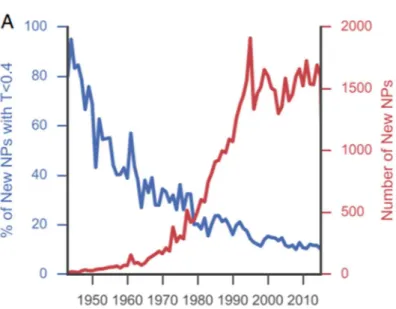
The implementation of NMR, high performance liquid chromatography, and mass spectrometry in the mid-1980s at natural product chemistry research accelerated the
discovery of new natural products (Figure 1-2) [9]. However, in contrast to this, the number of novel natural products, as judged by Tanimoto similarity scores, are declining year by year. In addition, no antibiotics based on natural products with novel molecular skeletons were approved over the near 30 years. To our disappointment, new antibiotics that have recently entered the market, such as daptomycin and tiacumicin B1, have their discovery origins back in the 1980s [9].
Figure 1-2. The relative data about the number of new compound isolation (red line) and the rate of novel compound isolation (blue line) were relisted.
The decline in the ratio of discovering new molecular scaffolds is mainly because the major traditional sources, fungi and actinomycetes, have been exploited so extensive that the room for discovering molecules with new skeletons becomes quite limited. Thus, to address this problem, exploring new resource would be a necessary option to be taken.
1-3 Source exploration of natural products
of the taxa should directly determine the output of natural product discovery. Exploring those closely related to the already proven taxa is a conceivable choice, which has especially been often attempt in plant product research [10].For example, the plants of producing alkaloids, terpenoids, sinapyl alcohols, and sinapyl alcohol derivatives were rather continuous at the morphological characteristics [10].
Another choice is to find taxa which are phylogenetically distant but have similar life history. So far, nearly three-quarters of the total antibiotics have been isolated from actinomycetes and fungi [11]. There are many traits that are common to the both groups, such as energy metabolism, filamentous growth, spore formation, ability to degrading persistent plant-derived biopolymers, etc. these traits are shared by bacteria of the class Ktedonobacteria within phylum Chloroflexi, and several new natural products were reported from this group [12].
Another important trait is the high incidence of pathogenic and symbiotic species in these microbes. For example, potato scab, a common and disfiguring disease of potato tubers that affect potatoes and other root vegetables, are caused by actinomycetes of the genus the Streptomyces. Green mold, a noxious postharvest disease of citrus, was caused by Penicillium digitatum within the kingdom Fungi. Furthermore, many of them also play important roles in nature as symbionts like ant symbiotic actinobacteria Pseudonocardia, which protected the ants’ fungal garden and plant root symbiotic fungus Arbuscular mycorrhiza, which enhanced host plant acquisition of mineral nutrients, as shown in Figure 1-3.
Figure 1-3. The pathogenicity and symbiotic life forms shared by actinomycetes and fungi.
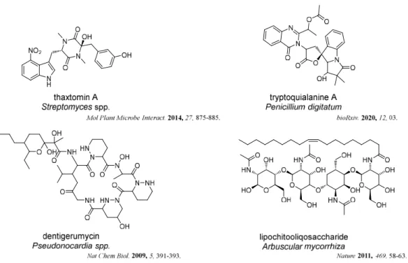
Recent studies have shown that these relationships are maintained through the production of low-molecular weight compounds (Figure 1-4). For example, thaxtomin A, produced by the potato pathogen Streptomycin spp.,showed a positive correlation between their pathogenicity and thaxtomin production. Tryptoquialanine A, isolated from the pathogen of citrus fruits Penicillium digitatum, was shown to completely inhibit the citrus seed germination. Dentigerumycin, a bacterial mediator, and lipochitooliqosaccharide, a symbiotic signal, were produced by the symbiotic actinobacteria Pseudonocardia and symbiotic fungus Arbuscular mycorrhiza, respectively.
Figure 1-4. Natural products from host-symbionts/pathogens interaction.
Considering that the evolutional origin of symbiosis is pathogenic infection [13], there is no wonder if symbiotic and pathogenic species are co-classified in the same taxon or a symbiotic species sometimes shows pathogenicity to other host organisms. Thus, bacteria in pathogenic lineages are expected to have the larger capacity to produce natural products than free-living organisms.
Most of the microbial natural products were found from Gram-positive bacteria, and chemical studies on Gram-negative bacteria has not been carried out extensively [14].Based on these backgrounds,Gram-negative proteobacteria in pathogenic lineages are expected to be promising, and two genera Burkholderia and Vibrio, were especially noted, as described below.
1-3-1 Burkholderia as a source of natural products
The genus Burkholderia, belonging to the class Betaproteobacteria, occupies a vast niche in terrestrial ecosystems as free-living organisms or in association with eukaryotic hosts such as humans, animals, plants, and fungi. In recent years,
Burkholderia has specifically grabbed our attention because some species in this genus are confirmed as human pathogens, such as B. cepacia complex causing lung infections in immunocompromised people and B. mallei leading to glanders [15-17].
Some Burkholderia species are beneficial to the host organisms. For instance, B. ambifaria and B. caribensis can promote the grain crop amaranth growth through nitrogen fixation[18]. In addition, some natural products have been discovered (Figure 1-5), which seem to be involved in the interaction with their hosts such as toxoflavin, one of the key virulence factors produced by the plant pathogen B. gladioli [19], malleiactone, a virulence factor from B. pseudomallei [20], and burkholdine, a potent antifungal peptide from B. ambifaria [21].
Figure 1-5. Natural products from hosts-Burkholderia/Vibrio interaction.
The gene mining technology has confirmed that Burkholderia genomes encode a plethora of natural products with potential therapeutic relevance and biotechnological applications. It is thought as one of the emerging, gifted producers of natural products because of the metabolically diverse multifaceted traits [22-24].
1-3-2 Vibrio as a source of natural products
The genus Vibrio, belonging to the class Gammaproteobacteria, is a genetically and metabolically diverse group of heterotrophic bacteria that are ubiquitously distributed in the oceans and represents a large fraction (0.5–5%) of the total bacterial community in major oceans around the world [25]. Vibrio is an emerging health threat linked to increasing seawater temperatures [26]. Although less well-known, the interactions between Vibrio and ocean planktons play a major role for the coastal ecology and human infection potential.
The genus Vibrio have cataloged 128 species at the time of writing and more than 12 are associated with food poisoning [27-29]. Among these species, Vibrio cholerae are the most feared and the most extensively studied pathogen [30, 31]. Thus, other related species such asV. parahaemolyticus, V. vulnificus, V. alginolyticus, V. damsela, V. fluvialis, V. furnissii, V. hollisae, V. metschnikovii, and V. mimicus have drawn special attention in the last decade [31]. It is reported that 70% food poisoning in Japan are caused by seafood such as fish and shellfish, contaminated with V. parahaemolyticus [32].
On the other hand, studies have shown that some symbiotic species of Vibrio have the capacity of fixing nitrogen/phototrophy/producing plant hormone [33-35]. Meanwhile, some natural products were produced because of the interaction between the genus Vibrio and host in Figure 1-5 like autoinducers, GQO97-T and cyclo (d-his-l-pro), which can regulate the virulence and fitness [36-38].
1-3-3 The objective of this thesis
As discussed already, exploitation of new drug resources, following to actinomycetes and fungi, are requested, and Gram-negative proteobacteria in pathogenic lineages were proposed as one of the promising candidates.
species, two from the genus Burkholderia and one from the genus Vibrio, each representing those of terrestrial and marine origins, were attempted in this thesis, which will be detailed in the following chapters.
References
1. United Nations Department of Economic and Social Affairs PD World Population
Prospects: The 2017 Revision, DVD Edition.
2. Foreman, K. J., Marquez, N., Dolgert, A., Fukutaki, K., Fullman, N., McGaughey, M.
Lancet. 2018, 392, 2052–2090.
3. Tabish, S. A. J Cardiol Curr Res. 2017, 9, 326.
4. Byrne, J. P. Encyclopedia of Pestilence, Pandemics, and Plagues: A–M. ABC-CLIO.
p. 99.
5. McConnell, T. H. The Nature of Disease: Pathology for the Health Professions. 2007,
p. 159.
6. WHO. 2017. Noncommunicable diseases. Fact Sheet.
7. Ali, M., Nelson, A. R., Lopez, A. L., Sack, D. PLOS. 2015, 9, 3832-3845.
8. Newman, D. J., Cragg, G. M. J. Nat. Prod. 2012, 75, 311–335.
9. Pye, C. R., Bertin, M. J., Lokey, R. S., Gerwick, W. H., Linington, R. G. PNAS. 2017,
114, 5601–5606.
10. Nagano, H., Hanai, R., Yamada, H., Matsushima, M., Miura, Y., Hoya, T., Kuroda, C.
Chem. Biodivers. 2012, 9, 789–805.
11. Ding, T., Yang, L. J., Zhang, W. D., Shen, Y. H. RSC Adv. 2019, 9, 21964–21988.
12. Igarashi, Y., Yamamoto, K., Ueno, C. Yamada, N., Xiaohanyao, Ye., Zhou, T.,
Harunari, E., Oku, N. J Antibiot. 2019, 72, 653–660.
13. Nishiguchi, M. K., Hirsch, A. M., Devinney, R., Vedantam, G., Riley, M. A., Mansky, L.
14. Buijs, Y., Bech, P. K., Vazquez-Albacete, D., Bentzon-Tilia, M., Sonnenschein, E. C.,
Gram, L., Zhang, S.D. Nat. Prod. Rep. 2019, 36, 1333-1350.
15. Parke, J. L., Gurian-Sherman, D. Annu. Rev. Phytopathol. 2001, 39, 225−258.
16. Mahenthiralingam, E., Baldwin, A., Dowson, C. G. J. Appl. Microbiol. 2008, 104,
1539−1551.
17. Larsen, J. C., Johnson, N. H. Mil. Med. 2009, 174, 647−651.
18. Parra-Cota, F. I., Peña-Cabriales, J. J., de los Santos-Villalobos, S.,
Martínez-Gallardo, N. A., Delano-Frier, J. P. PLoS One. 2014, 9, e88094.
19. Li, X., Li, Y., Wang, R., Wang, Q., Lu, L. Appl. Environ. Microbiol .2019, 3, 106-119.
20. Klaus, J. R., Deay, J., Neuenswander, B., Hursh, W., Gao, Z., Bouddhara, T.,
Chandler, J. R. J. Bacteriol. 2018, 200, 8-18.
21. Tawfik, K. A., Jeffs, P., Bray, B., Dubay, G., Falkinham, J. O., Mesbah, M., Schmidt,
E. W. Org. Lett. 2010, 12, 664–666.
22. Pidot, S. J., Coyne, S., Kloss, F., Hertweck, C. Int. J. Med. Microbiol. 2014, 304,
14−22.
23. Baltz, R. H. J. Ind. Microbiol. Biotechnol. 2017, 44, 573−588.
24. Liu, X., Cheng, Y. Q. J. Ind. Microbiol. Biotechnol. 2014, 41, 275− 284.
25. Takemura, A. F., Chien, D. M. Polz, M. F. Front Microbiol. 2014, 5, 38.
26. Diner, R. E., Rabines, A. J., Zheng, H., Steele, J. A., Griffith, J. F., Allen, A. E.
General Microbiology 2020 (in press). dol: 10.21203/rs.2.18876/v1
27. Parte, A. C. Int. J. Syst. Evol. Microbiol. 2018, 68, 1825–1829.
Al-saari, N., Mino, S., Sawabe, T. Aquaculture 2019, 512, 734289
29. Ceccarelli, D., Amaro, C., Romalde, J. L., Suffredini, E., Vezzulli, L. Food
Microbiology: Fundamentals and Frontiers. 2019, p. 347–388.
30. Kaper, J.B., Morris, G.J., Levine, M. M. Clin Microbiol Rev.1995, 8, 48–86.
31. Chakraborty, S., Nair, G. B., Shinoda, S. Rev Environ Health. 1997, 12, 63–80.
32. Kaneko, T., Colwell, R. R. J. Bacteriol. 1973, 113, 24-32.
33. Kang, S. R., Srinivasan, S., Lee, S. S. Int. J. Syst. Evol. Microbiol. 2015, 65, 3552–
3557.
34. Gómez-Consarnau, L., Akram, N., Lindell, K., Pedersen, A., Neutze, R., Milton, D. L.,
González, J. M., Pinhassi, J. PLoS Biol. 2010, 8, e1000358.
35. Gutierrez, C. K., Matsui, G. Y., Lincoln, D. E., Lovell, C. R. Appl. Environ. Microbiol.
2009, 75, 2253–2258.
36. Al-Zereini, W., Laatsch, H., Anke, H. Antibiotics 2010, 63, 297–301.
37. Sandy, M., Han, A., Blunt, J., Munro, M., Haygood, M., Butler, A. J. Nat. Prod. 2010,
73, 1038–1043.
CHAPTER 2
Two new 2-alkylquinolones, inhibitory
to the fish skin ulcer pathogen
Tenacibaculum maritimum, produced
by a rhizobacterium of the genus
2-1 Background
As discussed in Chapter 1, a higher capability of producing bioactive molecules is expected from bacteria in pathogenic lineages, which is exemplified by actinomycetes and fungi that include many pathogenic or symbiotic species. To examine this prospect, the genera Burkholderia and Vibrio, representing terrestrial- and marine-inhabiting pathogenic lineages, respectively, were chosen as the subjects of this study.
As the first project of this thesis, Burkholderia strains, of vegetable rhizosphere origin, were examined in this chapter. These strains were provided by Associated Professor Masafumi Shimizu at Gifu University, who studies the beneficial effect of rhizobacteria for Alliaceae plants on the growth Cucurbitaceae plants, which is applied to companion planting of Melon and Welsh onion in Hokkaido and calabash (L. siceraria) and the same in Tochigi [1-4].
Using Pseudomonas agar supplemented with C-F-C, Burkholderia strains were selectively collected from rhizosphere soils of Welsh onion (Allium fistulosum), Chinese chive (Allium tuberosum), and cucumber (Cucumis sativus). In total, 38 strains were cultured in four different media, which were designed by Ms. Atsumi Hasada of this laboratory based on the known recipes for fermentation of plant pathogenic bacteria, to give 144 broth extracts. These extracts were screened against a panel of 9 pathogenic microorganisms consisted of four Gram-negative bacteria, one yeast, and four fungi, which results in the detection of antimicrobial activity from most of the extracts. Among these, the fermentation product of strain MBAF1239, identified as Burkholderia sp. within the B. cepacia complex based on the 16S rRNA gene sequence, showed wide antimicrobial spectrum with high potency. Intrigued by this result, the responsible constituents were pursued in the following study.
Figure 2-1. Burkholderia cepacia MBAF 1239 on YM agar.
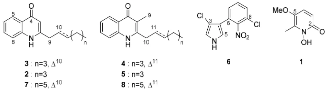
The antimicrobial testing was conducted throughout the whole isolation process. The strain MBAF 1239 was cultured in IMM-HS and purification was carried out as shown in scheme 2-1. As a result, two new 2-alkylquinolones 3 and 7 along with 6 known compounds were isolated by HPLC and their structures were determined by NMR and MS (Figure 2-2).
Figure 2-2. Structures of 1-8.
2-2 Results and Discussion
2-2-1 Fermentation and isolation
The seed culture V22 medium was used for the growth of the strain MBAF1239 for 2 days. Then the production medium IMM-HS, which was designed based on the composition of IMM [5] and HS media [6] for metabolite production, was prepared and cultured for 4 days. After fermentation, the n-BuOH was added to the fermented liquid cultures for extraction of metabolites. Then the combined butanol layer was dried and fractionated successively by solvent-partitioning to give n-hexane, 90% aqueous MeOH, and 60% aqueous MeOH-soluble fractions. The antimicrobial testing showed that the second fraction was the most potent against Rhizopus oryzae (fungal pathogen of rice seedling blight), Trichophyton rubrum (dermatophytosis pathogen), and Tenacibaculum maritimum (skin ulcer of marine fish). Then the second fraction was fractionated by ODS flash chromatography and purified by HPLC to give eight metabolites 1-8 (Scheme 2-1).
2-2-2 Structure Determination
Compound 7 was isolated as colourless liquid and gave a molecular formula of C18H23NO based on a HRESITOFMS ion peak ([M+H]+ m/z 270.1855, +0.3 mmu). The spectra of 1H, 13C, and HSQC showed that the existence of five aromatic (
H/C 8.34/126.3, 7.57/131.9, 7.32/123.6, 7.26/116.8, and 6.18/109.5) and two olefinic (H/C
5.79/137.7 and 5.54/123.0) methines, six aliphatic methylenes (H/C 3.37/37.4, 2.12/32.5, 1.43/29.1, 1.33/28.9, 1.31/22.6, and 1.30/31.7), and a methyl (H/C 0.89/14.1) group, leaving one carbonyl (C 179.0) and three aromatic resonances (C 149.9, 139.4, and 125.3) as quaternary carbons (Table 1). Based on the rest of two unsaturation degrees, two rings constituting a fused bicycle were confirmed by the number of available aromatic carbons (=eleven). In addition, UV spectrum showed an absorption around the 340-320 nm region, corresponding to a 4-quinolone substructure [7]. Additionally, 2D NMR spectra also supported this assignment. The 13C NMR spectrum showed weak resonances at the chemical shifts 157.6, 150.8, 134.6, and 118.4 ppm and the 1H NMR spectrum showed a broaden secondary amine proton signal ( 8.07). The HMBC correlation from H3 to C9 supported a 2-nonenyl group (C9~C17) at C2 (Figure 2-3).
Figure 2-3. Key COSY (bold line) and HMBC (arrow) correlations for 7.
The 1H NMR spectrum showed a blunt signal shape during the olefinic protons H10 and H11 and the geometry of double bond at C10 cannot be determined due to the amphibolous coupling constant. However, a literature survey found that an E-isomer, burkholone has a chemical shift of 32.5 ppm at C12 while a Z-isomer, haplacutine F exhibited the same at 27.7 ppm (Figure 2-4). Because C12 of 7 resonate at 32.5 ppm, Thus, the geometry at C12 was concluded as E and this compound 7 was determined to be (E)-2-(non-2-en-1-yl) quinolin-4(1H)-one.
Table 2-1: NMR data for compound 7 in CDCl3 (in ppm).
pos. 13C , mult. (J in Hz), integration COSY HMBC (1H to 13C)
1 8.07, br, 1H 2 149.9 3 109.5 6.18, s, 1H 2, 4a, 9 4 179.0 4a 125.3 5 126.3 8.34, d (7.9), 1H 6 4, 7, 8a 6 123.6 7.32, t, 7.5, 1H 5, 7 4a, 8 7 131.9 7.57, brs, 1H 6 8 116.8 7.26, ovla 8a 139.4 9 37.4 3.37, brs, 2H 10 10 123.0 5.54, m, 1H 9, 11 11 137.7 5.79, m, 1H 10,12 12 32.5 2.12 ddd (7.2, 6.5, 6.3), 2H 11, 13 10, 11, 13, 14 13 29.1 1.43, m, 2H 12, 14 14 28.9 1.33, m, 2H 15 31.7 1.30, m, 2H 16 22.6 1.31, m, 2H 17 17 14.1 0.89, t (6.5), 3H 16 15, 16
aSignal overlapped by a residual solvent peak.
Figure 2-4. Referential 13C chemical shifts of an allylic carbon in burkholone [8]
and haplacutine F [9].
Compound 3 was isolated as colorless solid and gave a molecular formula of C16H19NO based on the ESITOFMS ion peak m/z 242 and m/z 240 in the positive and negative modes, respectively. Compound 3 and 7 showed the similar 1H NMR spectra except for the intensity of the methylene resonances between 1.48 and 1.23 ppm: which amounted for eight protons in 7.This result implied that compound 3 is a congener of compound 7 having a two-methylene shorter alphatic chain. Further structure analysis by 2D NMR experiments confirmed the expected structure. The geometry of the double bond was similarly determined as discussed for 7. Based on the above analysis about
the double bond, compound 3 was determined as (E)-2-(hept-2-en-1-yl) quinolin-4(1H)-one.
The other isolated metabolites were determined to be known compounds based on 1D, 2D NMR data and MS spectra: 1 as 2-heptylquinolin-4(1H)-one [10], 2 as (E)-2-(hept-2-en-1-yl)-3-methylquinolin-4(1H)-one [6], 4 as PSC-C or 2-heptyl-3-methylquinolin-4(1H)-one) [10], 5 as PSC-D or (E)-3-methyl-2-(non-2-en-1-yl)quinolin-4(1H)-one) [11], pyrrolnitrin (6) [12], and BN-227 (8) [22] (Figure 2-2).
The biological source of 4-quinolone is abundantly distributed in nature, including Rutaceae plants [15], Gram-positive [16, 17] and Gram-negative bacteria [18-20], and a marine sponge [21]. In addition, 4-quinolone is a common core of synthetic antibactericides [14] and among of them, 2-alkyl-4-quinolones are the classics of antibiotics. Previous studies of bioactivity suggested that the substituted 4-quinolones play an important role as antibacterial, antifungal, iron-chelating, and autoinducer agents to improve the competition and survival of the producers [22]. Additionally, drug discovery assays have verified 5-lipoxygenase inhibitory activity [23], plant-growth promoting activity [22, 24], and IGF dependent cell-specific cytotoxicity [8]. Recent metabolomics approach revealed the presence of more than 50 analogs in this group [25] for 4-quinolone derivatives. Compounds 3 and 7 have been detected by MS spectrometry in previous studies [19, 26-28], although detailed structure characterization was not made. In this study, compounds 3 and 7 were isolated for the first time, which enabled rigorous structure characterization by spectroscopic method. In addition, their bioactivity (see below) was evaluated for the first time.
2-2-3 Bioactivity
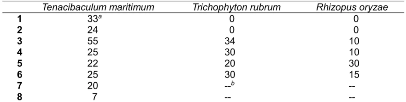
Antimicrobial test showed that some of the 2-alkyl-4-quinolones are inhibitory against fungal pathogens R. oryzae and T. rubrum. Compounds 1-6 have a stronger activity than compounds 7 and 8 for the test strain T. maritimum. In addition, 2-heptenyl-3-methyl congener 3 exhibited the strongest activity. Because T. maritimum
scaffolds to develop new medicines for the treatment of this economically devastating epizootic [29].
Table 2-2: Antimicrobial activity of 1-8 (10 g/6 mm disc)
Tenacibaculum maritimum Trichophyton rubrum Rhizopus oryzae
1 33a 0 0 2 24 0 0 3 55 34 10 4 25 30 10 5 22 20 30 6 25 30 15 7 20 --b -- 8 7 -- --
aSize of inhibitory zone in mm. bNot tested.
2-3 Conclusion
In this study, 9 human/fish/plant pathogenic microbes were used to screen 144 extracts from 38 Burkholderia strains in 4 different media. The result showed that most of the strains (35 out of 38) proved to be promising as a resource of antibiotics.
To date, more than 100 secondary metabolites have been isolated from Burkholderia strains. According to the large genome size recorded in NCBI genome database, much higher capacity in secondary metabolism is expected this bacterial genus. This study, two new alky quinolones were isolated along with six known compounds G1549 (1), 2-heptyl-4(1H)-quinolone (2), 2-heptyl-4(1H)-quinolone (4), PSC-D (5), pyrrolnitrin (6) and BN-227 (8) from strain B. cepacia MBAF1239, which confirmed this production capacity of the Burkholderia strains.
In the previous study, we have known that compounds 1, 2, 4-6, and 8 have a variety of bioactivities including antibacterial, antifungal, anticancer, antiallergenic activities and quorum-sensing signaling. This is the first report on the antibiotic property of alkyl quinolones against the fish pathogen Tenacibaculum maritimum, a main etiological agent of fish ulcer disease “tenacibaculosis” with high mortalities. This result demonstrated that alkyl quinolones are promising as a new treatment for fish
infective disease.
2-4 Experimental
2-4-1 General experimental procedures
The UV spectrum was recorded on a Hitachi U-3210 and IR spectrum was recorded on a Perkin Elmer Spectrum 100 spectrophotometer. NMR spectra were recorded on a Bruker AVANCE 500 spectrometer and using solvent CDCl3 (H/C 7.26/77.0 ppm) and CD3OD (H/C 7.26/77.0 ppm) as solvent. HR-ESI-TOFMS spectra were obtained on a Bruker micrOTOF focus mass spectrometer.
2-4-2 Collection of Burkholderia strains and broth screening
Collection of Burkholderia strains were done by Assoc. Prof. Masafumi Shimizu using Cfc-medium at Gifu University in 2010, while fermentation and broth screening of the collection bacteria were done by Ms. Atsumi Hasada in 2013. Burkholderia strains were isolated from the rhizosphere soil of Welsh onion and cucumber at an experimental farming of Mie University. Those collected strains were cultured in 4 different media to give 144 extracts and then tested against test microbes including 9 human/fish/plant pathogens (Edwardsiella ictaluri NBRC105724T T. maritimum NBRC16015, Trichophyton rubrum NBRC5467, Candida albicans NBRC0197, R. oryzae NBRC4705, Glomerella cingulata NBRC5907, Ralstonia solanacearum SUPP1541, Rhizobium radiobacter NBRC13263 and Athelia rolfsii NBRC30071). In this study, the strain MBAF1239 was considered as a candidate and identified as Burkholderia cepacia by an analysis of 16S rRNA gene sequence, demonstrated an impressively high incidence of antagonistic strains in this genus.
2-4-3 Fermentation, extraction, and isolation
The strain MBAF1239 was seed-cultured in 100 mL V22 (soluble starch 1%, glucose 0.5%, NZ-case 0.3%, yeast extract 0.2%, Tryptone 0.5%, K2HPO4 0.1%, MgSO4·7H2O 0.05%, and CaCO3 0.3%, pH 7.0) for 2 days. After seed-culture, a
three-mLresulting culture was transferred into 500 mL K-1 flasks each containing 100 mL production medium IMM-HS (glucose 1%, K2HPO4 0.36%, KH2PO4 0.41%, MgSO4·7H2O 0.02%, CaCl2·2H2O 0.01%, FeSO4·7H2O 0.002%, NH4Cl 0.1%, biotin 0.0001%, and L-histidine 0.4%) for metabolite production. After 4 days production culture at 30 °C, 1-butanol was added to each flask for the extraction of secondary metabolites as a ratio 1:1. Then the mixture was shake for 1 h and the 1-butanol layer was collected by centrifuge at 6000 rpm for 10 min. Evaporation of 1-butanol layer gave a crude extract 5.35 g from a 2 L production culture. The extraction was first partitioned between 60% MeOH (250 mL) and CH2Cl2 (250 mL x 3). Then CH2Cl2 layer was evaporated and was successively partitioned between 90% aqueous MeOH (150 mL) and n-hexane (150 mL x 3). in the antibacterial activity against Tenacibaculum maritimum, the most active MeOH layer was subjected toODS flash chromatography( 3 x 7 cm) eluted with a stepwise gradient of 25, 40, 55, and 85% (v/v) MeCN in 50 mM NaClO4. The third fraction showed the strongest activity for the test strain T. maritimum, which was subjected to the reversed phase HPLC on a Cosmosil AR-II column (1 x 25 cm), using a linear gradient elution program [eluents: MeOH (A), 1:1 CH3CN/H2O (B); 0–5 min 100% B, 5–45 min 100% B to 0% B, 45–65 min 0% B; flow 3 mL min-1 and UV detection at 210 nm to afford two new 2-alkyl quinolones 3 (0.5 mg) and 7 (0.7 mg), together with four known 2-alkyl quinolones, Pyo Ib or 2-heptylquinolin-4(1H)-one 1 (2.3 mg), (E)-2-(hept-2-en-1-yl)-3-methylquinolin-4(1H)-one 2 (2.5 mg), PSC-C or 2-heptyl-3-(E)-2-(hept-2-en-1-yl)-3-methylquinolin-4(1H)-one 4 (2.5 mg), PSC-D or (E)-3-methyl-2-(non-2-en-1-yl)quinolin-4(1H)-one 5 (0.8 mg), pyrrolnitrin 6 (0.5 mg), and BN-227 8 (0.8 mg).
(E)-2-(Hept-2-en-1-yl)quinolin-4(1H)-one (3): UV (MeCN) λmax, nm (ɛ): 328 (29887), 322 (27568), 316 (29828), 292 (10689), 288 (10901), 260 (6599), 240 (51173); IR ν max (ATR) cm1: 2927, 2873, 1636, 1595, 1553, 1505, 1473, 1445, 1355,1322, 1275, 1104, 1028, 969, 841, 762, 676; 1H NMR δ H (CD3OD): 6.21 (s, 1H, H3), 8.20 (d, J=8.1 Hz, 1H, H5), 7.39 ( brt, J=6.9 Hz 1H, H6), 7.68 (brs, 1H, H7), 7.57 (brs, 1H, H8), 3.42 (brs, 2H, H9),5.61 (brd, J=13.9 Hz, 1H, H10), 5.71 (m, 1H, H11),
2.09 (dt, J=6.3 and 6.2 Hz, 2H, H12),1.38 (m, 2H, H13), 1.34 (m, 2H, H14),0.90 (t, J=6.9 Hz, 3H, H15); 13C NMR δ
C (CD3OD): 155.7 (C2), 108.9 (C3), 180.8 (C4), 125.5(C4a), 126.0 (C5), 125.1 (C6), 133.5 (C7), 119.1 (C8), 141.6 (C8a), 37.8 (C9), 125.3 (C10), 136.5 (C11), 33.2 (C12), 32.6 (C13), 23.2 (C14), 14.2 (C15); HRMS-ESITOF (m/z) [M + H]+ calcd for C
16H20NO, 242.1539; found: 242.1539.
(E)-2-(Non-2-en-1-yl)quinolin-4(1H)-one (7): UV (MeCN) λmax, nm (ɛ): 328 (18600), 322 (16800), 316 (18200), 292 (8000), 288 (8400), 260 (5300), 240 (23,200); IR ν max (ATR) cm1: 2923, 2853, 1730, 1635, 1593, 1554, 1500, 1471, 1443, 1354, 1320, 1247, 1137, 1028, 965, 836, 759, 672; HRMS-ESITOF (m/z): [M + H]+ calcd for C18H24NO, 270.18524; found: 270.1855.
2-4-4 Evaluation of antimicrobial activity
In this study, 6 mm paper disk were first used to evaluate the activities. Then the 10 g/disc for each compound was prepared and a certain mount test pathogen contained: T. maritimum, R. oryzae and T. rubrum were used to detect the anti-activities of compounds 1-8 and the paper-disc agar diffusion method described in our previous study [30]. Flexibacter maritimus medium (0.5% peptone and 0.05% yeast extract in sea water) solidified with 10% agar was used to test against T. maritimum.
References
1. Igarashi, C., Asano, Y., Nishioka, T., Suga, H., Hyakumachi, M., Shimizu, M. Japanese
Jpn. J. Phytopathol. 2017, 83, 87–94.
2. Zhang, H., Mallik, A., Zeng, R. S. J Chem Ecol. 2013, 39, 243–252.
3. Kapoulas, N., Koukounaras, A., Ilić, Z. S. Scientia Horticulturae. 2017, 219, 310–318.
4. Suzuki, Y., Simizu, M. IOBC/WPRS Bull. Hyakumachi, M. 2013, 86, 187–188.
5. Ballio, A., Barra, D., Bossa, F., DeVay, E. J., Grgurina, I. Iacobellis, N. S., Marino, G.,
Pucci, P., Simmaco, M., Surico, G. Physiol. Mol. Plant Pathol., 33, 493-496.
6. Hoitink, H. A. J., Sinden, S. L. Phytopathol. 1970, 60, 1236-1237.
7. Hashimoto, M., Hattori, K. Chem. Pharm. Bull. 1967, 15, 718-720.
8. Mori, T., Yamashita, T., Furihata, K., Nagai, K., Suzuki, K., Hayakawa, Y., Shin-ya, K.
J. Antibiot. 2007, 60, 713-716.
9. Staerk, D., Kesting, J. R., Sairafianpour, M., Witt, M., Asili, J., Emami, S. A.,
Jaroszewski, J. W. Phytochem. 2009, 70, 1055-1061.
10. Wells, I. C. J. Biol. Chem. 1952, 196, 331-340.
11. Moon, S. S., Kang, P. M., Park, K. S., Kim, C. H. Phytochem. 1996, 42, 365-368.
12. Gerth, K., Trowitzsch, W., Wray, V., Höfle, G., Irschik, H., Reichenbach, H. J. Antibiot.
1982, 35, 1101-1103.
13. Itoh, J., Amano, S., Ogawa, Y., Kodama, Y., Ezaki, N., Yamada, Y. J. Antibiot. 1980,
33, 377-382.
14. Bisacchi, G. S. J. Med. Chem.2015, 58, 4874-4882.
2007, 24, 223–246; (c) Michael, J. P. Nat. Prod. Rep. 2005, 22, 627–646; (d) Michael,
J. P. Nat. Prod. Rep. 2003, 20, 476–493; (e) Michael, J. P. Nat. Prod. Rep. 2001, 18,
543–559.
16. Dekker K. A., Inagaki, T., Gootz, T. D., Huang, L. H., Kojima, Y., Kohlbrenner, W. E.,
Matunaga, Y., McGuirk, P. R., Nomura, E., Sakakibara, T., Sakemi, S., Suzuki Y.,
Yamauchi Y., Kojima N. J. Antibiot. 2008, 51, 145-152.
17. Cheng, C., Othman, E. M., Reimer, A., Grüne, M., Kozjak-Pavlovic, V. Tetrahedron
Lett. 2016, 57, 2786-2789.
18. Kunze, B., Höfle, G., Reichenbach, H. J. Antibiot. 1987, 40, 258-265.
19. Vial, L., Lepine, F., Milot, S., Groleau, M.-C., Dekimpe, V., Woods, D. E., Deziel, E. J.
Bacteriol. 2008, 190, 5339-5352.
20. Long, R. A., Qureshi, A., Faulkner, D. J., Azam, F. Appl. Environ. Microbiol. 2003, 69,
568-576.
21. Hertiani, T., Edrada, R., van Soest, R. W. M., Sudarsono, Proksch, P. J. Pharm. 2008,
19, 128-136.
22. Heeb, S., Fletcher, M. P., Chhabra, S. R., Diggle, S. P., Williams, P., Cámara, M. FEMS
Microbial. Rev. 2011, 35, 247-274.
23. Kitamura, S., Hashizume, K., Lida, T., Miyashita, E., Shirahata, K., Kase, H. J. Antibiot.
1986, 60, 1160-1166.
24. Moon, S. S., Myung, E. J., Cho, S. C., Park, J. B., Chung, B. J. Korean J. Pest. Sci.
2002, 6, 64-71.
15, 862-869.
26. Okada, B. K., Wu, Y., Mao, D., Bushin, L. B., Seyedsayamdostet, M. R. ACS Chem.
Biol. 2016, 11, 2124-2130.
27. Wang B., Waters, A. L., Sims, J. W., Fullmer, A., Ellison, S., Hamann, M. T. Microbial.
Ecol. 2013, 65, 1068-1075.
28. Depke, T., Franke, R., Broenstrup, M. J. Chromatogr. B, 2017, 1071, 19-28.
29. Avendaño-Herrera, R., Toranzo, A. E., Magariños, B. Dis. Aquat. Organ. 2006, 71,
255–266.
30. Oku, N., Matsumoto, M., Yonejima, K., Tansei, K., Igarashi, Y. Beilstein J. Org. Chem.
2-5 Spectral data
Table of contents (E)-2-(hept-2-en-1-yl) quinolin-4(1H)-one (3) 1H NMR spectrum (CH3OD, 500 MHz) 13C NMR spectrum (CH 3OD, 125 MHz)COSY spectrum (CH3OD, 500 MHz)
HSQC spectrum (CH3OD, 500 MHz) HMBC spectrum (CH3OD, 500 MHz) (E)-2-(non-2-en-1-yl) quinolin-4(1H)-one (7) 1H NMR spectrum (CDCl 3, 500 MHz) 13C NMR spectrum (CDCl 3, 125 MHz) COSY spectrum (CDCl3, 500 MHz) HSQC spectrum (CDCl3, 500 MHz) HMBC spectrum (CDCl3, 500 MHz)
1H NMR spectrum of (E)-2-(hept-2-en-1-yl) quinolin-4(1H)-one (3) (500 MHz, CH3OD) 1.0 1.5 2.0 2.5 3.0 3.5 4.0 4.5 5.0 5.5 6.0 6.5 7.0 7.5 8.0 ppm
13C NMR spectrum of (E)-2-(hept-2-en-1-yl) quinolin-4(1H)-one (3) (125 MHz, CH3OD) 20 30 40 50 60 70 80 90 100 110 120 130 140 150 160 170 180 ppm
COSY spectrum of (E)-2-(hept-2-en-1-yl) quinolin-4(1H)-one (3) (500 MHz, CH3OD) ppm 1.0 1.5 2.0 2.5 3.0 3.5 4.0 4.5 5.0 5.5 6.0 6.5 7.0 7.5 8.0 8.5 ppm 1 2 3 4 5 6 7 8
HSQC spectrum of (E)-2-(hept-2-en-1-yl) quinolin-4(1H)-one (3) (500 MHz, CH3OD) ppm 1.0 1.5 2.0 2.5 3.0 3.5 4.0 4.5 5.0 5.5 6.0 6.5 7.0 7.5 8.0 8.5 ppm 140 120 100 80 60 40 20 0
HMBC spectrum of (E)-2-(hept-2-en-1-yl) quinolin-4(1H)-one (3) (500 MHz, CH3OD) ppm 1.0 1.5 2.0 2.5 3.0 3.5 4.0 4.5 5.0 5.5 6.0 6.5 7.0 7.5 8.0 8.5 ppm 20 40 60 80 100 120 140 160 180
1H NMR spectrum of (E)-2-(non-2-en-1-yl) quinolin-4(1H)-one (7) (500 MHz, CDCl 3)
13C NMR spectrum of (E)-2-(non-2-en-1-yl) quinolin-4(1H)-one (7) (125 MHz, CDCl 3)
COSY spectrum of (E)-2-(non-2-en-1-yl) quinolin-4(1H)-one (7) (500 MHz, CDCl3) ppm 9 8 7 6 5 4 3 2 1 0 ppm 9 8 7 6 5 4 3 2 1 0
HSQC spectrum of (E)-2-(non-2-en-1-yl) quinolin-4(1H)-one (7) (500 MHz, CDCl3) ppm 9.0 8.5 8.0 7.5 7.0 6.5 6.0 5.5 5.0 4.5 4.0 3.5 3.0 2.5 2.0 1.5 1.0 0.5 0.0 ppm 140 120 100 80 60 40 20 0
HMBC spectrum of (E)-2-(non-2-en-1-yl) quinolin-4(1H)-one (7) (500 MHz, CDCl3) ppm 8 7 6 5 4 3 2 1 0 ppm 180 160 140 120 100 80 60 40 20 0
CHAPTER 3
4-Hydroxy-3-methyl-2(1H)-quinolone,
originally discovered from a
Brassicaceae plant, produced by a soil
bacterium of the genus Burkholderia
sp.: determination of a preferred
tautomer and antioxidant activity
3-1 Background
In Chapter 2, two new 2-alkylquinolones, 3 and 7, along with 6 known metabolites, were discovered from a Burkholderia strain isolated from a rhizosphere soil sample. To further explore the structural diversity of secondary metabolites from Burkholderia, a strain with a different isolation background was chosen as the subject of this chapter. The strain coded as 3Y-MMP (Figure 3-1), was isolated froma soil sample of rice pad collected in Toyama by Associate Professor Yukiko Shinozaki at National Institute of Technology, Toyama College, based on an enrichment culture technique using 1 mM ZnCl2 as a selection pressure. Strain 3Y-MMP showed a 99.9% similarity to Burkholderia cepacia N1_1_43 by a molecular phylogenetic analysis of 16S rRNA gene sequence.
Figure 3-1. Burkholderia sp. 3Y-MMP on YM agar.
Strain 3Y-MMP was cultured in three different media and resulting fermentation products were analyzed using an HPLC-DAD system, which exhibited the good production of the strain 3Y-MMP in medium King’s B. Among of them, three major peaks around 15-20 min were determined to be the known spoxazomicin C and pyochelin based on the UV analysis [1-3]. In addition, A new metabolite 9 without the similar UV spectrum in our in-house UV-database (Figure 3-2) were found.
Figure 3-2. HPLC-DAD analysis on the 3Y-MMP fermentation product.
Intrigued by this result, 3Y-MMP was subjected to production culture and chromatographic fractionation, which led to the isolation of 4-hydroxy-3-methyl-2 (1H)-quinolone (9, Scheme 3-1, Figure 3-3).
Figure 3-3. Structures of 9-13 [4-10].
3-2 Results and Discussion
3-2-1 Fermentation and isolation
The strain 3Y-MMP was recovered on YM agar then transferred with agar substratum directly into production King’s B medium (peptone 2%, glycerin 1%, K2HPO4 0.41%, MgSO4·7H2O 0.15%) for 4 days with reciprocal shaking at 200 rpm. The fermented products were extracted with 1-BuOH, and then partitioned between CH2Cl2 and 60% MeOH. Then 60% MeOH fraction was further fractionated by ODS flash chromatography and purified by elution with 16% aq. MeCN-0.1% HCOOH HPLC to give the 4-hydroxy-3-methyl-2 (1H)-quinolinone (9, 5.2 mg, Scheme 3-1).
3-2-2 Structure Determination
Compound 9 was isolated as white power and gave a molecular formula of C10H9NO2 based on a HRESITOFMS ion peak ([M+Na]+ obsd m/z 198.0525, calcd 198.0526). Broad absorption around 3364 cm-1 and strong absorption at 1580 cm-1 in the IR spectrum indicated the existence of hydroxy and amino groups.
The 1H and 13C NMR data in DMSO-d
6 are shown in Table 1.Analysis of the 1H NMR coupling constants and COSY and HSQC revealed the existence of four coupled aromatic methines (C 122.7/H 7.85 dd, J=7.9, 1.0 Hz; 121.2/7.12 ddd, J=7.9, 7.2, 0.7 Hz; 129.8/7.41 ddd, J=8.1, 7.2, 1.2 Hz; 115.0/7.23 d, J=8.1 Hz), two heteroatom-substituted non-protonated sp2 carbons ( 164.0 and 157.4), three sp2 non-protonated
carbons (C 137.4, 115.8, and 106.9), methyl proton at 1.98 ppm and a singlet exchangeable proton at 11.23 ppm; Further analysis of HMBC spectrum in DMSO displayed the correlations from H-5 to 8a, 4, and from H-7 to 8a, and H-6 to C-4a, and H-8 to C-4a to form a disubstituted benzene ring. However, the remaining parts were assembled into a C4 enol-amidyl or enol-imidic acyl unit because of the correlations from the proton H39 to the C-4, C-3 and C-2. In addition, the correlations of HMBC from the H3-9 to C-4a of the benzene ring implied the connection and the correlations from the exchangeable proton to C-4a and C-3 supported this connection as well as hydroxylation at the benzylic position.
Finally, by comparing the chemical shift with the known compounds 4-methoxy-1,3-dimethyl-2(1H)-quinolone 14 with a chemical shift of 138.4 ppm [11], N-methyl-2-pyridone 15 with a chemical shift of 139.5 ppm [12], 2,4-dimethoxy-3-methylquinoline 16 with a chemical shift of 147.0 ppm and 2-methoxypyridine 17 with a chemical shift of 147.2 ppm [13, 14], the structure of 9 was assigned to have a 2-quinolone form but not 2-quinolinol [15] (Figure 3-4). The same structure with 4-hydroxy-3-methyl-2 (1H)-quinolinone was synthesized from diethyl malonate and aniline and the assignment was further substantiated as below.
Figure. 3-4. COSY-deduced spin-system (bold lines) and key HMBC correlations (arrows) for 9, and structures of compounds 14-18 with a 13C chemical shift at C-8a position.
Table 3-1: NMR data of 9 in DMSO-d6 (297 K)
9
No. 13C 1H mult. (J in Hz), integration HMBC (1H to 13C)
1a 2 164.0 3 106.9 4 157.4 4-OH 11.30 brs, 1H 3, 4a 4a 115.8 5 122.7 7.85 dd (7.9, 1.0), 1H 4, 7, 8a 6 121.2 7.12 ddd (7.9, 7.2, 0.7), 1H 4a, 5, 7, 8, 8a 7 129.8 7.41 ddd (8.1, 7.2. 1.2), 1H 5, 8, 8a 8 115.0 7.23 d (8.1), 1H 4, 4a, 6, 7, 8a 8a 137.4 9 9.6 1.98 s, 1H 2, 3, 4, 4a
aSignal for amide proton not observed.
3-2-3 Synthesis of 9
In an effort to increase the amount of 9 to evaluate by bioassay, total synthesis was conducted according to the procedure (Scheme 3-2) established by Kawada and coworkers [16].
First, diethyl methyl malonate was hydrolyzed to 2-methyl-1-ethyl ester-propanedioic acid with the action of 0.25 M KOH and then converted to acid chloride by N, N-dimethylformamide, which was acylated aniline to give ethyl 2-methyl-3-oxo-3-(phenylamino) propanoate. Treatment with 0.5 M KOH, N-phenyl-2-methylmalonamic acid, which was cyclized with Easton’s reagent, gave 9 (Scheme
Scheme 3-2. Synthetic preparation of 9 [16].
Although compound 9 has been synthesized several times [17] and the chemical shifts of 1H and 13C resonances were enumerated [16],one-on-one assignments of the resonances to each structural part are first done in this work. In addition, the correlations of enol proton and the comparison of chemical shift with the known compounds confirmed 2-quinolone as a preferred tautomer. In addition, correction of the structures of known compounds 12 (139.2 ppm) and 13 (133.4 ppm) were made to 4-hydroxy-2(1H)-quinolone (4HQ, 11) and 4-O--D-glucopyranosyl-3,4-dihydroxy-2-quinolone (18) respectively, based on the chemical shift of C-8a [18,10].
Compared with the known compound 10, structural similarity between 9 and 11 implied that 9 also belongs to a member of 2-alkyl-4-quinolone class signaling molecules/antibiotics known from Pseudomonas aeruginosa and some Burkholderia species [19, 20]. Compound 9 with a 3-methyl group are exclusively produced by Burkholderia species [21]. These metabolites are biosynthesized by head-to-head condensation of anthranilate and -ketoacylate precursors, followed by a modification at C3 or nitrogen by putative monooxygenases or methyltransferase [20]. The biosynthesis of compound 11 is proposed based on malonate as the acylate precursor in this pathway. Thus, the same mechanism should be adopted by 9 with an additional methylation on C3.
3-2-4 Bioactivity
In the previous studies, the compound 9 showed inhibitory activities against Mycobacterium tuberculosis H37Ra and weak cytotoxicity against MRC-5 human lung-derived fibroblasts [22]. Meanwhile, an assay using RAW 264.7 murine macrophage-like cells showed that 9 could not inhibit the production of nitric oxide [23]. In this study, antimicrobial testing showed that 9 was inactive against Rhizobium radiobacter NBRC14554, Ralstonia solanacearum SUPP1541, Tenacibaculum maritimum NBRC16015 (Gram-negative bacteria), Staphylococcus aureus FDA209P JC-1 (Gram-positive bacterium), Candida albicans NBRC0197, and Saccharomyces cerevisiae S100 (yeasts).
The luminol chemiluminescence extinction assay was used to evaluate the antioxidant activity of 9 [24, 25] which quantifies the presence of the most detrimental ROS, hydroxy radical [26, 27], as intensity of luminescence emitted by oxidation of luminol. The result showed that 10 M of compound 9 decreased luminescence to 14% of the control reaction (Figure. 3-4). At first, the compound 9 was thought to entrap catalyst Cu2+ as the mechanism of chemiluminescence inhibitionbecause the Cu2+ was used to catalyze Fenton reaction to yield hydroxy radical. However, as far as checked by CAS assay [28], this speculation about entrapment of Cu2+ were overthrown based on the weak activity of metal-chelation in specified concentration. Thus, 9 was found to be another example of the antioxidants from Burkholderia.
Figure 3-4. Extinction of luminol chemiluminescence by 9.
3-3 Conclusion
In summary, 4-hydroxy-3-methyl-2(1H)-quinolone (9), an old synthetic molecule and recently discovered from a plant without providing abundant evidence to support the structure, was isolated from a fermentation extract of Burkholderia sp. 3Y-MMP. The structure of compound 9 was determined by the first full spectroscopic analyses using MS and NMR. According to the comparison of 13C chemical shift with the related compounds and synthetic compound 9, the compound 9 was determined as to have a 2-quinolone form.
Burkholderia strain 3Y-MMP, isolated in a Zn2+ enrichment culture, was chosen for metabolite analysis. While the activity of compound 9 was insignificant in the metal-chelating assay, the luminol chemiluminescence extinction assay showed that the compound 9 has a strong antioxidant activity. Even though the mechanisms of antioxidant are unclear, the compound 9 was considered as a new antioxidant from the
Burkholderia.
On the one hand, the discovery of compound 9 from the Burkholderia indicated the high production capacity of Burkholderia, on the other hand, even though more than 100 compounds isolated from this genus, new compound discovery has proven the genus Bukholderia as a promising resource of new bioactive metabolites.
3-4 Experimental
3-4-1 General experimental procedures
UV and IR spectra were collected on a Hitachi U-3210 and a Perkin Elmer Spectrum 100, respectively. NMR spectra were collected on a Bruker AVANCE 500 spectrometer in DMSO-d6 or CDCl3 using referencing solvent signals at δH/δC 2.5/39.5 and 7.27/77.0, respectively. HR-ESITOFMS were recorded on a Bruker micrOTOF focus mass spectrometer. Chemiluminescence was measured on a Molecular Devices SpectraMax M2 microplate reader.
3-4-2 Microorganism
Burkholderia sp. 3Y-MMP was isolated from a soil sample Toyama City, June 2015, by a similar procedure described in reference [29]. In the initial screening of strain, 1 mM of ZnCl2 was added to the medium as selection pressure for strains. Then taxonomy of strain 3Y-MMP was determined by a 16S rRNA sequence analysis using
a primer set 10F (5′-GTTTGATCCTGGCTCA-3′) and 800R
(5′-TACCAGGGTATCTAATCC-3′). The result showed a 99.9%similarly of to Burkholderia cepacia strain N1_1_43 based on the DNA Data Bank of Japan (DDBJ) website. This strain will be deposited in NBRC once it resumes services, which is currently suspended due to a nation-wide State of Emergency regarding COVID-19 declared on Apr. 16 by the Government of Japan.
3-4-3 Fermentation and isolation
Burkholderia sp. 3Y-MMP was recovered and the colony was transferred into 100 mL production medium King’s B medium composed of peptone 2%, glycerin 1%, K2HPO4 0.41%, and MgSO4·7H2O 0.15%. Then 500 mL K-flasks containing above medium were rotary shaken at 200 rpm at 30 ℃ for 4 days. After fermentation, 1-butanol was used to extracted the fermentation broth with the ratio 1:1 under the 6000 rpm centrifugation. The supernatant was collected and dried in vacuo to give a solid (2. 7g) from a 2 L culture. 60% MeOH layer separated from CH2Cl2 was subjected to ODS with a step gradient of MeCN-0.1% HCOOH (20%, 30%, 40%, 50%, 60%, 70% and 80%). Fraction 2 (30% MeCN) was evaporated to provide 69.4 mg, and then the purification was achieved by reversed-phase HPLC (Cosmosil AR-II column, 1 × 25 cm) with an isocratic elution of 16% MeCN-0.1% HCOOH, yielding 9 (5.2 mg,31.3 min) with sufficient purity for NMR-based structure characterization.
4-Hydroxy-3-methyl-2(1H)-quinolone (9): UV (MeOH) λmax nm (ε): 312 (2300), 226 (12000); IR (ATR) νmax 3268, 3186, 2958, 2927, 1595, 1486, 1387, 1354, 1243, 1026, 772, 761, 692, 664 cm-1; HR-ESITOFMS m/z 198.0525 [M+Na]+ (calcd for C10H9NNaO, 198.0526); 1H and 13C NMR data are shown in Table 1.
3-4-4 Evaluation of Fe
3+binding activity
The iron-binding activity was evaluated by the CAS assay developed by Schwyn and Neilands [21]. Two-point five mg of compound 9 was first dissolved in 20 μL DMSO. Then, add above mixture to the 50 μL blue-colored CAS stock solution and bring volume up to 100 μL with H2O (final concentration of 9: 160 mM). After incubation for 10 min, the solution changed color from deep blue to orange caused by the removed of Fe3+ from the indicator CAS dye and implied the compound 9 had chelating ability.
3-4-5 Antibacterial assay
Antibacterial and antifungal assay were performed against strains Tenacibaculum
maritimum (NBRC16015), Rhizobium radiobacter (NBRC14554), Ralstonia
solanacearum (SUPP1541), Staphylococcus aureus (FDA209P JC-1), Candida albicans (NBRC0197) and Saccharomyces cerevisiae (S100). The activity was evaluated by MIC method using 99-well plates with concentrations of 0.049, 0.098, 0.2, 0.4, 0.8, 1.6, 3.2, 6.4, 12.5, 25, 50, 100 μg/mL. After incubation at 30 ℃ for 2 days, the MIC value was the lowest drug concentration of no strain growth.
Antioxidant assay
The method of antioxidant activity was described in reference [30]. Ten M of luminol, 1000 M of H2O2 and a vehicle solvent with or without compound 9 were mixed in 50 mM boric acid-sodium hydroxide buffer at pH 9.0. Then 100 M of CuCl2 was added to the above mixture to initiate Fenton reaction. After incubation for 5 min, chemiluminescence at 500 nm was recorded on a microplate reader. The above experiments were repeated 3 times and the mean ratio of light extinction was expressed as the potency of antioxidant activity.
References
1. Inahashi, Y., Iwatsuki, M., Ishiyama, A., Namatame, M., Nishihara-Tsukashima, A.,
Matsumoto, A., Shiomi, K. J. Antibiot. 2011, 64(4), 303–307.
2. Cox, C. D., Rinehart, K. L., Moore, M. L., Cook, J. C. Proc Natl Acad Sci. 1981, 78,
4256–4260.
3. Sokol, P. A. FEMS Microbiol. Lett. 1984, 23, 313–317.
4. Shang, X. F., Susan, L., Natschke, M., Liu, Y. Q., Guo, X., Xu, X. S., Goto M., Li, J. C., Yang, G. Z., Lee, K. H. Med. Res. Rev. 2018, 38, 775–828.
5. Shang, X. F., Susan, L., Natschke, M., Yang, G. Z., Liu, Y. Q., Guo, X., Xu, X. S., Goto, M., Li, J. C., Zhang, J. Yu., Lee, K. H. Med. Res. Rev. 2018, 38, 1614–1660. 6. Arndt, F., Ergener, L. and Kutlu, O. Chem. Ber. 1953, 86, 951-957.
7. Hebanowska, E., Tempczyk, A., Łobocki, L., Szafranek, J., Szafranek, A., Urbanek, Z. H. J. Mol. Struct. 1986, 147, 351-361.
8. Luis García Ruano, J., Pedregal, C., Rodríguez, J. H. Heterocycles 1991, 32, 2151– 2159.
9. Lépine, F., Dekimpe, V., Lesic, B., Milot, S., Lesimple, A., Mamer, O. A., Rahme, L. G., Déziel, E. Biol. Chem. 2007, 388, 839–845.
10. Yang, X., Yang, J. Acta. Pharm. Sin. 2008, 43, 1116–1118.
11. Reisch, J., Mester, I. Arch. Pharm. (Weinheim, Ger.) 1980, 313, 751–755.
12. Still, I. W. J., Plavac, N., McKinnon, D. M., Chauhan, M. S. Can. J. Chem. 1976, 54, 280–289.
14. Chambers, R. D., Parsons, M., Sandford, G., Skinner, C. J., Atherton, M. J., Moilliet, J. S. J. Chem. Soc., Perkin Trans. 1 1999, 803–810.
15. Moon, B. S., Lee, B. S., Chi, D. Y. Bioorg. Med. Chem. 2005, 13, 4952–4959.
16. Kawada, M., Inoue, H., Ohba, S., Hatano, K., Abe, H., Hayashi, C., Watanabe, T., Igarashi, M. PCT Int. Appl. WO 2014/132902 A1.
17. Gabriel, S., Gerhard, W. Ber. Dtsch. Chem. Ges B. 1921, 54B, 1067–1078. 18. AIST Spectral Database for Organic Compounds. SDBS No. 15669.
https://sdbs.db.aist.go.jp/sdbs/cgi-bin/direct_frame_top.cgi (accessed Feb 23, 2020). 19. Heeb, S., Fletcher, M. P., Chhabra, S. R., Diggle, S. P., Williams, P., Cámara, M.
FEMS Microbiol. Rev. 2011, 35, 247–274.
20. Coulon, P. M. L., Groleau, M. C., Déziel, E. Front. Cell. Infect. Microbiol. 2019, 9, 33. 21. Vial, L., Lépine, F., Milot, S., Groleau, M. C., Dekimpe, V., Woods, D. E., Déziel, E. J.
Bacteriol. 2008, 190, 5339–5352.
22. de Maced, M. B., Kimmel, R., Urankar, D., Gazvoda, M., Peixoto, A., Cools, F., Torfs, E., Verschaeve, L., Lima, E. S., Lyčka, A., Milićević, D., Klásek, A., Cos, P., Kafka, S., Košmrlj, J., Cappoen, D. Eur. J. Med. Chem. 2017, 138, 491–500.
23. Aoki, S., Ye, Y., Higuchi, K., Takashima, A., Tanaka, Y., Kitagawa, I., Kobayashi, M. Chem. Pharm. Bull. 2001, 49, 1372–1374.
24. Paejo, I., Petrakis, C., Kefalas, P. J. Pharm. Toxicol. Methods 2000, 43,183–190. 25. Georgetti, S. R., Casagrande, R., Di Mambro, V. M., Azzolini, A. E., Fonseca, M. J.
AAPS PharmSci. 2003, 5, E20.
80.
27. Stadtman, E. R., Berlett, B. S. J. Biol. Chem. 1991, 266, 17201–17211. 28. Schwyn, B., Neilands, J. B. Anal. Biochem. 1987, 160, 47–56.
29. Shinozaki, Y., Kitamoto, H., Sameshima-Yamashita, Y., Kinoshita, A., Nakajima-Kambe, T. J. Gen. Appl. Microbiol. 2019, 65, 273–276.
30. Parejo, I., Petrakis, C., Kefalas, P. J. Pharmacol. Toxicol. Methods 2000, 43, 183–
3-5 Spectral data
Table of contents
4-Hydroxy-3-methyl-2(1H)-quinolone (9) UV spectrum of natural 9 IR spectrum of natural 9 UV spectrum of synthetic 9 IR spectrum of synthetic 91H NMR of natural (upper) and synthetic (lower) 9 (DMSO-d
6, 297 K, 500 MHz) 13C NMR of natural (upper) and synthetic (lower) 9 (DMSO-d6, 297 K, 125 MHz)
COSY spectrum of natural 9 (DMSO-d6, 297 K, 500 MHz)
HSQC spectrum of natural 9 (DMSO-d6, 297 K, 500 MHz)
HMBC spectrum of natural 9 (DMSO-d6, 298 K, 500 MHz)
COSY spectrum of synthetic 9 (DMSO-d6, 298 K, 500 MHz)
HSQC spectrum of synthetic 9 (DMSO-d6, 299 K, 500 MHz)
HMBC spectrum of synthetic 9 (DMSO-d6, 298 K, 500 MHz)
Ethyl 2-methyl-3-oxo-3-(phenylamino) propanoate UV spectrum IR spectrum 1H NMR spectrum (CDCl3, 500 MHz) 13C NMR spectrum (CDCl 3, 125 MHz) N-Phenyl-2-methylmalonamic acid UV spectrum IR spectrum 1H NMR spectrum (DMSO-d 6, 500 MHz) 13C NMR spectrum (DMSO-d 6, 125 MHz)
UV spectrum of natural 9
0
0.2
0.4
0.6
0.8
1
1.2
1.4
200
300
400
500
600
A
bs
Wavelength (nm)
UV spectrum of synthetic 9
0
0.2
0.4
0.6
0.8
1
1.2
1.4
200
300
400
500
600
A
bs
Wavelength (nm)
1H NMR spectra of natural (upper) and synthetic (lower) 9 (500 MHz, 297 K, DMSO-d 6)
13C NMR spectra of natural (upper) and synthetic (lower) 9 (125 MHz, 297 K, DMSO-d 6)
COSY spectrum of natural 9 (500 MHz, 297 K, DMSO-d6) ppm 2.0 2.5 3.0 3.5 4.0 4.5 5.0 5.5 6.0 6.5 7.0 7.5 8.0 ppm 2 3 4 5 6 7 8
HSQC spectrum of natural 9 (500 MHz, 297 K, DMSO-d6) ppm 2.0 2.5 3.0 3.5 4.0 4.5 5.0 5.5 6.0 6.5 7.0 7.5 8.0 ppm 20 40 60 80 100 120
HMBC spectrum of natural 9 (500 MHz, 298 K, DMSO-d6) ppm 2 3 4 5 6 7 8 9 10 11 ppm 20 40 60 80 100 120 140 160
COSY spectrum of synthetic 9 (500 MHz, 298 K, DMSO-d6) ppm 2.0 2.5 3.0 3.5 4.0 4.5 5.0 5.5 6.0 6.5 7.0 7.5 8.0 ppm 2 3 4 5 6 7 8
HSQC spectrum of synthetic 9 (500 MHz,299 K, DMSO-d6) ppm 2.0 2.5 3.0 3.5 4.0 4.5 5.0 5.5 6.0 6.5 7.0 7.5 8.0 ppm 20 40 60 80 100 120
HMBC spectrum of synthetic 9 (500 MHz, 298 K, DMSO-d6) ppm 2 3 4 5 6 7 8 9 10 11 ppm 20 40 60 80 100 120 140 160
UV spectrum of ethyl 2-methyl-3-oxo-3-(phenylamino) propanoic acid
0
0.5
1
1.5
2
2.5
200
300
400
500
600
A
bs
Wavelength (nm
)1H NMR spectrum of ethyl 2-methyl-3-oxo-3-(phenylamino) propanoic acid (500 MHz, CDCl 3)
13C NMR spectrum of ethyl 2-methyl-3-oxo-3-(phenylamino) propanoic acid (125 MHz, CDCl 3)
UV spectrum of N-phenyl-2-methylmalonamic acid
0
0.5
1
1.5
2
2.5
200
300
400
500
600
A
bs
Wavelenghth (nm)
1H NMR spectrum of N-phenyl-2-methylmalonamic acid (500 MHz, DMSO-d 6)
13C NMR spectrum of N-phenyl-2-methylmalonamic acid (125 MHz, DMSO-d 6)
CHAPTER 4
Three new
O-isocrotonyl-3-hydroxybutyric acid congeners
produced by a sea anemone-derived
marine bacterium of the genus Vibrio
4-1 Background
In Chapters 2 and 3, three new quinolone metabolites were isolated from two strains with different origins that belongs to the genus Burkholderia, a terrestrial taxon that contains many pathogenic species. The results substantiated the validity of targeting pathogenic bacteria for prospecting new bioactive molecules. To gain further support to this strategy, a strain from another pathogenic lineage, the genus Vibrio, indigenous to marine environments, was studies in this chapter.
In our laboratory, marine bacteria associated with invertebrates have been the focus of our research, because the number of new compounds from microorganisms associated with or symbiotic to the host invertebrates is quite small [1-4]. Recently, some new natural products were isolated from marine bacteria associated with invertebrates in our laboratory such as bulbimidazoles A–C, (6E,8Z)- and (6E,8E)-5-oxo-6,8-tetradecadienoic acids and (2Z,4E)-3-methyl-2,4-decadienoic acid [5-7]. The discovery further confirmed that marine bacteria associated with invertebrates are emerging source of new compounds. It is common knowledge that marine invertebrates, especially sedentary sea anemones, are evolved with rich sources of bioactive metabolites [8]. The related studies had clarified that the secondary metabolites produced from the associated microorganisms could transport into the sea anemone tissue [9]. Hence, exploitation of sea anemone-associated bacteria could solve the supply problem of raw materials. In this study, ten sea anemone-associated bacteria were collected.
UV spectra are one of the useful traits to find a new natural compound. In this study, HPLC-UV screening was used to directly detect secondary metabolites from ten sea anemone-associated bacteria cultured in three types of fermentation media. The screening result showed that a sea anemone-derived marine bacterium SI9 of thegenus Vibrio cultured in medium A16 was considered as promising because an extract isolated from SI9 had many absorption peaks at 210 nm and 230 nm. Even though this absorption spectrum of UV is not novel enough, a series of same absorption spectrum
appeared at the same time not identified in our laboratory.
Figure 4-1. Vibrio sp. SI9 on Marine Agar.
Thus, Vibrio SI9 was cultured, extracted and purified as described below to obtain three new O-isocrotonyl-hydroxybutyric acid 19-21 and one known 3-hydroxybutyric acid 22(Scheme 4-1 and Figure 4-2).
Figure 4-2. Structures of 19-22 and O-isocrotonyl-oligo(3-hydroxybutyrate).
4-2 Results and Discussion
4-2-1 Fermentation and isolation
The fermentation broth of strain SI9 was extracted with the same volume of n-BuOH, and then the concentrated butanol extract was purified by silica gel- chromatography, ODS chromatography and revered-phase HPLC to yield four acyloxy fatty acids 19 (1.5 mg), 20 (11.2 mg), 21 (4.3 mg) and 22 (135.5 mg) from the one of the mid-polar fractions.
4-2-2 Structure Determination
1H NMR spectra of 19-22 showed the same splitting patterns (see Supporting Information and Tables 4-1) and a pair of mutually coupled double-doublet resonances. It implied that 19-22 share the same core structure. In addition, 13C NMR spectroscopic data indicated the presence of two carboxy, two olefinic, and one oxygenated carbon resonances (see Supporting Information). Meanwhile, additional analysis of a HSQC spectrum confirmed the presence of two methyl groups and one to three aliphatic methylene(s). Analysis of ESITOFMS ion peaks [M+Na]+ revealed that the molecular formula was C9H14O4 for 19, C10H16O4 for 20 and 21, and C8H12O4 for 22. The structural differences of 19-22 turned out to be attributed to the presence of one to two methylene unit. Thus, 19-22 were confirmed to be a series of acyclic compounds with varying length of aliphatic chains.
Table 4-1: NMR data for 19-22 in CDCl3.
19
No. 13C 1H (J in Hz), integr. COSY HMBCa
3-hydroxy-4-methylbutyric acid 1 175.0 2 38.4 2.68, dd (7.3, 15.8), 1H 3 1, 3, 4 2.61, dd (5.5, 15.9), 1H 3 1, 3, 4 3 70.8 5.22, brqui (6.3), 1H 2, 4 1, 2, 4, 5, 1´ 4 26.9 1.71, m, 2H 3, 5 2, 3, 5 5 9.4 0.94, t (7.4), 3H 4 3, 4 isocrotonic acid 1´ 165.9 2´ 120.5 5.78, dq (11.5, 1.8), 1H 3´ 1´, 4´ 3´ 145.6 6.34, dq (11.5, 7.3), 1H 2´, 4´ 1´, 4´ 4´ 15.4 2.13 dd (7.3, 1.8)3H 3´ 1´, 2´, 3´ 20
No. 13C 1H (J in Hz), integr. COSY HMBCa
4-ethyl-3-hydroxybutyric acid 1 176.2 2 39.0 2.68, dd (7.2, 15.9), 1H 3 1, 3, 4 2.60, dd (5.6, 15.8), 1H 3 1, 3, 4 3 69.5 5.25, m, 1H 2, 4 1, 2, 4, 5, 1´ 4 36.1 1.61, m, 2H 3, 5 2, 3, 5, 6 5 18.4 1.35, m, 2H 4, 6 3, 4, 6 6 13.8 0.91, t (7.4), 3H 5 3, 4, 5 isocrotonic acid 1´ 165.8 2´ 120.5 5.74, qd (11.4, 1.8),1H 3´ 1´, 4´ 3´ 145.5 6.30, m,1H 2´, 4´ 1´, 4´ 4´ 15.4 2.10, dd (7.3, 1.6), 3H 3´ 1´, 2´, 3´


![Figure 2-4. Referential 13 C chemical shifts of an allylic carbon in burkholone [8]](https://thumb-ap.123doks.com/thumbv2/123deta/7672799.1212164/22.892.132.746.154.629/figure-referential-c-chemical-shifts-allylic-carbon-burkholone.webp)