Desorption and Ionization of Amino Acid Compounds Observed by Negative Ion Mode Surface-Assisted Laser Desorption/Ionization Mass Spectrometry (SALDI-MS) using Titanium Oxide Nanoparticles
9
0
0
全文
(2) Yonezawa et al., Desorption and Ionization of Amino Acid Compounds by Negative Ion Mode SALDI, Nano Biomed 12(2), 101-109, 2020. substrate for amino acids [16]. Nishikaze et al. investigated the key factors that dominate the ionization yield of MALDI-MS of amino acids and peptides [24,25]. In the positive ion mode, it was revealed that the ionization yield increase linearly as the proton affinity increases [24]. On the other hand, in the negative ion mode, the ionization yield increases as the gas phase acidity of amino acids increases [25]. Nitta et al. proposed the gas phase acidity is the key factor in negative ion modes [16]. However, these are the only report of the studies on the ionization yield of amino acids in SALDI-MS and no examination was carried out so far for other SALDI substrate materials. In the previous report [17], we have reported the total investigation of the positive ion mode SALDI-MS analyses of amino acids. The ion yield correlated linearly with the sodium ion affinity of the amino acids. In this paper, key factors affecting the ionization yields of various amino acids in negative mode using the commercial TiO2 nanoparticles as the SALDI-MS substrates will be discussed.. for forensic studies [5,6]. However, the obstructive peaks from matrix compounds discourage to apply MALDI-MS for forensic studies [7]. Inorganic nanoparticles for SALDI-MS were firstly applied by Tanaka et al. A glycerol suspension of 30-nm Co nanoparticles was applied to obtain mass spectra of proteins and polymers [2]. After his invention, various inorganic substrates were applied for SALDI-MS [8-11]. On the other hand, Sunner et al. used graphite as a matrix and they firstly used the word SALDI [12]. After that, nano-dimensional roughness of the porous silicon was used for SALDI. This system is called as DIOS (Desorption ionization from porous silicon) [13]. Various nanostructured substrates as well as nanoparticles have been used as the sample stage of SALDI-MS so far. Using SALDI-MS, the measurement sample molecules are ionized because no other organic component exists on the plate. Fragmented metal atoms/clusters may be found in the mass spectra of SALDI-MS [14, 15], but they can be easily identified especially when heavy precious metals were used for ionization. SALDI-MS is an excellent procedure that can measure the molecular weights in wide range from a very small molecule to high molecular weight polymers. For example, Pt nanoflower has been proposed as the effective ionization assisting materials for SALDI-MS for various molecules [14, 16]. Metal oxides are also good candidate for ionization assisting materials [8, 17, 18]. It is very important to discuss desorption process in LDI, which has not been clearly revealed yet [19,20]. Desorption process in SALDI is probably dominated by thermal mechanisms due to the laser-induced temperature increase of the nanoparticles or nanostructures with poor thermal conductivity [2,14,16]. However, non-thermal desorption process has also been proposed to enhance the desorption efficiency in SALDI-MS [21,22]. In order to understand the SALDI desorption process, the parameters controlling the desorption yields of various analytes is essential. Amino acids must be good models for this purpose [9,16,23]. Amino acids are one of the key organic compound series constituting various proteins. With the different types of their side groups, amino acids also show various physicochemical and biochemical properties. Therefore, amino acids have been used as model components for desorption/ionization of LDI. Nitta et al. used similar Pt nanoflower structures as SALDI-MS. Materials and Methods 1. Materials. Highly pure TiO2 NPs (Ishihara Sangyo Kaisha, PT401M, 99.99 % TiO2) were used for the DI assisting material. Their rutile ratio is 46.7 %. In our previous study, it showed a high SALDI performance. Acetonitrile (Wako, GR) was used without further purification. Water was purified with Mili-Q or Organo/ELGA Purelabo system (>18 MΩ·cm). Amino acids were purchased from Wako, Kanto or Junsei and used without further purification. 2. Preparation of SALDI Mass Measurement Samples of Various Amino Acids. Preparation of the samples was the same as previously described [17]. PT401M TiO2 NPs (5 mg) were dispersed in water/acetonitrile (1:1, v/v) mixed solvent by using sonication for 5 min. No additive was introduced in the dispersion. Aqueous solutions (1 nmol·dm-3) of 20 different amino acids were used as the samples. Salytilic acid as an internal DI standard was dissolved into water at the concentration of 1 nmol·mm-3. 3. SALDI-MS Measurement Mass spectra of the 20 amino acids were obtained with a Autoflex speed (Bruker Daltonics) mass spectrometer. The measurements have been done in the negative mode. 102.
(3) Yonezawa et al., Desorption and Ionization of Amino Acid Compounds by Negative Ion Mode SALDI, Nano Biomed 12(2), 101-109, 2020. Salicylic acid was used as the internal standard and it can be detected readily and show strong peaks. 355 nm laser was used for the desorption/ionization. Random walk mode (laser irradiation randomly on the sample plate) was used for the signal accumulation (5 spots, 1000 shots for each spot).. Table 1 Proton affinities [24,28], gas-phase acidities [24,25,29,30], and hydrophobicities [24,31] of various amino acids used in this study.. Results and Discussion 1. SALDI negative ion mode measurements of 20 common amino acids using TiO2 nanoparticles. We have selected a commercially available TiO2 NPs with a high purity (PT401M, Ishihara Sangyo Kaisha, Japan) for this study in order to avoid the effect of the contamination. TEM image (JEOL, JEM-2010, 200 kV) of PT401M TiO2 NPs is shown in Figure 1. The NPs seem to be well crystalized. Their average diameter is ca. 60 nm which was determined by the arbitrary chosen area of the TEM image although the web page of the company shows a rather larger size (~0.07 μm [26]). SALDI-MS measurements of 20 kinds of amino acid molecules with PT401M TiO2 NPs as the SALDI substrate were carried out in negative ion mode. Table 1 shows the various amino acids used in this study and their proton affinity, gas acidity and hydrophobicity are listed. The typical SALDI-MS spectra of the four amino acids. GAb/. Δfc/. 881. 1423. 3.39. 89.1. 896. 1423. 2.55. Val. 117.2. 906. 1417. -3.14. 4. Ile. 131.2. 915. 1419. -6.07. 5. Leu. 131.2. 910. 1419. -6.90. 6. Pro. 115.1. 929. 1426. -0.71. 7. Asp. 133.1. 905. 1319. 2.55. 8. Glu. 147.1. 936. 1331. 2.13. 9. Lys. 146.2. 986. 1416. 1.92. 10. Arg. 174.2. 1024. 1381. 2.89. 11. His. 155.2. 696. 1385. 2.89. 12. Asn. 132.1. 923. 1388. 3.72. 13. Gln. 146.2. 929. 1385. 4.06. 14. Ser. 105.1. 900. 1389. 1.76. 15. Thr. 119.1. 918. 1388. 1.21. 16. Trp. 204.2. 937. 1413. -5.02. 17. Tyr. 181.2. 924. 1413. -5.98. 18. Phe. 165.2. 920. 1420. -6.36. 19. Cys. 121.2. 895. 1393. 1.51. 20. Met. 149.2. 925. 1406. -2.76. No.. Amino. 1. Gly. 75.1. 2. Ala. 3. a. Mw. PAa/. PA: Proton affinity, bGA: Gas-phase acidity. cΔf: B&B Hydrophobicity.. Figure 1. TEM image and the size distribution of PT401M TiO2 NPs.. 103.
(4) Yonezawa et al., Desorption and Ionization of Amino Acid Compounds by Negative Ion Mode SALDI, Nano Biomed 12(2), 101-109, 2020. SALDI-MS with Pt nanoflowers coated by hydrophobic perfluorodecyltrichlorosilane (FDTS) [16]. In negative ion mode, the proton transfer reaction in MALDI is assumed to be major secondary charge transfer process as described in Eq. 1.. (Arg (10), Phe (18), His (11), and Cys (19)) are collected in Figure 2. TiO2 NPs showed excellent ionization ability of all amino acids as observed also in the positive mode detection [17]. All amino acid molecules could be detected in negative mode as proton detached molecular ion forms, [M-H]- but not as M-. In positive ion mode, the all amino acid molecules are detected as sodium adducted ion [M+Na]+ when TiO2 is used as the ionization assist material, but no molecular ion [M+H]+ was detected [17, 27]. These [M-H]- ion peaks could also be detected in. (M-H)- + A → M + (A-H)-. (1). Here, M stands for organic matrix molecule, and A stands for analyte molecule. However, in. Figure 2 SALDI-MS spectra of (a) arginine (10, MW = 174.2), (b) pheylalanine (18, MW = 165.2), and (c) histidine (11, MW = 155.2) in the negative ion mode. The laser power was fixed at LP = 40. Data were accumulated with 1000 shots at 500 Hz. 104.
(5) Yonezawa et al., Desorption and Ionization of Amino Acid Compounds by Negative Ion Mode SALDI, Nano Biomed 12(2), 101-109, 2020. NPs, Cys (19) could also be detected as [M-H]ion, not as a dimer. Cys was detected as a dimer when Pt nanoflowers were used as the SALDI substrate [16]. The dimer of Cys is its oxidation product. This indicates that the TiO2 nanoparticle substrate prevents the oxidation of the analytes. These favorable properties are probably due to the photo catalytic properties of TiO2.. SALDI, such charge transfer processes are not possible. The desorption/ionization process in SALDI-MS is considered as the physical process of the nanoparticle surfaces and the thermal effects due to the rapid, laser-induced temperature increase of the particle surfaces, respectively. In negative ion mode, lack of organic matrix molecules, which are usually acidic, the deprotonation of amino acids in the gas phase may be more favorable as compared to the negative ion MALDI. Reported by Nitta et al., with naked Pt nanoflowers, only Asp (3) an Glu (8) could be detected in the negative mode and Asp (3) and His (11) in the positive mode, even though all amino acids could be detected with FDTS-coated Pt nanoflower [16]. Nitta indicated that hydrophobic surface of FDTS-coated Pt nanoflower concentrated amino acids in small areas on the surface and laser-induced explosive evaporation of the coating fluorinated chains may help the desorption. On the contrary, using TiO2 NPs, all amino acids could be detected both in positive [17] and negative modes (Figure 3). These results strongly indicate that the desorption ability of TiO2 NPs is higher than Pt nanoflowers even the surface is not covered by fluorinated carbons. Moreover, by using TiO2. 2. Ion yields of various amino acids in SALDI-MS in the negative ion mode. The ion yields, which are corresponding to the relative peak intensities obtained from 20 amino acids in negative ion mode, are collected in Figure 3. The relative peak intensity was determined as the ratio of the peak intensity of the amino acid to that of salicylic acid (internal standard). Acidic amino acids: Asp (7) and Glu (8) gave very high peak intensities, which can be attributed to their strong acidic nature. The gas-phase acidities of these two amino acids are lower as shown in Table 1. Gas-phase acidity is expressed as a negative value of the Gibbs free energy change associated with acid dissociation. The stronger acid molecule has the smaller gas-phase acidity value. The deprotonation of amino acids in the negative ion mode is enhanced.. Figure 3 Ion yields of molecular ions [M-H]- of amino acids obtained by SALDI-MS in negative ion mode using TiO2 nanoparticles. The relative peak intensity was determined as the ratio of the peak intensity of the amino acid to that of salicylic acid (internal standard). The laser power was fixed at LP = 40. Data were accumulated with 1000 shots at 500 Hz.. 105.
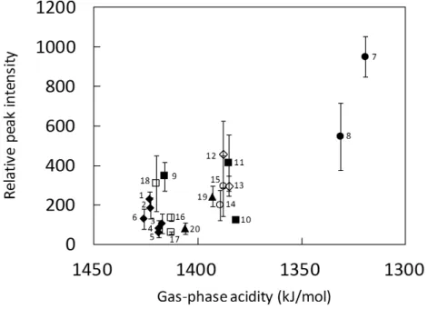
(6) Yonezawa et al., Desorption and Ionization of Amino Acid Compounds by Negative Ion Mode SALDI, Nano Biomed 12(2), 101-109, 2020. The relationship between the relative peak intensities of [M-H]- measured by negative ion mode and the gas-phase acidities of the 20 amino acids are plotted in Figure 4. The ion yields of the negative molecular deprotonated ion [M-H]- of amino acids were estimated at the ratio of the peak intensity of the amino acid to that of salicylic acid. In negative ion mode, we predicted that a negative molecular ion M- or a deprotonated molecule ion [M-H]- can be detected. In this SALDI-MS measurements with TiO2 nanoparticles, only the deprotonated molecule ions [M-H]- were observed. Deprotonation reactions are assumed to be major secondary charge transfer process according to Eq. 1 shown above. However, in SALDI-MS, no such charge transfer reaction is possible because no matrix molecules are added to the samples. The desorption/ionization process in SALDI-MS depends only on the physical properties of inorganic nanostructure surfaces. The physical properties can be considered as the thermal effects due to the laser-induced instant temperature increase of the surface of NPs. TiO2 is one of the best candidate for SALDI-MS because of its high UV absorbance and low thermal conductiv-. ity. Therefore, it can be considered that deprotonation of amino acids in gas phase dominates in the negative mode SALDI-MS. In Figure 4, high ion yields were observed Asp (7, acidic), Glu (8, acidic), Lys (9, basic), His (11, basic), and Asn (12, amidic). The high yields of Asp (7) and Glu (8) can be explained by their acidic nature, which enhances the deprotonation. His (11) and Asn (12) do not show high gas-phase acidities, but they show relatively high peak intensities. But His has an imidazolyl group. Proton attached to nitrogen of imidazolyl detached readily to form an imidazolyl anion. Lys (9) is a basic amino acid and has a high gas-phase acidity. In the case of FDTS-coated Pt nanoflower, Lys did not show a high peak intensity [16]. However, in our case, it shows relatively high intensity. Lys (9) is very hydrophobic. Therefore, it can be explained that the peak intensity of Lys with FDTS-coated Pt nanoflower is very low according to the hydrophobicity of the amino acid and the surface of the nanoflower. This indicates that the ion yield of amino acids depends on their ionization efficiency directly relating to gas-phase acidity. Figure 5 shows the relationship between the. Figure 4 Relationship between negative-ion relative intensity observed in SALDI-MS analysis using TiO2 nanoparticles and gas-phase acidity of 20 common amino acids obtained in negative-ion mode. The relative peak intensity was determined as the ratio of the peak intensity of the amino acid to that of salicylic acid (internal standard). The error bars were estimated from the data of five independent SALDI-MS samples. Inset numbers are corresponding to the numbers of amino acids shown in Table 1. The laser power was fixed at LP = 40. Data were accumulated with 1000 shots at 500 Hz.. 106.
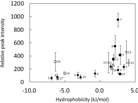
(7) Yonezawa et al., Desorption and Ionization of Amino Acid Compounds by Negative Ion Mode SALDI, Nano Biomed 12(2), 101-109, 2020. Figure 5 Relationship between negative-ion relative intensity observed in SALDI-MS analysis using TiO2 nanoparticles and hydrophobicity of 20 common amino acids obtained in negative-ion mode. The relative peak intensity was determined as the ratio of the peak intensity of the amino acid to that of salicylic acid (internal standard). The error bars were estimated from the data of five independent SALDI-MS samples. Inset numbers are corresponding to the numbers of amino acids shown in Table 1. The laser power was fixed at LP = 40. Data were accumulated with 1000 shots at 500 Hz.. relative peak intensity of [M-H]- molecular ion measured by the negative ion mode and their hydrophobicities. Although there is no linear relationship, amino acids with high hydrophobicities show relatively high [M-H]- molecular ion peak intensity in negative ion mode. On the contrary, there is no obvious relationship between the positive ion [M+H]+ peak intensity and hydrophobilicty [17]. An acidic amino acid Asp (7) shows a considerably higher peak intensity, and it is probably due to the extra amino acid which can show an interaction to the TiO2 surface. Phe (18) with a very low hydrophobicity also shows a high peak intensity. Phenyl group shows weak interaction with the proton of hydroxyl group on the metal oxide surface. The both amino acids show very low peak intensities when use FDTS-coated Pt nanoflower [16] This also supports the idea above. Trp(16) and Tyr(17) also have phenyl rings or hetero-rings, but the peak intensities are similar to other amino acids or Pt nanoflowers. Interaction with phenol and hetero-rings can be similar. These phenomena suggest that gas-phase acidity and the concentration on the TiO2 surface of amino acids is key factors of the intensity of mass signals.. Conclusions In this study, negative ion yields of [M-H]molecular ions of 20 amino acids obtained by SALDI-MS using TiO2 NPs as the assisting substrate. All amino acids could be detected by SALDI-MS using TiO2 NPs in negative mode (also in positive mode). A linear correlation between the peak intensity and the gas-phase acidity of amino acids was clearly observed. Hydrophobicity is also another factor of the peak intensity but some amino acids (Asp and Phe) which have interactions with the TiO2 surface also showed high peak intensities. We believe that this commercially available TiO2 NPs are strongly recommended for being used for routine analyses for common LDI users. TiO2 NP-based SALDI-MS will become a promising analytical technique of small molecules. Acknowledgements This work is partially supported by Hokkaido University and Grant-in-Aid for Scientific Research (A) from JSPS (to TY). Authors thank the fruitful discussions with Dr. Y. Ishida, Mr. H. Tsukamoto, Dr. M. Matsubara, Dr. S. Zhu (Hokkaido University) and Prof. S. Ishiuchi (Tokyo Inst. Tech.).. 107.
(8) Yonezawa et al., Desorption and Ionization of Amino Acid Compounds by Negative Ion Mode SALDI, Nano Biomed 12(2), 101-109, 2020. References 1) Karas M, Hillenkamp F. Laser desorption ionization of proteins with molecular masses exceeding 10,000 daltons. Anal Chem 1988; 60: 2299-2301. 2) Tanaka K, Waki H, Ido Y, Akita S, Yoshida Y, Yoshida T, Protein and polymer analyses up to m/z 100000 by laser ionization time‐of‐ flight mass spectrometry. Rapid Commun Mass Spectrom 1988; 2: 151-153. 3) Karas M, Kruger R, Ion formation in MALDI: The cluster ionization mechanism, Chem Rev 2003; 103: 427-440. 4) Fukuyama Y, MALDI matrix research for biopolymers, Mass Spectrom., 2015; 4: A0037. doi.org/10.5702/massspectrometry.A0037 5) Guinan T, Kirkbride P, Pigou PE, Ronci M, Kobus H, Voelcker NH, Surface ‐ assisted laser desorption ionization mass spectrometry techniques for application in forensics, Mass Spectrom Rev 2015; 34: 627-640. 6) Lim AY, Ma J, Boey YCF, Development of nanomaterials for SALDI ‐ MS analysis in forensics. Adv Mater 2012; 24: 4211-4216. 7) Mukai T, Nakazumi H, Kawabata S-I, Kusatanai M, Nakai S, Honda S, Direct identification of various copper phthalocyanine pigments in automotive paints and paint smears by laser desorption ionization mass spectrometry. J Forensic Sci 2008; 53: 107-115. 8) Kinumi T, Saisu T, Takayama M, Niwa N, Matrix ‐ assisted laser desorption/ionization time‐of‐flight mass spectrometry using an inorganic particle matrix for small molecule analysis. J Mass Spectrom 2000; 35: 417-422. 9) Tsuge T, Hoshina K, Investigation of protonation efficiency for amino acids in matrix-assisted laser desorption/ionization. Bull Chem Soc Jpn 2010; 83: 1188-1192. 10) Jing Z, Yang WH, Long GY, Amino acids analysis by MALDI mass spectrometry using carbon nanotube as matrix. Chin J Chem 2005; 23: 185-189. 11) Sonderegger H, Rameshan C, Lorenz H, Klauser F, Klerks M, Rainer M, Bakry R, Huck CW, Bonn GK, Surface-assisted laser desorption/ionization-mass spectrometry using TiO2-coated steel targets for the analysis of small molecules. Anal Bioanal Chem 2011; 401: 1963-1974. 12) Sunner J, Dratz E, Chen Y-C, Graphite surface-assisted laser desorption/ionization time-of-flight mass spectrometry of peptides and proteins from liquid solutions. Anal Chem 1995; 67: 4335-4342. 13) Wei J, Buriak JM, Siuzdak G. Desorption-ionization mass spectrometry on porous silicon. Nature 1999; 399: 243-246. 14) Kawasaki H, Yonezawa T, Watanabe T, Arakawa R. Platinum nanoflowers for 108. 15). 16). 17). 18). 19) 20) 21). 22). 23). 24). 25). 26). surface-assisted laser desorption/ionization mass spectrometry of biomolecules. J Phys Chem C 2007; 111: 16278-16283. Su C-L, Tseng W-L, Gold nanoparticles as assisted matrix for determining neutral small carbohydrates through laser desorption/ionization time-of-flight mass spectrometry. Anal Chem 2007; 79: 16261633. Nitta S, Kawasaki H, Suganuma T, Shigeri Y, Arakawa R, Desorption/ionizatoin efficiency of common amino acids in surface-assisted laser desorption/ionization mass spectrometry (SALDI-MS) with nanostructured platinum. J Phys Chem C 2013; 117: 238-245. Asano T, Yonezawa T, Desorption and ionization of amino acids by surface-assisted laser desorption/ionization mass spectrometry (SALDI-MS) using titanium dxide nanoparticles in positive ion mode. Nano Biomed 2018; 10: 83-90. Watanabe T, Kawasaki H, Yonezawa T, Arakawa R, Surface-assisted laser desorption/ ionization mass spectrometry (SALDI-MS) of low molecular weight organic compounds and synthetic polymers using zinc oxide (ZnO) nanoparticles. J Mass Spectrom 2008; 43: 1063-1071. Knochenmuss R. Ion formation mechanisms in UV-MALDI. Analyst 2006; 131: 966-986. Zenobi R, Knochenmuss R. Ion formation in MALDI mass spectrometry. Mass Spectrom Rev 1998; 17: 337-366. Song K, Cheng Q, Desorption and ionization mechanisms and signal enhancement in surface assisted laser desorption ionization mass spectrometry (SALDI-MS). Appl Spectrosc Rev 2020; 55: 220-242. Arakawa R, Kawasaki H, Functionalized nanoparticles and nanostructured surfaces for surface-assisted laser desorption/ionization mass spectrometry. Anal Sci 2010; 26: 1229-1240. Kish MM, Ohanessian G, Wesdemiotis C. The Na+ affinities of α-amino acids: side-chain substituent effects. Int J Mass Spectrom 2003; 227: 509-524. Nishikaze T, Takayama M. Cooperative effect of factors governing molecular ion yields in desorption/ionization mass spectrometry. Rapid Commun Mass Spectrom 2006; 20: 376-382. Nishikaze T, Takayama M. Study of factors governing negative molecular ion yields of amino acid and peptide in FAB, MALDI and ESI mass spectrometry. Int J Mass Spectrom 2007; 268: 47-59. Ishihara Sangyo Kaisha (ISK) Website: https://iskweb.co.jp/products/functional06.html (October 1, 2020, in Japanese)..
(9) Yonezawa et al., Desorption and Ionization of Amino Acid Compounds by Negative Ion Mode SALDI, Nano Biomed 12(2), 101-109, 2020. 27) Yonezawa T, Asano T, Matsubara M, Surface-assisted laser desorption ionization mass spectrometry (SALDI-MS) of low-molecular-weight medicines and toxic materials using commercial TiO2 nanoparticles. Bull Chem Soc Jpn 2016: 89: 346-353. 28) Harrison AG, The gas‐phase basicities and proton affinities of amino acids and peptides. Mass Spectrom Rev 1997; 16: 201-217. 29) Li Z, Matus MH, Velazquez HA, Dixon DA, Cassady CJ, Gas-phase acidities of aspartic acid, glutamic acid, and their amino acid amides. Int J Mass Spectrom 2007; 265: 213223. 30) O'Hair RA, Bowie JH, Gronert S. Gas phase acidities of the α amino acids. Int J Mass Spectrom 1992; 117: 23-36. 31) Bull HB, Breese K. Surface tension of amino acid solutions: a hydrophobicity scale of the amino acid residues. Arch Biochem Biophys 1974; 161: 665-670.. (Received: October 30, 2020/ Accepted: December 14, 2020) Corresponding author: Prof. Tetsu Yonezawa, Ph.D. Division of Materials Science and Engineering, Faculty of Engineering. Hokkaido University Kita13 Nishi 8, Kita-ku, Sapporo 060-8628, Japan Fax.: +81-11-706-7881 E-mail: [email protected]. 109.
(10)
図
![Table 1 Proton affinities [24,28], gas-phase acidities [24,25,29,30], and hydrophobicities [24,31] of various amino acids used in this study.](https://thumb-ap.123doks.com/thumbv2/123deta/6864062.1173242/3.892.424.806.195.735/table-proton-affinities-phase-acidities-hydrophobicities-various-amino.webp)

![Figure 3 Ion yields of molecular ions [M-H] - of amino acids obtained by SALDI-MS in negative ion mode using TiO 2 nanoparticles](https://thumb-ap.123doks.com/thumbv2/123deta/6864062.1173242/5.892.129.705.697.1015/figure-yields-molecular-amino-obtained-saldi-negative-nanoparticles.webp)
+2
関連したドキュメント
The mass error was confirmed using 200 μg/L Fusarium toxin standards in neat solvent, a corn sample spiked with 100 μg/kg standards of Fusarium toxins, and a reference corn
We intend to find the condition of the plasma in order to account for the observed flux ratio ( F RRC /F line ), which is free from specific modeling with iron abundance, the
A selective, sensitive and rapid method for determining 8-OHdG in human urine was developed using hydrophilic interaction chromatography- tandem mass spectrometry (HILIC-MS/MS)
Abstract Aims: The purpose of this study was to develop high-sensitivity analytical methods for the determination of lansoprazole and 5-hydroxy lansoprazole, glibenclamide and
In this study, a rapid, sensitive and selective LC-MS/MS method using deuterated 1-OHP-glucuronide as an internal standard and an effective pretreatment method for urine samples
All of the above data showed that bufogenin having the 3β-hydroxy-5β-structure is enzymatically metabolized to the inactive metabolite having the 3α-hydroxy-5β-structure (Nambara
21-28 In one of these studies, we reported that the mode of self-motion of a camphoric acid boat characteristically changes depending on the concentration of phosphate ion or
established ELISA, liquid chromatography tandem mass spectrometry (LC-MS/MS), and an automated high-throughput mass spectrometry (HT-MS/MS) system (RapidFire) to identify
