Introduction
Nε-carboxymethyllysine (CML) is a representative advanced glycation end product (AGE). Several studies related to CML are available in the literature. CML is rich in the skin epidermal layer of which cell cycles are rapid 1). Topical application of collagen modulated by CML induces apoptosis on human fibroblasts 2).
We have confirmed that, using the in vitro glucose/albumin reaction models, CML formation could be prevented by the extracts of banabá (Lagerstroemia speciosa), Chinese blackberry (Tien-cha) (Rubus suavissimus), persimmon leaf (Diospyros kaki), kuma bamboo (Kumazasa) (Sasa veitchii) 3), and the extract of water chestnut (Hishi) (Trapa bisponosa) inhibited the fluorescent AGEs 4).
CML, once modified by glycation, participates in inflammation mediated by white blood cells. The receptor for AGEs (RAGE), present on the membranes of macrophages, Original Article
Koichi Sato, Masayuki Yagi, Wakako Takabe, Yoshikazu Yonei
Anti-Aging Medical Research Center / Glycation Stress Research Center, Graduate School of Life and Medical Sciences, Doshisha University, Kyoto, Japan
KEY WORDS:
glycative stress, advanced glycation end products (AGEs), inflammation, receptor for AGE (RAGE), Nε-carboxymethyllysine (CML)Abstract
Objective: Proteins modified with Nε-carboxymethyllysine (CML), one of the advanced glycation end products (AGEs), activate macrophages and increase tumor necrosis factor-α (TNFα) production. In terms of preventive medicine, it is meaningful to suppress the production of TNFα which is involved in various lifestyle-related diseases. In this study, we examined the inhibitory effect of a plant extract on AGE-stimulated TNFα production of CML-stimulated cultured macrophages.
Methods: The mouse macrophage cell line RAW264.7 and, as a stimulator, human serum albumin (HSA) with CML modification (CML-HSA) was used. Test samples were extracts from the following 5 plants and also a mixture of the latter 4 plants: water chestnut (Trapa bispinosa), banabá (Lagerstroemia speciosa), Chinese blackberry (Tien-cha) (Rubus suavissimus), persimmon leaves (Diospyros kaki), and kuma bamboo (Kumazasa) (Sasa veitchii). TNFα concentration in the culture medium was measured by ELISA after CML-HSA stimulation with or without the pre-treatment of these test samples.
Results: TNFα production from RAW264.7 cells was significantly elevated 3 hours after adding CML-HSA to the medium (p < 0.001). Pretreatment with extracts of water chestnut, banabá, Chinese blackberry, persimmon leaf, and mixed plant extract inhibited the TNFα production by 14.6%-22.7%. Cell viability was not affected by the pre-treatment with these extracts.
Conclusion: The data that CML-HSA-stimulated TNFα production from macrophages was significantly reduced by pre- treatment with extracts of water chestnut, banabá, Chinese blackberry, persimmon leaf, or kuma bamboo, indicated that the components contained in these plants may have a favorable action against the glycative stress-induced inflammation.
Inhibitory effect of plant extract on tumor necrosis factor- α formation from carboxymethyllysine stimulated macrophages
can recognize and bind to the protein with modified CML.
RAGE activates the cellular signals, through the reactions of reactive oxygen species (ROS) formation, MAP kinase activation and nuclear factor-kappa B (NF-κB) activation, resulting in inflammatory cytokine formation 5). Tumor necrosis factor alpha (TNFα) is a major inflammatory cytokine. It is 17 kDa in molecular weight, forming a homotrimmer structure, circulating in the blood, and shows inflammatory actions 6). TNFα is an important biomarker in the early phase inflammation and participates in leukocyte chemotactic factor secretion from the endothelium and bio-reactions like vascular endothelial damage and apoptosis induction 7). In preventive medicine, it plays a significant role to inhibit TNFα formation since TNFα participates in the pathogenesis and progression of lifestyle-related diseases. Earlier studies have assessed the inhibitory effect of plant-extract (i.e. fruits, spices) on the inflammation induced by AGEs 8, 9).
In the present study, we observed TNFα formation when
Contact Address: Professor Yoshikazu Yonei, MD, PhD
Anti-Aging Medical Research Center / Glycation Stress Research Center, Graduate School of Life and Medical Sciences, Doshisha University 1-3, Tataramiyakodani, Kyotanabe-shi, Kyoto, 610-0394 Japan
cultured macrophages were stimulated by human serum albumin (HSA) modified with CML (CML-HSA), and assessed, using this model, the actions of 5 kinds of plant extracts (water chestnut, banabá, Chinese blackberry, persimmon leaf, and kuma bamboo), that are known to show a high activity preventing AGE formation in the in vitro reaction between glucose and albumin.
Methods
Plant extract preparation
Materials were provided from the following companies:
water chestnut extract from Hayashikane Sangyo (Yamaguchi, Japan) 10) and banabá, Chinese blackberry, persimmon leaf, kuma bamboo 11), and a mixture of these 4 leaves from Hikawa (Shimane, Japan) 3). Each leaf 1.0 g was immersed in 40 mL distilled water at 80°C for 1 hour, followed by centrifugation, and then extracts were prepared. Powered water chestnut extract 0.6 g was dissolved in 40 mL distilled water.
Cell culture
Cultured macrophage, RAW264.7, was provided from Prof. Yukiko Minamiyama, Kyoto Prefectural University. As previously reported 12), RAW264.7 was cultured in Dulbecco’s modified Eagle’s medium (DMEM; Sigma-Aldrich, St. Louis, MO, USA) added to 10% fetal bovine serum (FBS) (Nichirei Biosciences, Tokyo, Japan) containing penicillin 10,000 units/
mL, streptomycin 10 mg/mL and amphotericin B 25 μg/mL at 37°C under the condition of 5% CO2.
For the evaluation of CML-HSA-induced inflammation, cells, adjusted as 5 × 10⁵ cells/mL, were cultured for 24 hours in 24-well plate dishes, followed by stimulation by 0.5~1.0 μg/
mL CML-HSA (CircuLex, Nagano, Japan). For the assessment of plant extracts (water chestnut, banabá, Chinese blackberry, persimmon leaf, and kuma bamboo), each sample, diluted with DMEM to the final concentration of 1.0~10 μg/mL, was applied and incubated 24 hours before CML-HSA (0.5 μg/
mL) stimulation. The medium supernatant after CML-HSA stimulation was used for measurement of TNFα by enzyme- linked immunosorbent assay (ELISA).
Murine TNFα measurement
TNFα concentration in the cell culture medium was measured by ELISA as previously reported 12). Briefly, 0.8 μg/
mL mouse TNF-alpha antibody (R&D Systems, Minneapolis, MN, USA), diluted with 50 mM carbonate buffer (pH 9.6), was added to 96-well plates (Thermo Scientific, Waltham, MA, USA) and converted into solid phase. After 2-hour blocking of the reaction by 0.3% skim milk (Difco Laboratories, Detroit, MI, USA), each well was incubated for 1 hour with medium supernatant diluted with DMEM. Then wells were reacted with 0.1 μg/mL mouse TNFα biotinylated antibody (R&D Systems) for 2 hours, followed by 30-minute standing with application of Pierce Streptavidin Poly-HRP Conjugate (Thermo). After 15-minute coloring reaction with TMB One Component HRP Microwell Substrate (Surmodics, Eden
Prairie, MN, USA), the reaction was stopped by H2SO4 (Wako Pure Chemical Industries, Osaka, Japan), and absorbance at 450 nm was measured by SpectraMax Paradigm Multi-Mode Detection Platform (Molecular-Devices Japan, Tokyo, Japan).
TNFα concentration was calculated by the standard curve prepared by recombinant mouse TNF-alpha aa80-235 (R&D Systems) with known concentrations (15.6 pg/mL~1,000 pg/
mL).
Cell viability assessment
When CML-HSA and plant extracts were applied to the Kit-8 (CCK-8; Dojindo, Kumamoto, Japan), in which viable cell numbers can be counted by colorimetry determination of water-soluble formazan reduced by intracellular dehydrogenase. This assessment was conducted parallel with TNFα measurement.
Cells, adjusted to 5 × 10⁵ cells/mL, were inoculated into 96-well plates followed by application of 100 μL each test sample in the same manner as in the anti-inflammation evaluation. After 3 hours of incubation with CML-HSA, cells were reacted with 10 μL CCK-8 for 1 further hour. Absorbance at 490 nm was measured by Spectra Max Paradigm Multi-Mode Detection Platform (Molecular-Devices Japan). The wells were prepared as follows: test sample (S), no-reagent-applied well as the negative control (C), and no-cell-inoculated well as the blank (B). Viability was calculated using the following equation
Cell viability (%) =
(Abs. (S) – Abs. (B) ) / (Abs. (C) – Abs. (B) ) ×100
Anti-oxidative activity measurement
Anti-oxidative activity was evaluated by using i-Pack Oxystress Test TM (ARKRAY, Kyoto Japan) 13). Results of anti- oxidative activity was expressed as ascorbic acid equivalent (mg/L) using ascorbic acid as the standard reagent.
Statistical analysis
Data were expressed as mean ± standard deviation (SD).
All statistical analyses were performed using SPSS II Statistics 22 (IBM Japan, Tokyo, Japan). Dunnett’s test was used for intergroup comparison in the TNFα inhibition study and cell viability assessment. Differences were considered significant at a significance level of 5%.
Results
CML-HSA-stimulated TNFα formation in RAW264.7 cells
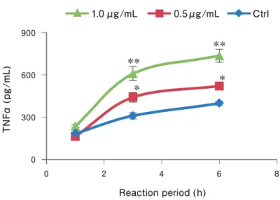
CML-HSA (0.5~1.0 μg/mL) was applied onto RAW264.7 for 1 ~ 6 hours and the release of TNFα was measured by ELISA. After 3 - and 6 - hour stimulation, TNFα (p = 0.001, 0.5 μg/mL and p < 0.001, 1.0 μg/mL) was significantly increased compared with control (Fig. 1).
0
0 2 4 6 8
300 600
Reaction period (h)
TNFα (pg/mL)
**
**
* *
0 500
250 1000
750
TNFα (pg/mL)
CML-HSA -
+ + + + + + +
Extract - - WC BN CB PL KB Mix
* **
** ** **
Inhibition by plant extracts on CML-HSA- induced TNFα formation
The following extracts were reacted with RAW264.7 for 24 hours and diluted with DMEM, medium: water chestnuts (1.0 μg/mL), banabá and Chinese blackberry (10 μg/mL), and persimmon leaf and kuma bamboo (5.0 μg/mL). The TNFα concentration was measured after 3-hour stimulation with CML-HSA (0.5 μg/mL). TNFα production was significantly reduced by the pretreatment with water chestnut (22.7%, p <
0.001), banabá (17.0%, p < 0.001), Chinese blackberry (14.6%, p = 0.001), persimmon leaf (16.8%, p < 0.001) and mixed
Fig 1. Changes of medium TNFα levels of the CML-HSA-stimulated macrophage.
Data are expressed as mean ± SD. * p < 0.01, ** p < 0.001 vs. control by Dunnett’s test, n = 4. The test culture cells, RAW264.7; stimulator, CML-HSA (0.5 μg/mL, 1.0 μg/mL); TNFα, tumor necrosis factor-α; CML, Nε-carboxymethyl- lysine; HSA, human serum albumin; SD, standard deviation.
Fig 2. Inhibition by plant extracts on CML-HSA-induced TNFα release.
Data are expressed as mean ± SD. * p < 0.01, ** p < 0.001 (1.0 μg/mL) vs. control (CML-HSA +, Extract – ) by Dunnett’s test, n = 4. The test culture cells, RAW264.7; stimulator, CML-HSA (0.5 μg/mL); WC, water chestnut; BN, banabá; CB, Chinese blackberry; PL, persimmon leaf; KB, kuma bamboo; Mix, mixture of BN, CB, PL and KB;
TNFα, tumor necrosis factor α; CML, Nε-carboxymethyllysine; HSA, human serum albumin; SD, standard deviation.
extract (17.2%, p < 0.001), when compared with CML-HSA- stimulation without pretreatment (Fig. 2). The mixed extract (Mix) consisted of banabá, Chinese blackberry, persimmon leaf, and kuma bamboo.
Cell viability assessment
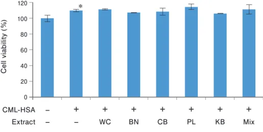
The influence on cell viability by CML-HSA and plant extracts was evaluated. The number of viable cells was significantly increased by CML-HSA stimulation compared with the control (p = 0.018). There was no significant change in viable cell numbers in the plant extract-pretreatment group
0 40 20 120
60 80 100
Cell viability (%)
CML-HSA -
+ + + + + + +
Extract - - WC BN CB PL KB Mix
*
compared with the CML-HSA-stimulated group without pretreatment (Fig. 3).
Anti-oxidative activity measurement
Anti-oxidative activity of 4 test samples were as follows:
banabá 2,243 mg/L, Chinese blackberry 1,882 mg/L, persimmon leaf 604 mg/L and kuma bamboo 200 mg/L. Each result is expressed as ascorbic acid equivalent.
Discussion
In this study, using cultured macrophage cells, RAW264.7, stimulated by CML-HSA as a glycative stress, TNFα formation was measured and the anti-inflammatory actions of plant extracts were assessed. The following receptors are known to bind to AGEs as a ligand on the cell surface in addition to RAGE: macrophage type-I and type-II class A scavenger receptors (MSR-A), CD36・SR-BI and LOX-1 (lectin-like oxidized low density lipoprotein receptor-1) which belong to the MSR-B family, galectin-3 complex, FEEL1・2 (fasciclin, EGF-like, laminin-type EGF-like, and link domain-containing scavenger receptor-1・2), megalin, and toll-like receptor 4 (TLR4) 14). These receptors play roles in inflammation reaction, degeneration and removal of AGEs, or cell proliferation.
RAGE participates in inflammation, recognizes proteins with CML modification or glyceraldehyde-derived AGEs (toxic AGEs; TAGE) which are known to be cytotoxic 15). Mediated by NF-κB activation, RAGE induces inflammatory cytokines (i.e. TNFα、interleukin [IL]-1β、IL-6), matrix metalloproteinase (MMP), vascular endothelial growth factor (VEGF) and adhesion factors (i.e. intercellular adhesion molecule-1 [ICAM-1], vascular cell adhesion molecule-1 [VCAM-1]) 16). The CML-HSA-stimulated TNFα formation observed in this study (Fig. 1) may be mainly due to the RAGE actions. Precise analysis is necessary for further investigations to determine
Fig 3. Evaluation of cell viability.
Data are expressed as mean ± SD. * p = 0.018, CML-HSA stimulation vs. control (CML-HSA –, Extract –), by Dunnett’s test, n = 5. The test culture cells, RAW264.7; stimulator, CML-HSA (0.5 μg/mL); WC, water chestnut; BN, banabá; CB, Chinese blackberry; PL, persimmon leaf; KB, kuma bamboo; Mix, mixture of BN, CB, PL and KB;
TNFα, tumor necrosis factor α; CML, Nε-carboxymethyllysine; HSA, human serum albumin; SD, standard deviation.
the specific contributions of each receptor.
CML accumulation induces chronic inflammation in tissue. Released inflammatory cytokines are reported to play a pathogenetic role in Alzheimer’s disease 17), age-related macular degeneration 18), cancer 19), and bronchial asthma 20), and also to inhibit activation of Sirtuin 1 21) which is known as a longevity gene.
In addition to these disorders, "locomotive syndrome,"
which has recently attracted attention in preventive medicine, has been suggested in relation to glycative stress, and glycative stress may reduce the bone quality by forming pathological cross-links between bone collagen fibers 22). Inflammatory cells infiltrated into the synovium induce synovitis, which plays a significant role in the pathogenesis of rheumatoid arthritis and knee osteoarthritis 23). The TNFα produced in the synovium induces the expression of the transcription factor CCAAT/
enhancer-binding protein (C/EBP) β, through the induction of inflammatory mediators (i.e. prostaglandins) and vascular endothelial growth factor (VEGF), and enhances inflammation and causes articular swelling. Furthermore, TNFα activates MMP-3 and MMP-13, cartilage matrix degrading enzymes, and finally causes the degeneration and destruction of cartilage 24). In addition, TNFα induces the expression of receptor activator of nuclear factor kappa-B ligand (RANKL), essential for the differentiation of osteoclasts, and accelerates bone resorption, thus increasing the risk of osteoporosis developing 23). On the other hand, since the half-life of cartilage collagen is long, CML-modified proteins are abundant in tissue 25). It has been suggested that AGE deposits in the joint cartilage tissue may participate in the pathogenesis and progress of osteoarthrosis of the knee 26). It is possible that the CML-modified protein-induced inflammatory substance in the cartilage may be involved in the pathogenesis of locomotive syndrome and in the hypofunction of locomotive organs in a similar manner as in the synovium.
The AGEs-RAGE interaction is related to various lifestyle- related diseases. Therefore it is important to prevent TNFα production, which is produced in the early phase of
action of the 5 plant extracts in this study has not yet been reported. The following actions are indicated by the polyphenol included in each extract. Tannin is included in water chestnut peel extract which inhibits rat hind limb edema 27). In a randomized controlled trial using banabá extract, production of the adhesion molecules ICAM-1 and VCAM-1, which are related to inflammation, was inhibited in patients with type 2 diabetes 28). The main polyphenol of Chinese blackberry (Tien- cha) is galloyl-oxygen-diphenyl (GOD) type ellagitannin; it is reported that Chinese blackberry extract prevents the release of leukotriene B4 from the rat peritoneal macrophage thus manifesting an anti-inflammation action 29). The persimmon leaf flavonoid is effective for inflammatory disturbance in the nervous system in cases of ischemia reperfusion injury 30). There is also a report that proanthocyanidin inhibits RAGE manifestation 31). In addition to its anti-inflammatory action, persimmon leaf proanthocyanidin improves hyperglycemia and dyslipidemia in the type 2 diabetes model mouse 32). Ellagitannin in banabá extract possesses an insulin-like action and activates the glucose transport in adipocytes 33). Therefore these components contribute to the improvement of glucose metabolism. Although the precise identification of the responsible ingredients was not undertaken in the present study, the results that these polyphenols prevent TNFα production and support disease prevention are considered to be worthy findings.
It's confirmed that the plant extracts used in this study indicate the inhibitory actions of in vitro AGE formation which is stronger than that of aminoguanidine (Fig. 2). It is not likely that each plant extract specifically inhibits the CML- HSA-stimulated TNFα formation. However, we can add the new action of AGE-induced inflammatory response prevention in addition to AGEs generation inhibition.
The anti-oxidative activity of the 4 extracts in the mixed plant extract, as measured by i-Pack Oxystress Test 13), showed an ascorbic acid equivalent of banabá 2,243 mg/L, Chinese blackberry 1,882 mg/L, persimmon leaf 604 mg/L and kuma bamboo 200 mg/L. According to "Standard Dietary Intake in Japan (2010)" guidelines announced by the Ministry of Health,
equivalent of the mixed plant extract is markedly large compared with the recommended value. Since the oxidative stress is known to promote the glycative reaction and increase the AGE production in the skin or other tissues 35), it is also important to control the oxidative reaction. These plant extracts may contribute to reduce glycative stress by acting as antioxidants.
Conclusion
In the present study model, employing TNFα formation from CML-HSA-stimulated macrophages as the index, some plant extracts inhibited TNFα formation, thus reducing inflammatory reaction. These actions of plant extracts may be useful since inflammation induced by glycative stress occurs in the body 34). As the next step, we plan to study the correlation between CML and TNFα in the body and also the inhibitory effect of these extracts on CML and TNFα in a clinical study.
Acknowledgement
The authors are indebted to Prof. Yukiko Minamiyama, Kyoto Prefectural University for providing the tissue culture cells. This work was supported by the Japanese Council for Science, Technology and Innovation (CSTI), Cross-ministerial Strategic Innovation Promotion Program (SIP Project ID 14533567) and JSPS KAKENHI Grant Number 26350917.
Conflict of interest statement
The authors have no conflict of interest related to this study to declare.
References
1) Mizutari K, Ono T, Ikeda K, et al. Photo-enhanced modification of human skin elastin in actinic elastosis by Nε-(carboxymethyl) lysine, one of the glycoxidation products of the Maillard reaction. J Invest Dermatol. 1997;
108: 797-802.
2) Alikhani Z, Alikhani M, Boyd CM, et al. Advanced glycation end products enhance expression of pro-apoptotic genes and stimulate fibroblast apoptosis through cytoplasmic and mitochondrial pathways. J Biol Chem. 2005; 280:
12087-12095.
3) Hori M, Yagi M, Nomoto K, et al. Inhibition of advanced glycation end product formation by herbal teas and its relation to anti-skin aging. Anti-Aging Medicine. 2012; 9:
135-148.
4) Ishioka Y, Yagi M, Ogura M, et al. Polyphenol content of various vegetables: Relationship to antiglycation activity.
Glycative Stress Research. 2015; 2: 41-51.
5) Tanaka N, Yonekura H, Yamagishi S, et al. The receptor for advanced glycation end products is induced by the glycation products themselves and tumor necrosis factor- alpha through nuclear factor-kappa B, and by 17beta- estradiol through Sp-1 in human vascular endothelial cells. J Biol Chem. 2000; 275: 25781-25790.
6) Wajant H, Pfizenmaier K, Scheurich P. Tumor necrosis factor signaling. Cell Death Differ. 2003; 10: 45-65.
7) Dimmeler S, Rippmann V, Weiland U, et al. Angiotensin II induces apoptosis of human endothelial cells. Protective Effect of Nitric Oxide. Circ Res. 1997; 81: 970-976.
8) Mueller M, Hobiger S, Jungbauer A. Anti-inflammatory activity from extracts from fruits, herbs and spices. Food Chem. 2010; 122: 987-996.
9) Ho SC, Chang PW. Inhibitory effect of several spices on inflammation caused by advanced glycation end products.
Am J Plant Sci. 2012; 3: 995-1002.
10) Takeshita S, Yagi M, Uemura T, et al. Peel extract of water chestnut (Trapa bispinosa Roxb.) inhibits glycation, degradesα-dicarbonyl compound, and breaks advanced glycation end product crosslinks. Glycative Stress Research.
2015; 2: 72-29.
11) Hori M, Yagi M, Nomoto N, et al. Effect of Kumaizasa (Sasa renansis Rehder) on the inhibition of advanced glycation end product (AGEs) formation. The Science and Engineering Review of Doshisha University. 2011; 52: 61- 67. (in Japanese)
12) Sato K, Yagi M, Umehara H, et al. Establishment of a model for evaluating tumor necrosis factor-α production by cultured RAW264.7 in response to glycation stress.
Glycative Stress Research. 2014; 1: 1-7.
13) Sato K, Yagi M, Yonei Y. A new method for measuring oxidative stress using blood samples. Glycative Stress Research. 2015; 2: 15-21.
14) Nagai R, Mori T, Yamamoto Y, et al. Significance of advanced glycation end products in aging-related disease.
Anti-Aging Medicine. 2010; 7: 112-119.
15) Yamamoto Y, Yonekura H, Watanabe T, et al. Short-chain aldehyde-derived ligands for RAGE and their actions on endothelial cells. Diabetes Res Clin Pract. 2007; 77: S30- 16) Yamagishi S, Imaizumi T. Diabetic vascular complications:40.
pathophysiology, biochemical basis and potential therapeutic strategy. Current Pharm Des. 2005; 11: 2279- 2299.
17) Takeuchi M, Yamagishi S. Possible involvement of advanced glycation end-products (AGEs) in the pathogenesis of Alzheimer's disease. Curr Pharm Des. 2008; 14: 973-978.
18) Yamada Y, Ishibashi K, Ishibashi K, et al. The expression of advanced glycation endproduct receptors in rpe cells associated with basal deposits in human maculas. Exp Eye Res. 2006; 82: 840-848.
19) Ohmori H, Luo Y, Kuniyasu H. Non-histone nuclear factor HMGB1 as a therapeutic target in colorectal cancer.
Expert Opin Ther Targets. 2011; 15: 183-193.
20) Kyoh S, Kanazawa H, Tochino Y, et al. Comparison of N-epsilon-(carboxymethyl)lysine levels and percentage of eosinophils in induced sputum for assessment of small airway involvements in asthma. Med Sci Monit. 2008; 14:
CR375-378.
21) Ishibashi Y, Nakashima S, Matsui T, et al. Rosuvastatin restores advanced glycation end product-induced decrease in sirtuin 1 (SIRT1) mRNA levels in THP-1 monocytic cells through its anti-oxidative properties. Int J Cardiol.
2013; 169: e102-103.
22) Saito M, Marumo K. New treatment strategy against osteoporosis: Advanced glycation end products as a factor for poor bone quality. Glycative Stress Research. 2015; 2:
1-14.
23) Okazaki K, Iwamoto Y. Gene regulation with cartilage degeneration in rheumatoid arthritis and osteoarthritis.
Fukuoka Acta Med. 2012; 103: 175-181.
24) Tsushima H, Okazaki K, Hayashida M, et al. CCAAT/
enhancer binding protein beta regulates expression of matrix metalloproteinase-3 in arthritis. Ann Rheum Dis.
2012; 71: 99-107.
25) Schleicher ED, Wagner E, Nerlich AG. Increased accumulation of the glycoxidation product Nε- (carboxymethyl) lysine in human tissues in diabetes and aging. J Clin Invest. 1997; 99: 457-468.
26) DeGroot J, Verzijl N, Jacobs KM, et al. Accumulation of advanced glycation endproducts reduces chondrocyte- mediated extracellular matrix turnover in human articular cartilage. Osteoarthritis Cartilage. 2001; 9: 720-726.
27) Patel AS, Patel NC, Shah MH, et al. Evaluation of anti- inflammatory activity of fruits of Trapa natans linn.
IJPRD. 2011; 3: 97-102.
28) Kim HJ, Yoon KH, Kang MJ, et al. A six-month supplementation of mulberry, Korean red ginseng, and banaba decreases biomarkers of systemic low-grade inflammation in subjects with impaired glucose tolerance and type 2 diabetes. Evid Based Complement Alternat Med. 2012; 2012: 735191. doi: 10.1155/2012/735191.
29) Takasugi M, Kitagawa T, Kato M, et al. Correlation among suppression of leukotriene B4 release, radical scavenging activity and polyphenol contents of commercial tea extract.
Nippon Shokuhin Kagaku Kogaku Kaishi. 2008; 55: 87- 94. (in Japanese)
30) Bei WJ, Xu AL, Li CY, et al. Flavonoids from Diospyros kaki inhibit the adhesion between lymphocyte and dorsal root ganglion. Zhong Yao Cai. 2009; 32: 740-774.
31) Lu M, Xu L, Li B, et al. Protective effects of grape seed proanthocyanidin extracts on cerebral cortex of streptozotocin induced diabetic rats through modulating AGEs/RAGE/NF-κB pathway. J Nutr Sci Vitaminol.
2010; 56: 87-97.
32) Jung UJ, Park YB, Kim SR, et al. Supplementation of persimmon leaf ameliorates hyperglycemia, dyslipidemia and hepatic fat accumulation in type 2 diabetic mice. PloS ONE. 2012; 7(11): Article ID e49030.
33) Hayashi T, Maruyama H, Kasai R, et al. Ellagitannins from Lagerstroemia speciosa as activators of glucose transport in fat cells. Planta Med. 2002; 68: 173-175.
34) Crespo A, Filla MB, Russell SW, et al. Indirect induction of suppressor of cytokine signalling-1 in macrophages stimulated with bacterial lipopolysaccharide: partial role of autocrine/paracrine interferon-α/β. Biochem J. 2000;
349: 99-104.
35) Ichihashi M, Yagi M, Nomoto K, et al. Glycation stress and photo-aging in skin. Anti-Aging Medicine. 2011; 8:
23-29.