Acta Med. Nagasaki 42 : 61-64
Endoscopic Polypectomy of Esophageal Leiomyomas; Report of Two Cases
Hiroshi NAGATA 1), Masuho HARAGUCHI 1) , Kunihiko MURASE 3), Katsuhisa OMAGARI 3), Yohei MIZUTA 3), Keisuke IWASAKI 2), Hidehiko FUKUDA 4), Sigeru KOHNO 3)
1) Department of Internal Medicine, Sasebo Municipal Hospital 2) Department of Pathology, Sasebo Municipal Hospital
3) The Second Department of Internal Medicine, Nagasaki University School of Medicine 4) Fukuda Clinic
We describe esophageal leiomyomas in two young patients (aged 35 and 32 years), who complained of dysphagia and epigastralgia, which were successfully treated by endoscopic polypectomy. Upper endoscopy showed a pedunculated polyp beneath the normal mucosa located at 28 cm from the incisor in the first case and 1 cm sessile 2.1 cm semiped- unculated polypoid lesion in the lower esophagus just above the esophageal-gastric junction in the second case. Both lesions were resected by snare polypectomy without any complication. Light microscopic examination and im- munohistochemistry of the tumor tissue confirmed the diagnosis of leiomyoma. Endoscopic polypectomy of eso- phageal leiomyoma is safe and should be considered as an optional treatment modality whenever possible.
Key wards : esophageal leiomyoma, endoscopic polypectomy
Introduction
Stromal cell tumors of the esophagus are relatively rare, and leiomyoma is the most common benign tumor of the esophagus. The frequency of leiomyoma in the upper gastrointestinal tract is 4% of all tumors of the esopha- gus'). Therefore, diagnosis and management of this tumor, which may not be straightforward, is important to endoscopists. Leiomyomas occur at all sites but in one extensive review') 11% were detected in the upper third segment of the esophagus, 33% in mid esophagus, and the remainder in the distal third segment. Of these, 18% were in the muscularis mucosa, 73% in the circular muscles, and 7% in the longitudinal muscles. The majority of tumors causing symptoms, usually dysphagia, are large measuring up to 5 cm in diameter, although they may be smaller at the lower esophageal sphincter. However, in small leio- myomas, 50% of the patients remain asymptomatic and the tumor is often discovered incidentally". Endoscopic polypectomy of esophageal leiomyoma is a very useful
procedure for both diagnosis and treatments). We report two cases of esophageal leiomyomas treated by endoscopic polypectomy. without any complication.
Case reports
Patient 1
A 35-years-old female was admitted to Sasebo Municipal Hospital for further management of sleep disturbances. As part of the diagnostic workup, she was referred to the gastrointestinal section of the Department of Internal Medicine for dysphagia. Barium meal showed an oval, well-defined filling defect in the lower esophagus (Fig. 1).
Endoscopy showed a white smooth round sessile 1 cm firm polypoid lesion in the lower esophagus just above the esophageal-gastric junction. A biopsy was performed (Fig.
2), but the specimens showed only a hyperplastic epithe- lium. Endoscopy was repeated in order to perform a snare polypectomy. The lesion was removed completely without any complications. The size of resected specimen was 1.0 x 1.0 x 0.8 cm. Histological examination of the polyp demon- strated spindle cell proliferation with bundle formation.
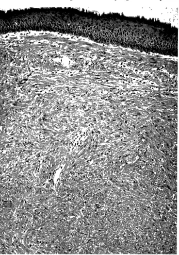
There was no alveolar pattern, nuclear pleomorphism or microscopic invasion of the lamina propria or adjacent blood vessels. Only rare mitotic figures were noted (Fig.
3). Immunohistochemically, a positive reaction with a - smooth muscle actin was noted, although no reaction was observed with S-100 protein. Since the endoscopic poly- pectomy of the tumor 1.5 year ago, no recurrence has developed and the patient continues to be free of symp- toms at the time of writing of this report.
Patient 2
A 32-years-old man was initially admitted to the Fukuda Clinic for epigastralgia, but was later referred to Sasebo
Address Correspondence : Dr. Kunihiko Murase, Second Department of Internal Medicine, Nagasaki University School of Medicine, 1-7-1 Sakamoto, Nagasaki 852-8501, Japan
Fig. 1 Case 1. Barium meal showing a filling defect with smooth edges in the lower esophagus.
Fig. 3 Case 1. Microscopic finding showing an interlacing pattern of spindle cells with only rare mitotic figures (x 100).
Fig. 2 Case 1. Endoscopic photograph showing a white smooth rounded sessile polypoid lesion in the lower esopha- gus.
Fig. 4 Case 2. Endoscopic photograph showing a semi- pedunculated polypoid lesion in the middle esophagus.
Municipal Hospital for further examination. Upper gastrointestinal imaging showed an oval, well-defined filling defect in the middle esophagus. A barium meal showed a mid-esophageal filling defect with smooth edges.
On endoscopy, a semipedunculated polyp was identified, which was covered with normal esophageal mucosa, 28 cm from incisors (Fig. 4). Endoscopy was repeated at a later stage in order to perform a snare polypectomy. His-
tological examination of the resected polyp demonstrated a leiomyoma. A follow up endoscopic examination of the esophagus one year later showed no residual tumor or other abnormalities.
Discussion
Stromal and smooth muscle tumors of the esophagus ; leiomyoma, schwanoma, granular cell tumor, vary widely in size from less than 1 cm to well over 20 cm, I). Important diagnostic findings pertaining to the nature of a sub- mucosal esophageal tumor are usually obtained from endoscopy, esophageal ultrasonography and computed tomography"'. However, it is difficult to distinguish these tumors in some cases. Histological examination of a biopsy material represent the most accurate and reliable method of a definite diagnosis of these tumors. Virtually all esophageal tumors have a submucosal component, and many merge imperceptibly with either the muscularis propria or the muscularis mucosaa 10' . Many of these tumors are submucosal or deeper and, accordingly, biop- sies may be inadequate for obtaining deep tissue samples necessary for an accurate diagnosis. Endoscopic biopsy usually do not allow to obtain tissue from the submucosal tumor, so endoscopic polypectomy has to performed for diagnosis of submucosal tumors"). The first report of transfiberscopic esophageal polypectomy was published in 1973"). Since then it has been recognized as a surgical procedure in selected patients"'). Benedetti et al'). pro- posed that in symptomatic cases, or when the lesions are more than 1 cm in diameter, the treatment of choice is local resection. Treating a much larger sample, Classen et al"). reported no complications in 104 esophageal polypec- tomies. On the other hand, Cox et al'). advocated a conservative approach of medical treatment in asympto- matic esophageal leiomyoma rather than surgical excision.
In Japan, surgical treatment of esophageal leiomyomas reported by Taniguchi et al"). and Izumi et all". indicated that thoracoscopic enucleation of the esophageal leio- myoma is safe and effective operation. Other therapeutic approaches have also been reported. For example, Lange et a118'. demonstrated that endoscopic laser ablation was a fast and successful treatment with no signs of recurrence at more than 2 years of follow-up. Moreire and Dani19' described a case of esophageal leiomyoma successfully treated by endoscopic injection of dehydrated ethyl alco- hol. There is no definite criteria for endoscopic polypec- tomy of esophageal submucosal tumors. Yu et al proposed that the criteria for endoscopic polypectomy of sub- mucosal tumors were presence of a pedunculated tumor, or presence of a sessile tumor with a base smaller than 2 cm which originated in the upper wall layers'). Histological examination of a typical leiomyoma shows interlacing fascicles of spindle cells in a herribone arrangement'.
Immunohistochemically, these tumors are positive for smooth muscle a -actin but negative for S-100 protein or PGP 9.522'. Recently, Miettinen et al'). proposed the term gastrointestinal stromal tumor (GIST) for mesenchymal
tumors that represent neither typical leiomyoma nor schwanoma. Most of GIST show limited or no reactivity for muscle markers and S-100 reactivity, and CD 34 reac- tivity between benign and malignancy. A variety of clinical, macroscopic and microscopic parameters need to be considered in evaluating the malignant potential of a
stromal tumor. Among the high risk-factors for metasta- sis are large size tumors, high frequency of mitosis with atypical mitosis, tumor necrosis, nuclear pleomorphism, dense cellularity, microscopic invasion of lamina propria
or blood vessels, and alveolar pattern'). In both cases reported here, no high risk factors were detected. If the patients with leiomyoma of esophagus were performed complete histological resection by endoscopic polypec- tomy, they do not need another treatment.
In summary, polypectomy of esophageal leiomyoma can be performed safely. Transfiberscopic polypectomy should be considered as a method of resection. However, surgery should be performed when neoplastic changes or when the tumor is too large to be removed by endoscopy.
References
1) Wenger FA, Jacobi CA, Zieren HU: Diagnosis and therapy of leiomyoma of the upper gastrointestinal tract. Langenbecks Archiv
Chir 381: 221-4, 1996
2) Seremetis MG, Lyons WS, deGuzman VC : Leiomyoma of esophagus.
An analysis of 838 cases. Cancer 38: 2166-77, 1976
3) Takubo K, Nakagawa H, Tsuchiya S, Mitino Y, Sasajima K, Shirota A : Seeding leiomyoma of the esophagus and esophagogastric junction
zone. Human Pathol 12: 1006-10, 1981
4) Marmuse JP, Johanet H, Louvent F, Benhamou G. Leiomyomas of the lower third of the esophagus : Value of transhiatal enucleation. Ann
Chirurgie 48: 446-51, 1994
5) Benedetti G, Sablich R, Bonea M, Mariuz S : Fiberoptic endoscopic resection of symptomatic leiomyoma of the upper esophagus. Case
report. Acta Chir Scand 156: 807-8, 1990
6) Solomon MP, Rosenblum H, Rosato FE : Leiomyoma of the esopha- gus. Ann Surg 199: 246-8, 1984
7) Dancygier H, Nattermann C. Endoscopic ultrasonography-indications
and results in diseases of the esophagus and stomach : Schweiz Rund Medizin Praxis 83: 1281-7, 1994
8) Kalantzis N, Laoudi F, Kallimanis G, Gabriel P, Farmakis N : The role of endoscopic ultrasonography in diagnosis of benign lesions of the
upper GI tract. Eur J Surg Oncol 19: 449-54, 1993
9) Cox MA, Cooper BT, Sagar G : Endoscopy and computed tomography
in the diagnosis and follow up of esophageal leiomyoma. Gut 37:
288-91, 1995
10) Rose JD : Myoma of the esophagus. Brit J Surg 24: 297-308, 1936 11) Cornish D, Feinstat T, Schneider P, Ruebner B, Trudeau W. Esopha-
geal granular cell tumor removed by endoscopic polypectomy : Am J
Gastroenterol 80: 950-3, 1985
12) Seifert E, Elster K : Value of endoscopic polypectomy in the esophageal and gastric tumors. Leber Magen Garm 3 : 155, 1973
13) Deyhle P : Results of endoscopic polypectomy in the gastrointestinal
tract. Endoscopy (suppl) : 35-46, 1980
14) Jacobs W, Bruns D : Endoscopic electrosurgical polypectomies the upper gastrointestinal tract. Am J Gastroenterol 68-241, 1977
15) Classen M, Hagenmuller F, Wurbs D : Klinischer wer der Endoskopis-
chen Polypektomie in Oesophagus, Magen and Duodenum unter Beruckisichtigung der Spatergebnisse. Akt Gastrol 8-495, 1979 16) Taniguchi E, Kamiike W, Iwase K, Nishida T, Akashi A, Ohashi S,
Matsuda H : Thoracoscopic enucleation of a large leiomyoma located
on the left side of the esophageal wall. Surg Endoscopy 11: 280-2,
1997
17) Izumi Y, Inoue H, Endo M : Combined endoluminal-intracavitary
thoracoscopic enucleation of leiomyoma of the esophagus. A new method. Surg Endoscopy 10: 457-8, 1996
18) Lange V, Wenk H, Schildberg FW : Laser vaporization of leiomyoma of the esophagus. Lasers Surgery Med 9 : 74-6, 1989
19) Moreira LS, Dani R : Treatment of granular cell tumor of the
esophagus by endoscopic injection of dehydrated alcohol. Am J Gastroenterol 87: 659-61, 1992
20) Lewin KJ, Riddell RH, Weinstein WM : Gastrointestinal pathology and its clinical complication. New York. Igaku-Shoin : 284-341, 1992 21) Yu JP, Luo HS, Wang XZ : Endoscopic treatment of submucosal
lesions of the gastrointestinal tract. Endoscopy 24: 190-3, 1992 22) Saul SH, Rast ML, Brooks JJ : The immunocytochemistry of gastroin-
testinal stromal tumors. Evidence supporting an origin from smooth
muscle. Am J Surg Pathol 11: 383-6, 1987
23) Miettinen M, Virolainen M, Rikala MS: Gastrointestinal stromal tumor-Value of CD 34 antigen in their identification and separation
from true leiomyomas and schwanomas. Am J Gastroenterol 19:
207-16, 1995