Acta Med. Nagasaki. 23:63-81
Histometrical Approach of Aging Processes on Rat Aorta Using Low Power Magnification: Photographs of
Electron Microscopy
Takayoshi TODA*
Department of Pathology, Scientific Data Center of atomic Bomb Disaster, Nagasaki University School of Medicine,
Nagasaki, Japan
Received for publication, September 15, 1978
A Part of this work was presented at the 18th Annual Meeting of the Japanese College of Angiology, Nov. 19, 1977.
72 Wistar Strain rats ranging in age from new born to age of 3 years were used to clarify specificity in growth of aorta in aging processes of vascular system. Developmental transformation of aortic media was investigated using low power magnification photographs of electron microscopy (x 850), because cell membrane can be traced easily. Number and size of smooth muscle cells, density of intercellular material and elastic fiber in a given unit area were histometrically estimated. In addition the fine structural study of each components of aorta was performed.
In growing rat aorta, atypical biphasic pattern with double peak was observed in total intercellular material density around postnatal 5th and 35th day. Number of smooth muscle cell of aorta decreased with aging, and size of smooth muscle cell of aorta showed peak around 50th day. Smooth muscle cell of aortic media from newborn rat showed secretary function with organella-rich cytoplasm.
Around 50th day after birth, transitory smooth muscle cell from organella-rich secretary cells to contractile cells with a large amount of myofibril was seen in proportion of one tenth in a given unit area. Rat aortic media maintained high level in density of elastic fiber per unit area from 30th to 60th day.
During involutional period, fluctuation of total intercellular material density in rat aortic media was not obvious. However, detailed analyis of each component of aortic media revealed that decrease in number of smooth muscle cells and density of elastic fiber density developed, but cell debris increased. From the view point of acceleration of pubescence, it was considered to be of interest finding that atypical biphasic pattern was observed in alteration of total intercellular material density of rat aortic media during growth.
*戸 田 隆i義
INTRODUCTION
It is well known that arteriosclerosis develops with aging. B) There are many reports about senescence of vascular system microscopically and ultramicroscopically, but few reptots about growth and developmental process of vascular system.
The present study is to clarify property of aorta as organ on the following bases ; 1) proper structure of aorta itself can be enough kept. 2) results obtained are to be described objectively. Moreover histometrical method utilizing low power magnification photographs of electron microscopy was carried out to detect attitude of rat aorta in growing period.
MATERIAL and METHOD
72 Wistar Strain rats ranging from newborn to age of 3 years were used in this study.
The rats were fed by pellet diet (Oriental Yeast Industry). Growth of rat is considered to be influenced by environment and nutrition, so constant condition was kept as possible.
Body weight, length of head, body and tail were examined as the landmark for systemic growth rate. Aorta was obtained from at least three rats at each of the following ages : at birth, 5th, 10th, 30th, 40th and 50th day, and 2nd, 3rd, 4th, 6th, 12th and 36th month after birth. Specimens of aorta obtained from thoracic and abdominal portion were sectioned into ring shaped small pieces respectively. Specimens were fixed with 1.5% glutaraldehyde in pH 7.3, postfixed 1% osmium acid, dehydrated with alcohl series and embeded in Eopn 812.
Thick sections for light microscopy were oriented so as to give transverse sections of the aorta and minimize deviation of histometrical results. The prepared thick sections for light microscopy were stained with toluidin blue. Upon triming of these blocks, ultrathin sections for electron microscopy were cut with Porter-Blum Ultra-Microtome MT-1, followed by double staining of uranyl acetate and lead citrate.
The aortae were observed with JEM-7A type of electron microscope, and was photo- graphed. Developmental changes of each component with aging, namely total intercellular material density, elastic fiber density, and number and size of smooth muscle cell were analized in 10 given unit area of 208.5 cm2 of photographs in middle layer of aortic media.
Also their fine structural observation was exhibited.
RESULT
1) Alteration of Average Body Weight and Body Length in Growing Rats
In present study, it is necessary to define growth and developmental period, and involutional period. 66 Wistar Strain rats under the same condition, ranging from newborn to age of 4 months were measured about alteration of average body weight and body length
(from head to root of tail).
The details of obtained data are shown in Figure 1-1, 1-2. In this study, the concept that growth is increase in quantity in life system was accepted as definition of growth.
Growth of body weight and length reach constant high level, and ability to deliver in female
Fig. 1-1 Alteration of Average Body Weight in Growing Rats
Fig. 1-2 Alteration of Average Body Length in Gowing Rats
rat is accomplished around 3 months after birth. Hence, the term of 3 months after birth is considered to be critical period between growth and involutional period.
In general, growth is classified into the following two types, 1) Relative Growth (Allometry), which exhibits the relationship between the whole animal tissue and some part of animal tissue (eg. body and brain weight), 2) Absolute Growth which shows chronogical increase in quantity. Via allometric formula, y=axa (y: body weight, x : body
length, a : initial growth constant, a : relative growth coefficient), were computed the all- ometric relation of body weight to body length in Rat. The results obtained were the following : in male rat, y=0.089x2.686, in female rat, y=0.031x3.156. This allometric equation obtained in rat, is considered to reveal good fittness to well balanced growth and developmen- tal style in relation to (body weight) -- (Body length)'. Accordingly, it is reasonable to consider 3 month after birth is a critical period between growth and involution.
2) Rat Aorta during Growth and Development (0-3 month)
The results obtained of the following aortic components are described via the absolute growth, and the difference in results between male and female rat was not investigated.
a) Alteraion of Total Intercellular Material and Elastic Fiber Densities per Unit Area of Middle Layer of Aortic Media. The mean value obtained from 10 samples of thoracic aorta and abdominal aorta was summarized in Table 1. Based on Table 1, total intercellu- lar material density including collagen fiber, elastic fiber and matrix was compared between thoracic and abdominal aorta. It was observed double peaks of total intercellular material around both newborn to 5th day and 30th to 40th day after birth. Thoracic aorta was superior in quantity of it than abdominal aorta, which was testified by t-test. (p<0.05)
(Fig. 2)
In the elastic fiber density of thoracic aorta and abdominal aorta, there was no difference between thoracic aorta and abdominal aorta by the age of postnatal 5th day, however thereafter thoracic aorta was superior in the elastic fiber density than abdominal aorta by the age of 50th day after birth. (Fig. 3)
On sum of both matrix and collagen fiber, thoracic aorta shows predominant increase than abdominal aorta from newborn to postnatal 5th day, but thereafter abdominal aorta shows predominant increase than thoracic aorta by the age of 60th day. (Fig. 4)
b) Size of a Smooth Muscle Cell (Fig. 5-1) and Total Number of Smooth Muscle Cell (Fig. 5-2) per Unit Area of Middle Layer of Aortic Media.
Table. 1
Results of Aortic Components per Unit Area of Aortic Media in Growing Rats*
Day 0 5 10 30 40 50 60 90
Component
28.9 48.4 45.5 46.0 63.1 57.0 64.5 60.9
TICM** ± ± ± ± ± 4- ± 4 -
Thoracic 1.6 3.0 2.4 3.9 1.6 1.8 1.0 0.5
Aorta Elatic 10.2 18.0 24.2 26.3 45.9 30.1 45.7 35.6
± ± ± ± ± ± ± ~-.
Fiber 1 .2 1.5 0.2 1.3 0.2 5.5 0.2 4.9
28.4 42.0 43.4 31.4 51.3 50.3 64.2 52.5
TICM** ± ± ± ± ± ± 4-
Abdominal 2.2 0,8 1.8 2.4 1.7 1.7 3.3 3.6
Aorta Elastic 9.1 17.7 18.1 13.1 27.2 28.2 41.7 32.2
± ± ± ± ± ± ± 4 -
Fiber 1.4 1.1 1.4 1.0 3.2 3.0 5.1 3.0
* Mean±Standard Error
** TICM: Total Intercellular Material per Unit Area of Aortic Media
Fig. 2 Altertion of Total Intercellular Material Density per Unit Area of Middle Layer of Aortic Media
Fig. 3 Ateration of Elastic Fiber Density per Unit Area of Middle Layer of Aortic Media
Fig. 4 Alteration of Density of Collagen Fiber and Intrercellular Matrix per Unit Area of Middle Layer of Aortic Media
Fig. 5-1 Alteration of Size of a Smooth Muscle Cell of Rat Aorta
Fig. 5-2 Alteration of Total Number of Smooth Muscle Cells per Unit Area of Middle Layer of Aortic Media
On the occasion of appearing of several nuclei in single smooth muscle cell for cutting direction, several nuclei were calculated as single nucleus. The shape of smooth muscle cell was supposed to be almost an ellipse and the simple formula a x b was applied as a parameter in estimation of smooth muscle cell size (a : longest length of longitudinal diameter, b : intermediate length of transverse diameter)
There was no particular difference between thoracic aorta and abdominal aorta in size and number of smooth muscle cell at the birth time. Around 5th days, smooth muscle cell of thoracic aorta showed larger appearance in size and decrease in number, on the other hand smooth muscle cell of abdominal aorta showed smaller appearance in size and increase in number. In general, during growth period, number of smooth muscle cell decreased gradually and size of smooth muscle cell also increased with peak around postnatal 50th day.
c) Fine Structure of Intima and Media in Growing Rat Aorta
It is very difficult to find out fine structural differences between thoracic aorta and abdominal aorta qualitatively. In this report general ultrastructural findings of either thoracic aorta or abdominal aorta were described.
The intima of 5-day-old rats showed no particular structure in subendothelium. An extensive amount of rough endoplasmic reticulum was present in endothelial cells. (Fig. 6) Smooth muscle cells of media connected each other with appearance of syncitial cell and contained plentiful rough endoplasmic reticulum and glycogen particle, but myofilaments were not well developed.
Fig. 6 : Intima of 5-day-old rat. An extensive amount of rough endoplasmic reticulum is present in endotherial cells. No reticular basement membrane is seen in
subendothelium. X 4,200
Fig. 7 : Media of 5-day-old rat. Smooth muscle cells of media with syncytial cell appearance contain plentifull rough endoplasmic reticulum and glycogen
particles. In intercellular space, granular or rodshaped elastic fibers are
arranged incontinuously, and a small amount of formation of collagen
fibers is noted. X 4,200
In intercellular space, granular or rod-shaped elastic fibers were arranged incontin- uously, and a small amount of collagen fiber formation was observed. (Fig. 7) Thoracic aorta of 50-day-old rats showed numerous cystic space in intima and irregularly shaped smooth muscle cells which were seperated each other owing to enhanced production of intercellular material. (Fig. 8) Inset revealed flat endothelial cell and granular to rod-shaped tiny elastic fiber in subendothelium.
Fig. 8 : Thoracic aorta of 50-day-old rat. This Photograph represent numerous cystic spaces in intima and irregularlly shaped smooth muscle cells
due to enhanced production of intercellular material. x 2,100
Inset : Higher magnification. Note flat endothelial cells and granular to rod-shaped tiny elastic fibers in subendothelium. x 5,000
Fibrillar and organella-rich regions in the cytoplasm were seen in smooth muscle cell of media. These smooth muscle cells were considered to be transitory cell from secretary cell to contractile cell, which was observed in proportion of one-tenth of total smooth muscle cells. (Fig. 9)
Fig. 9 : Higher magnificaton of Fig. 8. Both fibrils and organellae are seen in smooth muscle cell of media. x 7,500
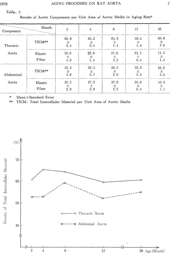
Table, 2
Results of Aortic Components per Unit Area of Aortic Media in Aging Rats*
Month 3 4 6 12 36
Component
60.9 65.3 64.3 59.4 60.6
TICM** ± ± + ± ±
Thoracic 0.5 0.4 1.4 1.4 2.0
Aorta Elastic 35.6 23.6 27.6 25.1 11.5
+ + + + ±
Fiber 4.9 1.4 3.2 0.4 1.4
i
52.5 52.5 59.5 52.3 55.0
TICM** ± ± ± ± ±
Abdominal 3.6 0.7 2.0 2.5 3.0
Aorta Elastic 32.2 27.2 27.8 20.9 10.3
+ + -+ --I- +
Fiber 2.8 2.8 2.2 0.4 1.1
* Mean±Standard Error
** TICM: Total Intercellular Material per Unit Area of Aortic Media
Fig. 10 Alteration of Total Intercellular Material Density per Unit Area of Middle Layer of Aortic Media
Fig. 11 Alteration of Elastic Fiber Density per Unit Area of Middle Layer of Aortic Media
Fig. 12 Alteration of Density of Collagen Fiber and Interclleular Matrix per Unit Area of Middle Layer of Aortic Media
By the age of 3 months, in subendothelial space, which was covered with endothelial coils having a sparse amount of organella, reticular basement 'membrane appeared, in addition, very irregularly outlined smooth muscle cells with large amount of contractile myofibrils, elastic fibers and collagen fibers were developed.
3) Rat Aorta in Involutional Period (from 4 months to 3 years)
a) Alteration of Total Intercellular Material and Elastic Fiber Densities per Unit Area of Middle Layer of Aortic Media
The results obtained on total intercellular material and elastic fiber densities about thoracic and abdominal aorta were summarized in Table 2.
Fig. 13-1 Alteration of Size of a Smooth Muscle Cell of Rat Aorta
Fig. 13-2 Alteration of Total Number of Smooth Muscle Cells per Unit Area of Middle Layer of Aortic Media
In general, total intercellular material density showed mild fluctuation with age, however, difference between thoracic and abdominal aorta seemed to disappear with age.
(Fig. 10) Transiently elastic fiber density showed reversal relation between thoracic aorta and abdominal aorta around 4 months, but significant difference of the two was not detected statistically by t-test (p>0.05) . It was obvious that elastic fiber density decrease with senescence in both thoracic aorta and abdominal aorta. (Fig. 11)
Superficially, alteration of total intercellular material density was considered to be relatively constant in quantity. When sum of matrix, collagen fiber and cell debris (rest of total intercellular material excluded by elastic fiber) was investigated, both thoracic aorta and abdominal aorta showed gradual decrease by the age of 12 months, thereafter, increase by the age of 3 years. (Fig. 12) This finding was considered to express progressive decrease of smooth muscle cells and elastic fiber with senescence.
b) Size of a Smooth Muscle Cell (Fig. 13-1) and Number of Smooth Muscle Cells (Fig. 13-2) per Unit Area of Middle Layer of Aortic Media
Number of smooth muscle cell decreased with advancement of age, and smooth muscle cell size increased by the age of 12 months with peak, thereafter decreased gradually with senescence.
c) Fine Structure of Intima and Media of Rat Aorta in Aging Rats
Reticular basement membrane structure was continuously observed in the intima of
Fig. 14 : Aorta of 6-month-old rat. Reticular basement membrane is well developed in subendotheium. Irregularly shaped smooth muscle cells of media have
indented unclei, abundant collagen fibers and thickened elastic fibers with branching in intercellular space of aortic media x 2,100
rat aorta after the age of 4 months, and smooth muscle cells of media appeared of irregular shape and contained myofibril rich cytoplasm. In aorta of 6-month-old rats, well developed reticular basement membrane structure was found in intima.
Smooth muscle cell of aortic media in the same age, was irregularly shaped and contained prominently an indented nucleus. Abundant collagen fibers and thickened elastic fibers with branching were observed in intercellular space of aortic media. (Fig. 14)
In subendothelium of 12-month-old rat aorta, there appeared reticular basement membrane structure, cell debris and fragments of internal elastic fiber, but smooth muscle cell hyperplasia was not observed at all. Smooth muscle cells of aortic media were atrophic with relatively smooth outline and obscure nuclei, and contained numerous degenerative products such as dense bodies, myelin figures and vacuoles. A large amounts of cell debris and collagen fibers accumulated in intercellular space, furthermore, branching of atrophic elastic fiber became more obvious. (Fig. 15)
In this way, rat aorta showed degenerative or necrotic change in smooth muscle cell of media, however cellular reaction to these necrotic foci was never detected. In intima, reticular basement membrane structure with cell debris developed with age, but intimal thickening with smooth muscle cell hyperplasia never developed during a whole life of rat.
Fig. 15 : Aorta of 12-month-old rat. Intima contains well developed reticular basement membrane, cell debris and fragments of internal elastic fibers. Atrophic smooth
muscle cell with numerous degenerated products such as dense bodies, myelin
figures, and vacuoles are noted. A large amount of cell debris and fibers
accumulate in intercellular space. x 2,100
DISCUSSION
It is generally accepted that aging phenomena in organs of animal are involved i n fertilization, growth and involution"). In the various organs, the vascular system strongly reflects the aging process. The thickening of the subendothelial layer is necessary for deve- lopment and progression of atherosclerosis of the aorta, which is closely related to the aging process. Many reports have suggested species differences in development of the intimal thic- kening ; the human aorta easily bring about it, on the other hand, the rat aorta does it not so easily. No reasons of the species differences in development of atherosclerosis have been clearly explained, although there are some structural explanations that it would be due to the structure differences such as wall thickeness of vessels wall and vasa vasorum supplying aortic media23>24>. Schwartz, S.M. 14) and Cliff, W. J. 1)2) reported the ultrastructural studies on the aging process of the rat aorta, but they made few interpretation on relationship between function and structure of rat aorta in aging process. In the present study, therefore , the author tried to investigate relationship between function and structure of rat aorta in aging process, using histometrical method. Scammon divided") the growth pattern of the organ into four main types ; 1) lymphoid tissue type, 2) nervous system type, 3) general type, and4) genital organ type, and the growth pattern of both body weight and height was classified as general type, whose character is to indicate double spurt around the infantile period and puberty. Auther et al20. analized the growth pattern of the human aorta with a parameter of total intercellular material in the aortic media, and found the character of growth pattern of human aorta as general type, too. It was reported that developmental change of body weight of rat is monophasic, and spurt in puberty is not seen 18). The finding reported herein is consistent with the previous report, as shown in Fig. 1-1. In the present study, it further appears that developmental change of body length is monophasic, as shown in Fig. 1-2. The concept of relative growth which represents the balance between the growth of whole body and the growth of each organ was applied to the relationship between body weight and body length"), the cardiac weight and the coronary artery 16) , and the body weight and main pulmonary artery". It was reported that the growth rate indicating the relationship between body weight and body length in human beings is 2.1996 in male and 2.2459 in female"). The growth rate of rats caliculated from the data obtained in the present study is 2.686 in male and 3.156 in female, which is far different from the data obtained from human beings. In the human beings, the developmental change of total intercellular material in a given unit area of aortic media showed typical biphasic pattern due to spurt in puberty, otherwise, in the rat it showed atypical biphasic pattern. This finding suggests that in the rat, whole body growth process would be reflected in the rat aorta. It should be noted that in the rat participation of accelerated process in aortic growth during puberty was suggested by the finding ; in which atypical biphasic pattern in the developmental change of total intercellular material was recognized around the neonatal period and puberty. It is unknown why growth pattern is different between in the rat in the human beings, but there would be two possibilities ; one is difference between short and long duration of growth period, another is difference
of metabolism including hormonal biomechanics. According to progress of radioimmunoassy for numerous hormones, recently some investigators have reported the relationship between sex hormone dynamics and puberty. Also, Takeda et al"). reported participation of sex hormone on growth process of the connctive tissues, utilizing collagen sythesizing ability of fibroblast. There is no agreement about participation of sex hormone on growth process of the elastic fiber, collagen fiber and matrix, mainly due to problems of assay methods.
Vascular system in broad sense is included in the connective tissue. It should be reasonable that the smooth muscle cell responses to sex hormone, since both fibroblast and smooth muscle cell as the substantial cell of vascular wall originate from the same cell fine').
Many investigators have indicated that the character of the smooth muscle cells is different in the various organs. For example, the smooth muscle cell of the uterus is strongly dependent on sex hormone"). In that of the aorta, administartion of estrogen produces the morphological change 4) and diminishes development of atherosclerosis11), furthermore, hypertension and administration of sex hormone or growth hormone promote production of intercellular materia121) . On the basis of these evidences, it is required to consider participation of many factors including hormonal environment or hemodynamics on growth pattern on the aorta. Long period is required to accomplish the whole course of life history for aorta, and in which period very complexed phenomera are developed. Various papers have been appeared from philosophical to biochemical fields on the problem of senecence.
However, at the present time, few data on detailed mechanism of senescence are reported.
Morphologically, essential changes for senescence are pointed out that the parenchymal cells show decrease in number and a slight increase in volume19). As to the aorta, data which support the above mentioned hypothesis is reported 22) . This study also showed gradual decrease in number and slight increase in volume of smooth muscle cells of aortic media by the age of 12th month. Degree of alteration in intercellular material density show more severe change in human aorta than in rat aorta. The reason on this differences is explained by the following possibilities ; 1) life span is longer in human being than in rat and destruction of smooth muscle cell progresses. 2) Life span of smooth muscle cell itself differs in various species. As to the alteration in character of collagen fiber and elastic fiber with aging, there exist two different reports ; 1) Both elastic fiber and collagen becomes insoluble and biochemically inactive with aging6 . 2) Elastase in pancreas or smooth muscle cell of vascular wall participate in metabolism of elastic fider and relates to the development of arteriosclerosis5).
The present study shows that elastic fiber becomes thinner and decrease in quantity, histometrically, but absolute quantity of elastic fiber can not be estimated owing to advancing branching of elastic fiber with aging.
CONCLUSION
Histometrical studies using low power magnification of electron microscopy and fine structural studies were carried out on aging phenomenon of the rat aorta. The results obtained is the following :
1) In growing period, alteration of total intercellular material of rat aortic media showed two phasic pattern, which was observed around newborn (0 to 5th day) and puberty (30 to 40th day). However, in involutional period, no particular pattern in alteration of total intercellular material of aortic media was found.
2) In growing period, especially around 1 to 2 month, elastic fiber density of rat aortic media was highest. In involutional period, especially in 12 month over, elastic fiber density of rat aorta showed rapid decrease.
3) Decrease in number with aging was observed in smooth muscle cell of aortic media.
Size of smooth muscle cell of aortic media showed double peak around 50th day and 12th month, thereafter gradual decrease with aging.
4) Morphologically, reticular basement membrane-like structure appeared in suben- dothelium of rat aorta by the age of 3 month, but intimal thickening with smooth muscle cell never be observed in whole life course of rat.
5) Smooth muscle cell of rat aortic media accomplished morphological changes from secretary cell-like appearance with abundant organella to contractile cell-like appearance with well developped myofilament, in proportion of one-tenth, around 50th day.
6) Aging phenomenon of rat aorta developed polyphasic pattern and especially in growing period, mechanism of acceleration of purberty was considered to be reflected in rat aorta.
ACKNOWLEDGMENT
The author wishes to thank Prof. Issei NISHIMORI and Assistant Prof. Nobuo TSUDA in the Department of Pathology, Atomic Disease Institute, Nagasaki University School of Medicine for their teaching and guidance in this study.
REFERENCES
1) CLIFF, W. J.: The Aortic Tunica Media in Growing Rats Studied with the Electron Microscope, Lab. Invest. 17 (6) : 599-615, 1967
2) CLIFF, W.J.: The Aortic Tunica Media in Aging Rats, Exp. Mol. Path. 13 : 172- 189, 1970
3) FEINER, H. and KAYE, G.I.: Ultrastructural Evidence of Myofibroblasts in Circumscribed Fibromatosis, Arch. Pathol. Lab.Med. 100 : 265-268, 1976
4) GOSTIMIROVICH, D.: Effects of Different Doses of Estrogen on the Aortic wall of Young Immature Rabbits, Virchows Arch. A. Path. Anat. and Histol. 349:93-97,1970
5) HORNEBECH, W. and ROBERT, L.: Isolation and Properties of an Aortic Elastase, Reprinted from Protide of the Biological Fluids 23RD Colloquium, Edited by H.
Peeters Pergamon Press-Oxford and New York, 1976, 205-209
6) KOHN, R. R. and ROLLERSON, E.: Aging of Human Collagen in Relation to Susceptibility to the Action of Collagenase, J. Geront. 15 : 10, 1960
7) KOKUBO, K.: Histometrical Study of the Pulmonary Artery in Congenital Heart Disease, 1. Main Pulmonary Artery, J. Tokyo. wom. Med. Coll. 37 (9) : 569-573,
1967 (in Japanese)
8) NAKASHIMA, T. and TANIKAWA, J.: A Study of Human Aortic Distensibility
with Relation to Atherosclerosis and Aging, Angiology 22 :447-490, 1971
9) NEGRO, V.A., KRULICH, L, and MCCANN, S. M: Changes in Serum Prolactin and Gonadtropins During Sexual Development of the Male Rat, Endocrinology 93 (3) :
660-664, 1973
10) NOGUCHI, H.: Histological Studies on Development of Elastic Fibers in Human Beings, I. Development of Elastic Fibers in Human Fetuses, J. Keio. Med. Soc. 42 : 537-564,
1965 (in Japanese)
11) PICK, R., STAMLER, J., RODBARD, S. and KATZ, L. N.: The Influence of Coronary Atherosclerosis in Cholesterol-fed Chickens Receiving Estrogen, Circ. 4 : 468, 1951 12) Ross, R. and KIEBANOFF, J. S.: Fine Structural Changes in Uterine Smooth
Muscle and Fibroblasts in Response to Estrogen, J. Cell Biol. 32 : 155-167, 1967 13) SCAMMON, R. E.: in "The Measurement of Man" (ed. Harrison, J. A. et al.), Univ.
of Mineapolis press, Mineapolis, 1930. cited by Yamagishi, H. : in "Seichoh No
Seibutsugaku", Kohdansha, Tokyo, 1977, p. 165 (in Japanese)
14) SCHWARZ, S. M. and BENDITT, E.P.: Postnatal Development of the Aortic Subendothelium in Rat, Lab. Invest. 26(6) : 778-786, 1972
15) SHIMIZU, M. and INOUE, T.: Studies on the Allometric Growth of Japanese School Boys and Girls, Shinshu Med. J. 5(4) : 251-256, 1965 (in Japanese)
16) SuwA, N.: Kikanbyohrigaku, Asakurashoten, Tokyo, 1977, p. 165 (in Japanese) 17) TAKEDA, T., SUZUKI, Y., YAO, C. S. and YAMADA, S.: Experimental Studies on
the Effect of Aging and Sex on Collagen in Mice, Connective Tissue 5(2) :29-34, 1973
(in Japanese)
18) TANNER, J. M.: Growth at Adolescence, 2nd ed. Blackwell, 1962, p. 229
19) TAUCHI, H. On the Fundamental Morphology of the Sinile Changes, Nagoya, J. Med.
Sci. 24 : 97-132, 1961
20) TODA, T., TSUDA, N . , KISHIKAWA, M. and MISHIMORI, I.: Histometrical
Approach of Aging Processes on Matrix of Human Aortic Media Using Low Power Electron Microscopic Photographs, J. Clin. Electron Microscopy (in press)
21) YAMAGISHI, H.: Seichoh No Seibutsugaku, Kohdansha, Tokyo, 1977 (in Japanese) 22) YANO, H.: Morphological Changes of Aging in the Human Aortic Media. An Electron-
Microscopical Study, J. KUR. M.A. 36(12) :1042-1095, 1973 (in Japanese)
23) WOLINSKY, H. and GLAGOV, S.: A Lamellar Unit of Aortic Medical Structure and Function in Mammals, Circ. Res. 20:99-111, 1967
24) WOLINSKY, H. and GLAGOV, S.: Nature of Species Differences in the Medial Distribution of Aortic Vasa Vasorum in Mammals, Circ. Res. 20 :409-421, 1967 25) WOLINSKY, H.: Mesenchymal Response of the Blood Vessel Wall. A Potential Avenue
for Understanding and Treating Atherosclerosis, Circ. Res. 32(5) :543-549, 1973