Pathologic and biological assessment of lung tumors showing ground‑glass opacity
著者 Ohta Yasuhiko, Shimizu Yosuke, Kobayashi Takeshi, Matsui Osamu, Minato Hiroshi, Matsumoto Isao, Watanabe Go
journal or
publication title
Annals of Thoracic Surgery
volume 81
number 4
page range 1194‑1197
year 2006‑04‑01
URL http://hdl.handle.net/2297/2812
Pathological and Biological Assessment of Lung Tumors Showing Ground-glass
Opacity
Yasuhiko Ohta,
aMD; Yosuke Shimizu,
aMD; Takeshi Kobayashi, MD;
bOsamu Matsui, MD;
bHiroshi Minato, MD,
cIsao Matsumoto, MD;
aand Go Watanabe, MD
aa
Department of General and Cardiothoracic Surgery, Kanazawa University School of Medicine,
Kanazawa, Japan
b
Department of Radiology, Kanazawa University School of Medicine, Kanazawa, Japan
c
Department of Pathology, Kanazawa University School of Medicine, Kanazawa, Japan
Correspondence to Yasuhiko Ohta, MD, Department of General and Cardiothoracic Surgery,
Kanazawa University School of Medicine, Kanazawa 920-8641, Japan.
e-mail: [email protected]
TEL: +81-76-265-2353, FAX: +81-76-222-6833
Abstract
Background: We evaluated the pathological and biological aspects of lung tumors 3.0 cm or less in
diameter with the appearance of ground-glass opacity (GGO).
Patients and Methods: Of 988 patients with non-small cell lung cancer who underwent operations
at our institute between January 1994 and December 2004, 87 resected lung tumor specimens that
showed GGO appearance on helical CT were obtained from 81 patients. Forty-four lesions were
pure GGO with no solid component in the tumor and 43 lesions were mixed GGO consisting of
areas of attenuation with a solid component. Together with histological features, MIB1 and nm23
expression within tumors were examined immunohistochemically.
Results:
The mean tumor size in the pure GGO group was significantly smaller than that in the
mixed GGO group. The composition of pathological subtypes and biological characteristics were
clearly different between the two groups. Although atypical adenomatous hyperplasia and localized
bronchioloalveolar cell carcinoma of Noguchi’s A and B were the predominant pathological subtypes
and Nm23 negativity was rare in the pure GGO group, a high score for expression of MIB1 was
often found in pure GGO tumors even though the tumors were less than 10 mm in diameter.
Conclusions: If the tumor is 2 cm or less in diameter, the ability of invasion and metastasis appears
to be low in pure GGO tumors. However, the proliferation ability of these tumors suggests the
necessity of a careful follow-up schedule if the tumor is greater than 5 mm in diameter. For mixed
GGO tumors, surgical resection instead of observation is justified.
Key words: lung cancer; ground-glass opacity; bronchioloalveolar carcinoma, nm23, MIB1
Abbreviation: GGO, ground-glass opacity
Introduction
With the advent of radiology, i.e., helical computed tomography (CT) mass-screening systems, our
thoracic surgeons have often encountered tiny or small lung nodules with the appearance of
ground-glass opacity (GGO). Interestingly, some recent investigators have begun to address the
possibility of lung parenchymal sublobar limited resection for this specific subgroup of small lung
cancers with GGO appearance [1-7]. Although operative procedures are generally dependent on size,
number, and location of lesions, limited resection procedures, such as wedge resection, are a
well-recognized form of operative procedure for small-sized pure GGO. The basis of this surgical
tactic is the observation that non-invasive localized bronchioloalveolar carcinoma (LBAC) and
atypical adenomatous hyperplasia (AAH) are the dominant pathological types in tumors with pure
GGO appearance and the risk of regional nodal metastasis is very low [8,9]. On the other hand, in
the management of pure GGO, the timing of the operation is also controversial. In the management
of small-sized pure GGO, while some authors advocate a positive stance for VATS biopsy, others
recommend a careful follow-up schedule in Japan. Here, although surgery remains the main form of
treatment for localized non-small cell lung cancer, indications for surgical treatment for pure GGO
remain obscure. To address this issue, more information is required, including determination of the
biological aspects of this specific subgroup of lung tumors. The present study was performed to
evaluate both pathological and biological aspects of small lung tumors with GGO appearance.
Patients and methods
Between January 1994 and December 2004, a total of 988 patients with non-small cell lung cancer
underwent operations at Kanazawa University Hospital. Among these cases, 87 resected lung tumor
specimens measuring 3.0 cm or less in diameter that showed GGO appearance were obtained from
81 patients (33 men and 47 women; mean age, 63.6±1.4; range, 36–86 years). GGO appearance
which showed a diffuse increase in attenuation without obscuring the underlying vascular markings
was reviewed on helical CT by 2-3 independent observers including a radiologist who are diagnostic
experts in chest radiology. Pure GGO was defined as a homogeneous GGO with no solid
components, and mixed GGO was defined as a GGO consisting of areas of attenuation with a solid
component. Forty-four lesions obtained from 39 patients were pure GGO and 43 lesions from 42
patients were mixed GGO. For pure GGO, we performed thoracoscopic wedge resection after
CT-guided marking if the tumors were not diminished after several months of follow up. For mixed
GGO, we performed VATS biopsy via wedge resection instead of follow-up by CT. Generally, the
final operative procedures for lung parenchymal resection were determined by the location of the
tumor and intraoperative frozen section diagnosis. For patients with definite diagnosis of LBAC of
Noguchi’s type A/B [10] or AAH 1.0 cm or less in diameter, we completed the operation by wedge
resection with a clear surgical margin of more than 1 cm. For patients with invasive carcinoma or
uncertain intraoperative pathological diagnosis with regard to Noguchi’s classification, we
performed standard resection, i.e., lobectomy plus systemic lymphadenectomy. If the area of pure
GGA was 1.0–2.0 cm in diameter and the location was definitely restricted to the left upper lobe or
S6 segment, we generally performed segmentectomy instead of standard lobectomy. We performed
lobectomy in patients with pure GGO measuring more than 2.0 cm in diameter. Written informed
consent was obtained from all of patients included in the present study.
Immunohistochemical assessment of nm23 and MIB1
In this study, we performed immunohistochemical assessment of proliferative activity using the
monoclonal antibody MIB1, which detects the proliferation-associated antigen Ki-67. In addition to
this marker of proliferative activity, we also explored the metastatic ability by assessment of nm23
expression. This metastasis-associated marker because we selected because we previously confirmed
its association with nodal micrometastasis in non-small cell lung cancer patients in the early stages
of disease [11].
The primary antibodies used in the present study were an anti-nm23 monoclonal antibody
(Dako Corporation, Carpinteria, CA) diluted 50-fold and an anti-MIB1 monoclonal antibody (Dako)
diluted 50-fold. After reviewing the hematoxylin and eosin-stained slides of the tumor specimens,
we selected blocks of the edge of the tumor area. Paraffin-embedded tumor tissues were cut into
sections 4 µm thick, deparaffinized, and immunohistochemical staining was performed using the
labeled streptavidin-biotin method, as described previously [11].
For assessment of nm23 protein expression, tumors were considered positive if all the epithelial
cells in the lesion showed cytoplasmic staining. If any of the epithelial cells were unstained, they
were considered negative [12]. Evaluation of MIB1 staining was carried out within areas with a high
degree of cellularity [13,14]. After all fields of the sections were scanned at low (×40) and high
(×400) power, we selected the three most strongly stained areas and color photographs were taken in
high power fields. More than 1000 tumor cells were counted on the photographs, and proliferative
activity was scored as the percentage of MIB1-positive tumor cells [13,14].
Statistics
Associations between variables were analyzed with the χ
2test. The Mann-Whitney U test for
differences in mean values was used for comparison of nominal data. Mean values are shown ± the
standard error.
Results
The basic clinicopathological background characteristics are shown in Table 1. There were no
significant differences in gender, age, or tumor location (right vs. left, upper lobe vs. lower lobe)
between pure GGO and mixed GGO groups (Table 1). The mean tumor size in the pure GGO group
was significantly smaller than that in the mixed GGO group (9.2±0.5 mm vs. 15.5±0.8 mm,
P<0.0001). In the pure GGO group, 5 patients with GGO more than 10 mm in diameter selected
initial operation and 39 patients underwent follow-up work before operation. Of 40 lesions in these
patients, 6 lesions increased in size in the mean period of 8.3±3.0 months (range, 2–22 months) and
the remaining 33 lesions showed no change in size in the mean period of 7.2±1.5 months (range,
2–45 months). The operative procedures used for pure GGO tumors were wedge resection in 30
cases, segmentectomy in 2 cases, and lobectomy in 6 cases, while those for mixed GGO tumors were
wedge resection in 8 cases, segmentectomy in 2 cases, and lobectomy in 32 cases. Pathological
subtypes of the tumors of 2.0 cm or less in diameter with pure GGO appearance were atypical
adenomatous hyperplasia (AAH) in 7 cases, localized bronchioloalveolar carcinoma (LBAC) of
Noguchi’s type A in 25 cases, LBAC of Nuguchi’s type B in 9 cases, and invasive adenocarcinoma
of greater than Noguchi’s type C in 2 cases. The pathological type of one pure GGO measuring 2.2
cm in diameter was LBAC. Tumors with mixed GGO appearance were AAH in 0 cases, LBAC of
type A in 5 cases, type B in 12 cases, and invasive adenocarcinoma in 26 cases. The composition of
pathological subtypes was clearly different between the two groups.
With respect to the two biological markers, nm23 staining was found in the epithelial component
and was mainly cytoplasmic in tumor cells, while MIB1 protein showed nuclear staining. There were
significant differences in both nm23 and MIB1 expression between the pure GGA group and the
mixed GGA group (Table 2). That is, nm23 expression was greater and MIB1 expression score was
lower in tumors with pure GGO appearance as compared to tumors with mixed GGO appearance.
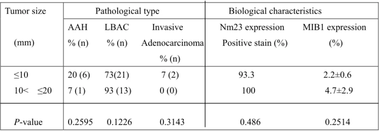
The pathological distribution and biological characteristics of tumors 2.0 cm or less in diameter with
pure GGO appearance are summarized in Table 3. While AAH and non-invasive LBAC were
predominant pathological types of pure GGO tumors, invasive adenocarcinoma of Noguchi’s C type
was found in only 2 lesions (4.5%) among 44 pure GGO tumors. There were no significant
differences in nm23 or MIB1 expression between pure GGO tumors 10 mm or less in diameter and
those 10–20 mm in diameter. Among these three pathological and biological factors, i.e., invasive
adenocarcinoma, nm23negativity, and high MIB1 score, none of the tumors had multiple factors
simultaneously if the tumor size was 1.0 cm or less in diameter.
At less than the median follow-up period after surgery of 18 months (2–127 months), 2 patients
died of diseases other than lung cancer and one patient in the mixed GGO group who underwent
partial resections for multiple lesions developed bone metastasis 18 months after the operation. The
pathological type of this patient with recurrent disease was Noguchi’s type C adenocarcinoma 10
mm in diameter. This type C lesion showed negativity for nm23 and MIB1 expression rate of 10%.
Comment
Although clinical roentgenographic data on the natural history of small lung tumors with pure GGO
appearance are sparse, a previous study showed that lung cancer nodules with pure GGO appearance
do not only increase in size or density, but also decrease in size with the appearance of a solid
component [15]. Therefore, while an increase in size and/or density suggests the absolute necessity
of surgical removal, a decrease in size does not exclude the requirement of surgery. In our series,
excluding 5 patients with initial operation, 34 patients with 39 pure GGO lesions went through
observation with a mean follow-up period of 7 months. Six lesions increased in size, 33 lesions
showed no change in size or density, and no lesions were found to have decreased or diminished in
size. With respect to the operative indications in this study, as described in the Patients and methods
section, we performed VATS operation for mixed GGO. Cases of pure GGO 2 cm or less in diameter
were observed for several months to exclude inflammatory changes. As the result of pathological
examination of resected specimens, 87 lesions with GGO appearance were all found to be tumors.
Pathologically, the Noguchi’s classification has prevailed in Japan as a useful indicator of
postoperative outcomes that would serve as a pathological basis for the selection of patients who
would benefit from limited surgery. Interestingly, several cases of non-invasive LBAC of so-called
Noguchi’s A and B types revealed pure GGO appearance. In our series, consistent with previous
studies, a large number of pure GGO tumors were included in Noguchi’s A or B adenocarcinoma or
AAH despite the tumor size. As lung cancers of Noguchi’s type A and B are free from nodal
metastasis, including micrometastasis [16], this observation appears to support the validity of limited
operation for pure GGO measuring 2.0 cm or less in diameter.
In this study, we further assessed the expression of two biological markers by
immunohistochemical analysis. MIB1 is a marker of tumor proliferation and nm23 is a putative
anti-metastatic gene representing a metastasis-associated marker. Previously, we confirmed
thatnm23 expression in early-stage non-small cell lung cancers is inversely correlated with nodal
micrometastasis. In the present study, using these two novel markers that mirror biological aspects of
the tumors, we found significant differences in their expression between pure and mixed GGO
groups. These findings support the hypothesis that mixed GGO tumors represent relatively
high-grade malignancy with faster growth and greater metastatic ability in comparison with pure
GGO tumors. These results also compare well with the observation that the mean tumor size in the
mixed GGO group was significantly greater than that in the pure GGO group. In pure GGO tumors,
a low MIB1 expression score and negativity of nm23 expression were found regardless of the size of
the tumors. If we look at the critical diameter of pure GGO tumor less than that at which any factors
among 1) pathologically invasive type (Noguchi’s C≤), 2) high score of MIB1 expression (>5%),
and 3) negativity of nm23 expression were not identified, pure GGO less than 5 mm in diameter
satisfied the criteria (data not shown). Although further studies in larger numbers of clinical cases
should be performed, we concluded that pure GGO 5 mm or less in diameter does not require
treatment and observation over a long period by CT is the best option. Based on our observation
that several pure GGO tumors showed high MIB1 scores even though pathological examination
revealed non-invasive Noguchi’s A/B type, we concluded that a careful follow-up schedule would
be needed for tumors in this category measuring more than 5 mm in diameter.
In conclusion, based on the pathological features and expression of nm23, the invasive and
metastatic potential appears to be low in pure GGO tumors. In addition, this tendency was retained
irrespective of tumor size in tumors less than 2 cm in diameter. On the other hand, the tumor
proliferative ability assessed by MIB1 expression seems not to be necessarily low, and careful
observation is needed in cases in which the tumor is more than 5 mm in diameter. However, surgical
resection is justified instead of observation for mixed GGO tumors.
References
1. Kodama K, Higashiyama M, Yokouchi H, et al. Natural history of pure ground-glass opacity
after long-term follow-up of more than 2 years. Ann Thorac Surg 2002; 73: 392-3.
2. Asamura H, Suzuki K, Watanabe S, et al. A clinicopathological study of resected subcentimeter
lung cancers: a favorable prognosis for ground glass opacity lesions. Ann Thorac Surg 2003; 76:
1016-22.
3. Nakata M, Sawada S, Saeki H, et al. Prospective study of thoracoscopic limited resection for
ground-glass opacity selected by computed tomography. Ann Thorac Surg 2003; 75: 1601-6.
4. Nakamura H, Saji H, Ogata A, et al. Lung cancer patients showing pure ground-glass opacity on
computed tomography are good candidates for wedge resection. Lung Cancer 2004; 44: 61-8.
5. Okada M, Nishio W, Sakamoto T, et al. Correlation between computed tomographic findings,
bronchioloalveolar carcinoma component, and biologic behavior of small-sized lung
adenocarcinomas. J Thorac Cardiovasc Surg 2004; 127: 857-61.
6. Yamada S, Kohno T. Video-assisted thoracic surgery for pure ground-glass opacities 2 cm or less
in diameter. Ann Thorac Surg 2004; 77: 1911-5.
7. Matsuguma H, Nakahara R, Anraku M, et al. Objective definition and measurement method of
ground-glass opacity for planning limited resection in patients with clinical stage IA
adenocarcinoma of the lung. Eur J Cardiothorac Surg 2004; 25: 1102-6.
8. Nakata M, Saeki H, Tanaka I, et al. Focal ground-glass opacity detected by low-dose helical CT.
Chest 2002; 121: 1464-7.
9. Matsunaga H, Yokoi K, Anraku M, et al. Proportion of ground-glass opacity on high-resolution
computed tomography in clinical T1N0M0 adenocarcinoma of the lung: A predictor of lymph
node metastasis. J Thorac Cardiovasc Surg 2002; 124: 278-84.
10. Noguchi M, Morikawa A, Kawasaki M, et al. Small adenocarcinoma of the lung. Histologic
characteristics and prognosis. Cancer 1995; 75: 2844-52.
11. Ohta Y, Nozawa H, Tanaka Y, et al. Increased vascular endothelial growth factor and vascular
endothelial growth factor-c and decreased nm23 expression associated with microdissemination
in the lymph node in stage I non-small cell lung cancer. J Thorac Cardiovasc Surg 2000; 119:
804-13.
12. Royds JA, Stephenson TJ, Rees RC, Shorthouse AJ, Silicocks PB. Nm23 protein expression in
ductal in situ and invasive human breast carcinoma. J Natl Cancer Inst 1993; 85: 727-31.
13. Cooper LS, Gillett CE, Smith P, et al. Cell proliferation measured by MIB1 and timing of
surgery for breast cancer. Br J Cancer 1998; 77: 1502-7.
14. Bottini A, Berruti A, Bersiga A, et al. Relationship between tumour shrinkage and reduction in
Ki 67 expression after primary chemotherapy in human breast cancer. Br J Cancer 2001; 85:
1106-12.
15. Ryutaro K, Hironobu O, Masahiro K, et al. Progression of focal pure ground-glass opacity
detected by low-dose helical computed tomography screening for lung cancer. J Comput Assist
Tomogr 2004; 28: 17-23.
16. Wu J, Ohta Y, Minato H, et al. Nodal occult metastasis in patients with peripheral lung
adenocarcinoma of 2.0 cm or less in diameter. Ann Thorac Surg 2001; 71: 1772-8.
Table 1. Clinicopathological background characteristics of 73 patients with ground-glass opacity (GGO)
Variables Pure GGO Mixed GGO P-value
Total number of patients 39 (44) 42 (43)(Total number of lesions)
Sex 0.9482
Male 17 17 Female 22 25
Mean age 63.5±1.8 64.0±1.6 0.9096
Mean tumor size (mm) 9.2±0.5 15.5±0.8 <0.0001
Location (1) 0.7380 Right 23 25
Left 21 18
Location (2) 0.7693 Upper lobe 25 30
Middle lobe 7 2
Lower lobe 12 11
Operative procedure <0.0001 Partial resection 30 8
Segmentectomy 2 2 Lobectomy 7 32
Pathology <0.0001 AAH 7 0
LBAC 35 17 Invasive adenocarcinoma 2 26
Table 2. Biological appearance between pure and mixed ground-glass opacity (GGO) tumors 3.0 cm or less in diameter
GGO pattern Nm23 expression MIB1 expression (%)
Positive stain (%)
Pure GGO (n=44) 97.7 2.7±1.0
Mixed GGO (n=43) 79.1 8.4±1.5
P-value
0.0064 <0.0001
Table 3. Pathological results of pure ground-glass opacity (GGO) tumors 2.0 cm or less in diameter Pathological type Biological characteristics
Tumor size
(mm)
AAH LBAC Invasive Nm23 expression MIB1 expression
% (n) % (n) Adenocarcinoma Positive stain (%) (%) % (n)
≤10
10< ≤20
P-value