Fukushima Medical University
福島県立医科大学 学術機関リポジトリ
This document is downloaded at: 2021-11-08T00:04:57Z
Title Low serum ferritin levels as a clue to colonic cancer detection in two patients with coronary artery disease: a case report
Author(s) Yaoita, Hiroyuki; Ohkawara, Hiroshi; Uekita, Hironori;
Mitsugi, Minoru; Tajima, Hiroko; Kaneko, Hironori; Hoshino, Yutaka; Otani, Satoshi; Gotoh, Mitsukazu; Maruyama, Yukio Citation Fukushima Journal of Medical Science. 51(2): 95-104
Issue Date 2005-12
URL http://ir.fmu.ac.jp/dspace/handle/123456789/175
Rights © 2005 The Fukushima Society of Medical Science
DOI
Text Version publisher
Fukushima J. Med. Sci., Vol. 51, No.2, 2005
[Case Report]
LOW SERUM FERRITIN LEVELS AS A CLUE TO COLONIC CANCER DETECTION IN TWO PATIENTS WITH CORONARY ARTERY DISEASE:
A CASE REPORT
HIROYUKI YAOITN), HIROSHI OHKAWARN), HIRONORI UEKITN), MINORU MITSUGP), HIROKO T AJIMNl, HIRONORI KANEKO!), YUTAKA HOSHIN02 ), SATOSHI OTANP), MITSUKAZU GOTOH2 )
and YUKIO MARUYAMN)
')Department of Internal Medicine I, Fukushima Medical University, Fukushima, 960-1295, Japan
2)Department of Surgery L Fukushima Medical University, Fukushima, 960-1295, Japan (Received September 20, 2005, accepted November 10, 2005)
Abstract : We diagnosed colonic cancer using low serum ferritin levels as a clue in two patients with cardiac or cardiopulmonary disease. In the course of the follow- up, the serum ferritin levels decreased to less than 18 ng/mL without significant appearance of iron-deficiency anemia. One patient showed positive immunological fecal occult blood test results whereas the other not. Both patients rejected further colonoscopy because of their concern for stress in relation to their cardiac or cardiopulmonary diseases, but instead agreed to positron emission computed tomog- raphy (PET) using a F -18 deoxyglucose at their own expense. In both patients, PET documented abnormal tracer accumulation in the colon. From the results of PET imaging, they eventually agreed to colonoscopy. A colonic adenocarcinoma was detected at the site of the positive PET finding in each patient. Both patients underwent curative resection of the cancer. The detection of the levels of serum ferritin may be available for the screening colonic cancer in patients declining colonoscopic examination.
Key words: heart disease, cancer, screening, ferritin
5R:1€.:tliiJ'e·-¥, :f;:VlJi*f.!f, _UtiiffiRt, ~;/Jz ~, E8~f.!fT, ~T1W~, £!l!f :R, :f;:~ JU!j,
~ilili!ill-, 1Li1I-¥::1i.:
Correspondence to: Yukio Maruyama, Department of Internal Medicine I, Fukushima Medical University, Fukushima City 960-1295, Japan.
E-mail: [email protected]
95
96 I-I. Y AOIT A el al.
Right coronary artery (RAO) Tc-99m perfusion SPEeT
stress
rest
comparison with normal profile comparison with normal profile
Fig.1. Coronary artery disease condition of patient no. 1 before surgery in 2004.
Cineangiographic findings of right anterior oblique view (RAO) (left upper and lower panels) are shown. In left upper and lower panels, small, large, and dotted arrows show collateral vessels, site of chronic total occlusion, and left anterior descending artery filled by collateral flow, respectively. Right panels show BULL'S EYE (polar map) images of Tc-99m myocardial perfusion scintigraphy on stress and at rest, and their comparison images with normal profiles obtained from healthy volunteers. Arrow-indicated black areas show by mean perfusion defects.
CASE PRESENTATION Case 1 (78-year-old male)
A 64-year-old male was admitted to our Department of the Internal Medicine I for effort angina in 1991. Cardiac catheterization documented severe triple-vessel coronary artery disease, in which chronic total occlusion of the proximal portion (segment no. 7 by American Heart Association classification!)) of the left anterior descending artery and 90% stenosis of the branch (segment no. 12) of the left circumflex artery (arrows not shown in Fig. 1) were observed. Percutaneous coro- nary intervention for this severe lesion (segment no. 7) in 1991 proved unsuccessful.
Since he declined to undergo coronary artery bypass graft surgery, medical therapies
CANCER DETECTION BY LOW SERUM FERRITIN 97
were continued. In 1996, myocardial perfusion scintigraphy documented new silent ischemia in the myocardial area of the right coronary artery in addition to that of the 'jeopardized' left anterior descending artery. Coronary stent implantation was performed in the 90% stenotic right coronary artery successfully. His final follow- up cineangiographic findings in September 2004 (before surgery as mentioned later) showed total occlusion at the proximal portion of the left anterior descending artery (left upper and lower panels of Fig. 1, large solid black arrows), and fair collateral flow from the right (left upper pane of Fig. 1, small solid black arrows) and left circumflex (left lower panel of Fig. 1, narrow arrows) coronary arteries to the site of the post-chronic total occlusion of the left anterior descending artery (left upper and lower panels of Fig. 1, dotted black arrows).
Hemoglobin (g/dL) or
ferritin (ng/mL) 25 - r - - - ,
20
15
- 0 - Hb - ferritin
CF refused by patient CF
1
GFPrT1
cancer
MCV (fL) or 10+--...--...--...--...--...--...----1 Fe (J.lg/dL) 2 0 0 , . - - - ,
- 0 - MCV
150 - Fe
100
50
O+--.--.---r--.-~-~-~
Mar Jul 2004
Sep middle late Dec Oct Oct
Fig. 2. Blood data and clinical course of patient no. 1. In July 2004, the serum ferritin level decreased (15 ng/mL, normal range, 18-440 ng/mL) without any symptom and significant changes in the Fe and hemoglobin levels and the mean corposcular volume (MCV) of red blood cells. At this time point, the patient continued to refuse colonoscopy (CF). Gastric fiberscopy (GF) findings in July 2004 were normal. In response to the results of positron emission computed tomography (PET) performed in September 2004, the patient agreed to CF in October 2004. He underwent curative partial resection of sigmoid colonic cancer in October of this year.
98 H. Y AOIT A et at.
At this time, myocardial Tc-99m perfusion scintigraphy on stress and at rest showed large (in the anterior wall) and small (in the lateral wall) perfusion defects (right panel of Fig. 1) and the 'fill-in,' a finding of inducible ischemia, in the anterior wall (left panel of Fig. 1, white arrows). In July 2004, the laboratory data showed a low ferritin level (15 ng/mL, normal range 18-440 ng/mL in our hospital) with a bottom level of Hb [Hb 12.9 g/dL, mean corpuscular volume (MeV) 93 fL, Fe 73 j.Lg/
dL] (Fig. 2). In August 2004, the immunological fecal occult blood, which detects human Hb, was negative, no hemorrhagic or tumor lesions and no esophageal varix were detected by gastric fiberscopy, and abdominal ultrasound showed no abnormal findings. The patient rejected colonoscopy or colonic fluoroscopy since they were anxious that either examination including its pretreatment (transient discontinua- tion of medicines including anti-platelet agent in preparation for biopsy and use of a purgative) might be stressful to his ischemic heart. Instead, he agreed to positron emission computed tomography (PET) using F -18 deoxyglucose (FDG) at his own expense in September. PET images documented abnormal accumulation of FDG in the sigmoid colon (left panels of Fig. 3). Based on the results of PET, he agreed to colonoscopy. The colonoscopy documented sigmoid colon cancer (Borrmann type
FOG-PET images resected sigmoid colon anterior view left lateral view outer view inner view
oral side
anal side
,-
Fig. 3. Positron emission tomography (PET) images before surgery (left panels) and macroscopic findings of resected sigmoid colon (right panel) of patient no. l.
PET documented abnormal accumulation of F-18 deoxyglucose (FDG) at sigmoid colon. FDG has also physiologically accumulated in brain, heart, kidney, and bladder. Borrman type 1 cancer was resected successfully. There was no inva· sian to serosa (outer view of right panel).
CANCER DETECTION BY LOW SERUM FERRITIN 99
1) with histopathology of well-differentiated adenocarcinoma. There were no metastatic lesions by ultrasound, PET, computed tomography (CT), and colonic fluoroscopy. In October 2004, curative resection of sigmoid colon cancer was performed (right panel of Fig. 3). Until September 2005, there are no findings of a recurrence of colon cancer by ultrasound, enhanced CT scan, and tumor markers.
Case 2 (76-year-old male)
A 65-year-old male patient was admitted to our Department of Internal Medicine I due to chest pain on effort in 1994. He had a previous history of total left pneumonectomy due to lung cancer at 46 years of age (left panel of Fig. 4) and type C viral hepatitis related to blood transfusion. Cineangiography documented chronic total occlusion of the proximal portion (segment no. 7) of the left anterior descending artery. Percutaneous coronary intervention was performed on segment no. 7 of the left anterior descending artery. We assumed that cardiac catheteriza- tion may not be best-suited for future assessment of coronary artery disease in this patient. During follow-up after discharge, he did not complain of anginal pain, but showed shortness of breath (Hugh Jones grade III) due to the pneumonectomy. In 2005, myocardial Tc-99m perfusion scintigraphy revealed only small perfusion
Chest X ray
comparison with normal profile rest comparison
with normal profile
Fig. 4. Chest roentogenography (left panel) and BULL'S EYE (polar map) images of Tc-99m myocardial perfusion scintigraphy (right panel) of patient no.2 before surgery in 2004. This patient had prior total left pneumonectomy due to curable lung cancer. Myocardial scintigraphy showed small perfusion defects in the anterior and lateral walls.
100 H. YAOITA et al.
Hemoglobin (g/dL) or ferritin (ng/mL)
25.---~---_,
20
15
CF refused by patient
!
cancer resection10 - 0 - Hb
I
- ferritin
MCV (fL) or 5 -t---.-...---r-...---r-.----r--r----r---i Fe (~g/dL) 200 - y - - - ,
150
100
50 - 0 - MCV - - - - FE
O+-~r__r--~~--_r--~~--_r--~~
Dec Apr Jul Nov Mar May Jul Aug Sep 2003 2004 2005
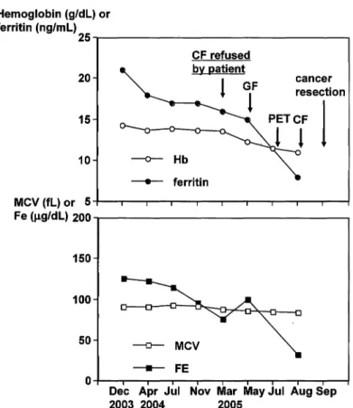
Fig. 5. Blood data and clinical course of patient no. 2. In July 2004, the serum ferritin level was slightly below the lower normal limit (17 ng/mL, normal range 18-440 ng/mL) without any abdominal symptom, along with a decrease in hemo- globin levels, and mean corpuscular volume (MeV) of red blood cells. The serum ferritin level did not change in November compared to July of that year, but further decreased to 16 ng/mL in March 2005. The patient refused colonoscopy at the time. In May 2005, the findings of gastric fiberscopy (GF) were normal. In response to the results of positron emission tomography (PET) of July 2005, colonoscopy was performed in August of the same year. He received curative partial resection of ascending colon in September. Serum Fe levels tended to decrease between July 2004 and March 2005, although absolute values were still within normal range (normal range, 55-190 ,ug/dL). Finally, serum Fe level absolutely decreased in August 2005.
defects in the anterior wall (related to chronic total occlusion) of the left anterior descending artery and lateral wall (right panel of Fig. 4) as previously observed in 1994 (data not shown), but there was no inducible ischemia. His serum ferritin level was within the normal range (21 ng/mL) with no findings of Fe-deficiency anemia (Hb 14-3 g/dL, MCV 91 fL, Fe 126 j.lg/dL) in December 2003. In April 2004, his serum ferritin level reached the lower normal limit (18 ng/mL) without anemia (Hb 143 g/dL, MCV 91 fL, Fe 76 j.lg/dL) (Fig. 5). In May 2005, gastric fiberscopy was performed and there were no hemorrhagic lesions in esophagus and stomach. The immunological fecal occult blood was positive, although hemorrhoids were present.
CANCER DETECTION BY LOW SERUM FERRITIN 101
FDG-PET images resected sigmoid colon anterior view left lateral view inner view
outer view
- .
•
Fig. 6. Positron emission tomography (PET) images before surgery (left panels) and macroscopic findings of resected sigmoid colon (right panel). PET documented abnormal accumulation of F-1S deoxyglucose (FDG) at ascending colon. FDG has accumulated in brain, heart, kidney, and bladder physiologically. There was no abnormal accumulation suggesting metastases by PET. Cancer invasion was limited to the submucosal layer.
He refused colonoscopy, feeling the examination and pretreatment might be stress- ful to his ischemic heart. However, he agreed to PET using FDG at his own expense in July 2005. PET images documented abnormal accumulation of FDG in the ascending colon close to the caecum (left panels of Fig. 6). He finally understood his disease and agreed to colonoscopy in August. Colonoscopy documented the colonic cancer with a histopathological finding of moderately-differentiated adenocarcinoma. There were no metastatic lesions by ultrasound, fluoroscopy of the colon, PET, computed tomography, gastric fiberscopy, and his curative surgery for colonic cancer was underwent (right panels of Fig. 6). Histopathologically, operation was turned out to be curative.
DISCUSSION
Patients with coronary artery disease are increasing in number2>. While the prognosis has been improved by the recent progress in cardioprotective anti -is- chemic therapies, aging is inevitably in progress. Since cancer is the most common death among the elderly3>, such elder patients with coronary artery disease might
102 H. Y AOIT A et at.
have some kinds of cancer in their life. Endoscopic examination for detecting cancer of the gastro-intestinal tract is generally safe. However, cardiac or car- diopulmonary morbidity may limit even such a relatively non-invasive examination for screening the cancer4), and may also limit the surgical indications if the cancer is much advanced. To screen several kinds of cancers, PET using FDG is one of the choices5 ).
We reported here two cases of coronary artery disease who evidenced colonic cancer in the long-term follow-up of cardiac or cardiopulmonary disease. The low serum ferritin level in the absence of significant Fe-deficiency anemia was the basis for screening and diagnosing the cancer in both patients. One may think that our way of cancer diagnosis reported here is unusual. In the 1990s, there were clinical reports based on the hypothesis that low serum ferritin levels preceding the appear- ance of Fe-deficiency anemia may be useful as a non-invasive screening test for detecting malignant cycle of ulcer or erosion associated with cancer of the upper6,7) and lowerS) gastrointestinal tracts. We assume that the anti-platelet or anti- coagulant therapy in patients with coronary artery disease may facilitate oozing from cancer. In addition, there may be a high incidence of colonic cancer in patients with coronary disease9,IO) although it is not known whether it relates to dietary excess in cholesterol uptake. Thus, there may be a relation between low serum ferritin levels and the presence of colonic cancer. However, low serum ferritin levels result, not only from gradual loss of blood from a malignant ulcer, but also from the low intestinal uptake of ferritin associated with hypo- and a-chlorhy-
drial l -13). Although such abnormal stomach acidity is also known to be a pre-
gastric cancer state, the ferritin method with such pseudo-positivity on bowel cancer screening and required blood sampling were considered disadvantageous compared to the perfectly non-invasive (no needle) method of detecting the fecal occult blood and lost favor as a screening testl l -13). For this reason, the fecal occult blood test became the general standard for colonic cancer screening.
The immunological fecal occult blood test has been established as a screening test for colonic cancer with a high relative risk (3.45, 95% confidence interval, 2.76 to 4.35).14) Among several concerns regarding this test, first, there are factors which can cause pseudo-negative findings. The cancer may not bleed continuously or may bleed only a little. In addition, degeneration of human Hb, if such occurs, causes a loss of antigeneity against the antibody used in this test. Second, there are pseudo- positive findings represented by the presence of bleeding from nonmalignant lesions such as oral cavity bleeding, benign gastric ulcer and hemorrhoidsI5>. Therefore, even if the result of this test is positive, it does not necessarily mean cancer, whereas even if a negative result is obtained in the follow-up fecal test, the possibility of having a neoplasmic digestive disease cannot be altogether denied. The subjects usually judged to be positive for this test in healthy cardiac condition may agree with the diagnostic examination by colonoscopy while aware of its limitations. How- ever, subjects with coronary artery disease may hesitate to undergo colonoscopy
CANCER DETECTION BY LOW SERUM FERRITIN 103
even if a fecal occult blood test is positive, because of their concerns regarding inducible heart attacks due to physical stresses. Admittedly, the guidelines for endoscopy note an increased risk of complications in patients with coronary artery disease4 ). Thus, strong motivation may be needed for patients to undergo colonos- copy in order to rule out the presence of the cancer when they have cardiac or cardiopulmonary disease. Therefore, based on our experience with these two patients, assessment of serum ferritin levels in combination with Fe and peripheral blood data (Hb and MeV etc.) and the fecal occult blood test several times a year may be beneficial in patients with significant coronary artery disease. If serum ferritin levels decrease, one may also check the fecal occult blood again. The combination of a low ferritin level and a positive result on fecal occult blood test may well encourage patients to undergo colonoscopy. In patients who are suspected of cancer but rejecting colonoscopy, PET using FDG at their expense may be an alternative. However, to confirm the validity of this cancer screening process in patients with cardiac risks, more experience, a large sample and randomised trials are necessary.
REFERENCES
1. Ryan TJ, Faxon DP, Gunnar RM, Kennedy JW, King SB 3rd, Loop FD, Peterson KL, Reeves TJ, Williams DO, Winters WL Jr, et al. Guidelines for percutaneous trans·
luminal coronary angioplasty. A report of the American College of Cardiology/Amero ican Heart Association Task Force on Assessment of Diagnostic and Therapeutic Cardiovascular Procedures (Subcommittee on Percutaneous Transluminal Coronary Angioplasty). Circulation, 78(2): 486-502, 1988.
2. Hiroshi I, Atsushi I, Ryuichi K, Osamu Y, Shin-Ichiro U, Yoshikazu Y, Osamu K, Minoru H, Keishi K. Trends over the last 20 years in the clinical background of young Japanese patients with coronary artery disease. Circ J, 68(3): 186-91, 2004.
3. Kobayashi M, Kudomi H. Analysis of accelerated death benefit claims at a Japanese life insurance company. J Insur Med, 34(2): 94-9, 2002.
4. DiSario JA, Waring JP, Talbert G, Sanowski RA. Monitoring of blood pressure and heart rate during routine endoscopy: a prospective, randomized, controlled study. Am J Gastroenterol, 86(8): 956-60, 1991.
5. Bar-Shalom R, Valdivia AY, Blaufox MD. PET imaging in oncology. Semin Nucl Med, 30(3): 150-85, 2003.
6. Akiba S, Neriishi K, Blot WJ, Kabuto M, Stevens RG, Kato H, Land CEo Serum ferritin and stomach cancer risk among a Japanese population. Cancer, 67(6): 1707-12, 1991.
7. Nomura A, Chyou PH, Stemmermann GN. Association of serum ferritin levels with the risk of stomach cancer. Cancer Epidemiol Biomarkers Prev, 1(7): 547-50, 1992 8. Nelson RL. Elevation of serum ferritin is superior to fecal occult blood testing as a
screening test for colonic adenoma... and not only because patients do not have to handle their own stool. Dis Colon Rectum, 39(12): 1441-2, 1996.
9. Neugut AI, Jacobson JS, Sherif G, Ahsan H, Garbowski GC, Waye J, Forde KA, Treat MR. Coronary artery disease and colorectal neoplasia. Dis Colon Rectum, 38(8): 873- 7, 1995.
10. Kee F, Collins B]. Colorectal cancer and ischaemic heart disease: an uncommon inheritance! Eur J Cancer Prev, 1(2): 149-52, 1992.
104 H. Y AOIT A et at.
11. Scholefield JH, Robinson MH, Bostock K, Brown NS. Serum ferritin. Screening test for colorectal cancer? Dis Colon Rectum, 41(8}: 1029-31, 1998.
12. Seery JP. Achlorhydria and gastric carcinogenesis. Lancet, 338(8781}: 1508-9, 1991.
13. Kabuto M, Imai H, Tsugane S, Watanabe S. Does high gastric cancer risk associated with low serum ferritin level reflect achlorhydria-An examination via cross-sectional study. Jpn J Cancer Res, 84(8}: 844-51, 1993.
14. Lieberman DA, Weiss DG; Veterans Affairs Cooperative Study Group 380. One-time screening for colorectal cancer with combined fecal occult-blood testing and examina- tion of the distal colon. N Engl J Med, 345(8}: 555-60, 200l.
15. Karnam US, Felder LR, Raskin JB. Prevalence of occult celiac disease in patients with iron-deficiency anemia: a prospective study. South Med J, 97(1}: 30-4, 2004.