九州大学学術情報リポジトリ
Kyushu University Institutional Repository
Relevance of calcification and contrast
enhancement pattern for molecular diagnosis and survival prediction of gliomas based on the
2016 World Health Organization Classification
道脇, 悠平
http://hdl.handle.net/2324/4060040
出版情報:九州大学, 2019, 博士(医学), 課程博士 バージョン:
権利関係:© 2019 Elsevier B.V. All rights reserved.
Contents lists available atScienceDirect
Clinical Neurology and Neurosurgery
journal homepage:www.elsevier.com/locate/clineuro
Relevance of calcification and contrast enhancement pattern for molecular diagnosis and survival prediction of gliomas based on the 2016 World Health Organization Classification
Yuhei Michiwaki
a, Nobuhiro Hata
a,⁎, Masahiro Mizoguchi
a, Akio Hiwatashi
b, Daisuke Kuga
a, Ryusuke Hatae
a, Yojiro Akagi
a, Takeo Amemiya
a, Yutaka Fujioka
a, Osamu Togao
c,
Satoshi O. Suzuki
d, Koji Yoshimoto
e, Toru Iwaki
d, Koji Iihara
aaDepartment of Neurosurgery, Graduate School of Medical Sciences, Kyushu University 3-1-1 Maidashi, Higashi-Ku, Fukuoka 812-8582, Japan
bDepartment of Molecular Imaging & Diagnosis, Graduate School of Medical Sciences, Kyushu University 3-1-1 Maidashi, Higashi-Ku, Fukuoka 812-8582, Japan
cDepartment of Clinical Radiology, Graduate School of Medical Sciences, Kyushu University 3-1-1 Maidashi, Higashi-Ku, Fukuoka 812-8582, Japan
dDepartment of Neuropathology, Graduate School of Medical Sciences, Kyushu University 3-1-1 Maidashi, Higashi-Ku, Fukuoka 812-8582, Japan
eDepartment of Neurosurgery, Graduate School of Medical and Dental Sciences, Kagoshima University 8-35-1 Sakuragaoka, Kagoshima 890-0075, Japan
A R T I C L E I N F O Keywords:
Glioma
Molecular diagnosis Enhancement Calcification Survival
World Health Organization Classification
A B S T R A C T
Objectives:The significance of conventional neuroimaging features for predicting molecular diagnosis and pa- tient survival based on the updated World Health Organization (WHO) classification remains uncertain. We assessed the relevance of neuroimaging features (ring enhancement [RE], non-ring enhancement [non-RE], overall gadolinium enhancement [GdE], and intratumoral calcification [IC]) for molecular diagnosis and sur- vival in glioma patients.
Patients and methods:We evaluated 234 glioma patients according to the updated WHO classification. Isocitrate dehydrogenase (IDH),H3F3A,BRAFhotspot mutations,TERTpromotor mutation, and chromosome 1p/19q co- deletion were examined. RE, non-RE, GdE, and IC were evaluated as significant neuroimaging findings. Kaplan-Meier analyses were performed to evaluate overall survival (OS) and the correlations of prognostic factors were evaluated by log-rank tests. Univariate and multivariate analyses were performed to detect prognostic factors for OS.
Results:A total of 207 patients were eligible. In 110 patients presenting RE, 102 (93%) were glioblastoma (GBM), IDH-wild type. In 97 patients without RE, presence of GdE or IC were not significantly different between IDH-mutant and -wild type tumors, whereas presence of GdE was a significant indicator of higher WHO grades.
IC was the only significant finding for 1p/19q co-deleted tumors.TERTpromoter mutation was observed in 7/17 patients with diffuse astrocytic glioma, IDH-wild type; recently-defined as “molecular GBM.” IC, RE, and GdE were observed with lower prevalence in molecular GBMs. While presence of RE, GdE, and absence of IC were significant factors of OS in overall cohort, presence of GdE was not significant in OS in cases without RE, and IDH-mutant tumors. IC was a significant predictor of favorable OS in cases without RE and IDH-wild type tumors. Multivariate analysis also validated these findings.
Conclusion:GdE alone is not a significant predictor of IDH mutation status, but the pattern of enhancement is a significant predictor with RE demonstrating high sensitivity and specificity for GBM, IDH-wild type. Predicting
“molecular GBM” by conventional neuroimaging is difficult. Moreover, GdE is not a significant factor of survival analyzed with pattern of enhancement or molecular stratifications. IC is an important radiographic finding for predicting molecular diagnosis and survival in glioma patients.
https://doi.org/10.1016/j.clineuro.2019.105556
Received 28 June 2019; Received in revised form 30 September 2019; Accepted 6 October 2019
⁎Corresponding author.
E-mail addresses:wayside.bamboo@gmail.com(Y. Michiwaki),hatanobu@ns.med.kyushu-u.ac.jp(N. Hata),mmizoguc@ns.med.kyushu-u.ac.jp(M. Mizoguchi), hiwatasi@radiol.med.kyushu-u.ac.jp(A. Hiwatashi),kuga@ns.med.kyushu-u.ac.jp(D. Kuga),ryhatae@ns.med.kyushu-u.ac.jp(R. Hatae),
yakagi@med.kyushu-u.ac.jp(Y. Akagi),takeo.amay.0330@gmail.com(T. Amemiya),yutakafujioka19830816@yahoo.co.jp(Y. Fujioka),
togao@radiol.med.kyushu-u.ac.jp(O. Togao),sosuzuki@np.med.kyushu-u.ac.jp(S.O. Suzuki),kyoshimo@m.kufm.kagoshima-u.ac.jp(K. Yoshimoto), iwaki@np.med.kyushu-u.ac.jp(T. Iwaki),kiihara@ns.med.kyushu-u.ac.jp(K. Iihara).
Available online 12 October 2019
0303-8467/ © 2019 Elsevier B.V. All rights reserved.
T
1. Introduction
The precise classification and diagnosis of gliomas are essential for their clinical management and outcome prediction. Conventional di- agnoses for gliomas have been based primarily on histopathological findings. However, in response to the recent innovative development of molecular diagnosis of gliomas by the accumulation of molecular ge- netic analysis data, the updated World Health Organization Classification of Tumors of the Central Nervous System (2016 CNS WHO) includes molecular genetic diagnoses, in addition to the classical histopathological diagnosis [1–3]. Molecular characteristics of gliomas have been shown to be superior to conventional histological grading in the estimation of patient prognosis [4–6]. In the pre-molecular era, ring-shaped gadolinium enhancement (RE) on magnetic resonance imaging (MRI) was well-known as a typical feature of glioblastoma (GBM), and gadolinium enhancement (GdE) was a strong indicator of malignancy [7,8]. However, the correlation between conventional neuroimaging features and the molecular diagnosis of gliomas ac- cording to the 2016 CNS WHO has not yet been sufficiently in- vestigated. Additionally, while the presence or degree of GdE has been reported as one of the prognostic factors of patient survival for gliomas in the pre-molecular era [8–12], the significance of radiographic fea- tures, such as RE, GdE, and intratumoral calcification (IC), for outcome prediction based on the molecular characteristics according to the 2016 CNS WHO remains uncertain. Herein, we aimed to assess the sig- nificance of the presence or pattern of GdE and IC on preoperative images for the prediction of molecular diagnosis and survival in glioma patients in the modern molecular era.
2. Materials and methods 2.1. Patients and genetic analyses
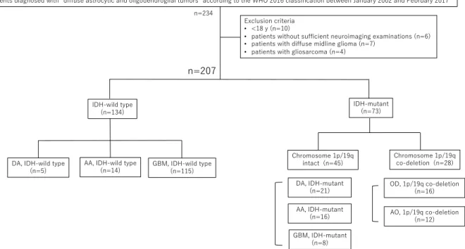
We retrospectively reviewed the clinical, radiographic, and mole- cular-pathological data of our case series of 234 consecutive patients who were treated at our university hospital (Fukuoka, Japan), between January 2002 and February 2017, and whose tumors had been diagnosed as “diffuse astrocytic and oligodendroglial tumors” according to the 2016 CNS WHO [1]. Snap-frozen tissue samples of the tumors of enrolled cases were used to detect isocitrate dehydrogenase (IDH),BRAF, andH3-G34R hot spot mutations,TERTpromoter mutation, and chromosome 1p/19q co-deletion as described previously [13–16]. The flow diagram of mo- lecular work-up is described inFig. 1. Subsequently, molecular diagnoses were categorized as diffuse astrocytoma IDH-mutant (DAmut); diffuse astrocytoma IDH-wild type (DAwt); anaplastic astrocytoma IDH-mutant (AAmut); anaplastic astrocytoma IDH-wild type (AAwt); oligoden- droglioma IDH-mutant and 1p/19q co-deleted (OD1p/19q); anaplastic oligodendroglioma IDH-mutant and 1p/19q-co-deleted (AO1p/19q); GBM IDH-mutant (GBMmut); and GBM IDH-wild type (GBMwt). Patients without sufficient neuroimaging examinations via computed tomography (CT) and MRI were excluded. In addition, patients diagnosed with diffuse midline gliomas, gliosarcomas, or aged < 18 years old were also ex- cluded. We only included patients with newly-diagnosed gliomas, and recurrent or re-treatment cases were excluded.
2.2. Neuroimaging findings
Preoperative neuroimaging examination was performed via con- trast-enhanced, thin-slice CT and gadolinium-enhanced MRI for all patients. GdE was evaluated in a post-contrast enhanced T1-weighted image and categorized into RE and non-ring enhancement (non-RE). RE was defined as the presence of gadolinium enhancement at the tumor margin and a lack of enhancement in the center of the tumor. Cases that presented with GdE but lacked RE were defined as non-RE. IC was also defined as intratumoral lesions presenting a very high-density level, similar to that presented by the skull in pre-contrast-enhanced, thin-
slice CT, including both coarse and minute calcification.Fig. 2shows typical cases with each neuroimaging finding. All neuroimages were evaluated and reported by experienced neuroradiologists before sur- geries. In this study, neuroimaging findings were evaluated based on the radiology reports and confirmed by at least one neurosurgeon.
All CT scans were performed by single detector (X Vigor; Toshiba, Tokyo, Japan), or multi-detector row CT (Aquilion; Toshiba). Typical imaging parameters were as follows: collimation, 4 mm; tube voltage, 120 kVp; tube current, 150–200 mA; field-of-view (FOV), 240 x 270 mm;
and matrix, 512 × 512. All MRI studies were performed using 1.5 T (Vision or Symphony; Erlangen, Germany or Achieva; Philips, Best, the Netherlands) or 3 T system (Achieva or Ingenia; Philips). For 1.5 T system, the typical imaging parameters were as follows: axial T1- weighted spin-echo imaging using repetition time (TR)/echo time (TE) = 522–612/12–14 ms, field-of-view (FOV) = 240 x 240 mm, ma- trix = 256 × 256, and slice thickness/gap = 5/1–2.5 mm. The contrast material was injected at 0.1 mmol/kg (gadopentetate dimeglumine, (Magnevist) Bayer, Osaka, Japan). Axial postcontrast T1-weighted spin- echo imaging using TR/TE = 558–593/17 ms, FOV = 240 x 240 mm, matrix = 256 × 256, and slice thickness/gap = 5/1-2.5. For 3 T system, the typical imaging parameters for the brain were as follows: axial T1- weighted spin-echo imaging using TR/TE = 450/9 ms, FOV = 240 x 240 mm, matrix = 256 × 256, slice thickness/gap = 5/1 mm, and ac- quisition time =2 min 39 s. The contrast material was injected at 0.1 mmol/kg (gadopentetate dimeglumine, (Magnevist) Bayer, Osaka, Japan). Axial postcontrast T1-weighted spin-echo imaging using TR/
TE = 465/21 ms, FOV = 256 x 256 mm, matrix = 256 × 256, and slice thickness/gap = 5/1 mm.
2.3. Statistical analysis
Chi-square tests were assessed to investigate the relationship be- tween each neuroimaging finding and molecular characteristics or WHO grades. Sensitivity and specificity were also evaluated for each radiographic finding. Kaplan-Meier analysis was performed to evaluate overall survival (OS), and a log-rank test was used to compare the survival distributions. Univariate and multivariate Cox proportional hazards regression models were performed to evaluate putative prog- nostic factors for OS. The level of statistical significance was set at p < 0.05. JMP Pro version 13 (SAS Institute Inc., NC, USA) was used for all statistical analyses.
3. Results
A total of 207 patients were eligible for evaluation based on the criteria of the present study (Fig. 1). The patient characteristics, mo- lecular diagnoses, and neuroimaging findings of participants are sum- marized inTable 1. Correlations between radiographic findings and molecular characteristics or WHO grades are described inTable 2.
3.1. Correlations between neuroimaging findings and molecular diagnoses or WHO grades
Among 110 tumors presenting with RE, 102 (93%) were diagnosed as GBMwtand 106 (96%) were categorized as IDH-wild type tumors (Table 1). In all patients, the presence of RE, overall GdE, and lack of IC (non-IC) were significant features of GBMwtcompared to other glioma subgroups (p < 0.0001) (Table 2). As most cases of GBMwtpresented with RE, non-RE was significant in tumors other than GBMwt
(p < 0.0001). In 97 tumors without RE, the presence of GdE (= non- RE) and IC were not significant findings between IDH-mutant and -wild type tumors. However, the presence of GdE was a significant feature in WHO grade Ⅳ tumors compared to grade Ⅱ/Ⅲ tumors (p < 0.0001).
Similarly, in the lower-grade glioma (grade Ⅱ/Ⅲ) subgroup, the pre- sence of GdE was a significant feature in WHO grade Ⅲ gliomas (p = 0.0344). Therefore, GdE was a significant indicator of WHO
Y. Michiwaki, et al. Clinical Neurology and Neurosurgery 187 (2019) 105556
2
grades, but was not a significant predictor of IDH mutation status.
Presence of IC was the only significant feature of 1p/19q co-deleted tumors in both the lower-grade glioma (p = 0.0008) and IDH-mutant tumor subgroups (p = 0.0002).
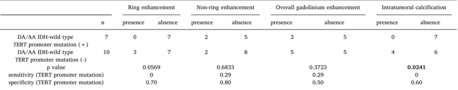
3.2. GBM subtypes, DAwt, and AAwtwith TERT promoter mutation Among 8 patients with GBMmut, 5 (63%) lacked RE and 1 presented with IC. In addition, 1 and 2 patients with GBMwtpresented withH3- G34R (GBMH3G34R) andBRAF (GBMBRAF) hotspot mutations, respec- tively. In these tumors, 1 GBMH3G34Rlacked RE, and 1 GBMBRAFlacked RE and presented with IC. Accordingly, GBMmut, GBMH3G34R, and GBMBRAFcould demonstrate different radiographic features from those of a typical GBMwt. Among the 17 patients with DAwtand AAwt(ex- cluding 2 cases with radiation induced glioma),TERTpromoter muta- tion was observed in 7 patients, who can be diagnosed as having the recently-defined “molecular GBM” [17]. Absence of IC was significant in such molecular GBM cases (p = 0.0241), and presence of RE and GdE was observed with lower prevalence in patients with molecular GBMs (Table 3).
3.3. Survival analyses
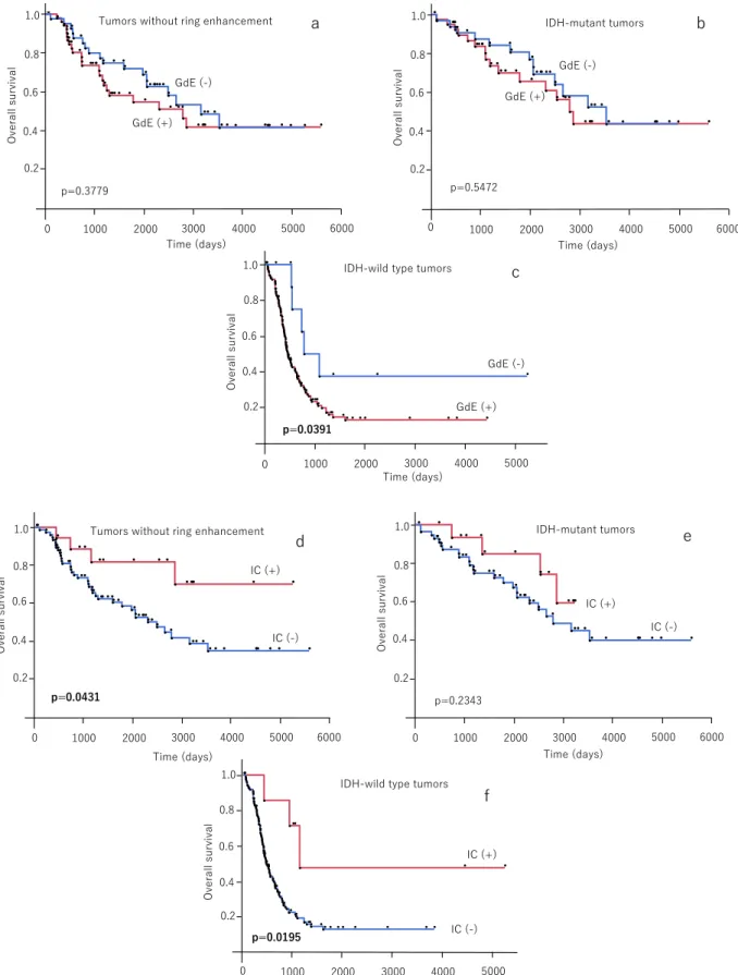
Kaplan-Meier analyses revealed that the presence of RE (p < 0.0001), GdE (p < 0.0001), and non-IC (p = 0.0003) were sig- nificantly associated with unfavorable OS in the overall patient cohort (Fig. 3a–c). However, presence or absence of GdE was not a significant factor of OS in cases excluding ring enhanced tumors (p = 0.3779, Fig. 4a) and IDH-mutant tumors (p = 0.5472,Fig. 4b). On the other hand, presence of IC was significantly associated with favorable OS in cases excluding those with ring enhanced tumors (p = 0.0431,Fig. 4d) and IDH-wild type tumors (p = 0.0195,Fig. 4f), whereas IC was not significant for OS among IDH-mutant tumors (p = 0.2343 Fig. 4e).
Multivariate Cox regression analysis revealed poor preoperative Kar- nofsky Performance Status (KPS) score, IDH-wild type tumors, and lack of calcification were significant prognostic factors for unfavorable OS, though GdE was not significant (Table 4).
4. Discussion
Since the 2016 CNS WHO was recently enacted, accurate correlation Fig. 1.Patient flow diagram according to molecular diagnosis. DA, diffuse astrocytoma; AA, anaplastic astrocytoma; OD, oligodendroglioma; AO, anaplastic oli- godendroglioma; GBM, glioblastoma; IDH, isocitrate dehydrogenase.
Fig. 2.Typical cases with ring enhancement (a), non-ring enhancement (b), and intratumoral calcification (c).
between conventional neuroimaging features and molecular diagnosis, and survival prediction based on molecular stratifications of gliomas, has not been thoroughly determined. The present study revealed the correlations between neuroimaging features and molecular stratifica- tion or WHO grades based on the 2016 CNS WHO. In addition, we discovered the relevance of RE, GdE, and IC as prognostic factors of survival in glioma patients in the modern molecular era.
The presence of contrast-enhancement, derived from either the de- struction or permeability of the blood-brain barrier, is known to be a representative radiographic finding of malignant gliomas in the pre- molecular era [7,8]. RE, which reflects internal tumor necrosis due to increased tumor volume, is a distinctive feature of GBM [8]. IC, which reflects high focal differentiation of the tumor components, is occa- sionally observed in certain lower-grade tumors, such as oligoden- droglioma or ganglioglioma [18–22]. Therefore, we hypothesized that the presence or pattern of GdE and IC are correlated with the molecular characteristics of gliomas and are also associated with patient prognosis in the molecular era.
4.1. Significance of RE
In this study cohort, 93% of ring enhanced tumors were GBMwtand 96% were categorized as IDH-wild type tumors. Moreover, ring en- hanced tumors exhibited unfavorable OS (Fig. 3a). Thus, gliomas with RE can be predicted as GBMwtwith high sensitivity (0.89) and speci- ficity (0.91), and ring enhanced tumors present an unfavorable course, even in the modern molecular era. Therefore, it is considered clinically beneficial to treat non-ring enhanced tumors separately from ring en- hanced tumors to predict their diagnosis and survival.
4.2. Significance of GdE and IC
Among tumors without RE, there were no significant differences between the presence or absence of GdE or IC and IDH mutation status, whereas presence of GdE was a strong indicator of higher WHO grades.
Although previous studies showed higher prevalence of GdE in IDH- wild type gliomas than in IDH-mutant gliomas [23–25], our study re- vealed that the presence of GdE could no longer be considered a pre- dictor of IDH-wild type tumors when ring enhanced tumors, which were most cases of GBMwt, were excluded.
IC is a well-known characteristic of oligodendroglial tumors, espe- cially in those with 1p/19q co-deletion [18–22]. Our study also re- vealed that IC was the only significant neuroimaging feature of 1p/19q co-deleted tumors. In our cohort, the presence or absence of GdE was not significant in 1p/19q co-deleted tumors, supporting previous re- ports that have described the insufficient specificity of GdE for the prediction of OD1p/19q [23,26]. However, contradictory reports have been published. While Reyes-Botero et al. reported that OD1p/19qwas associated with no or non-measurable enhancement [27], Sonoda et al.
reported that OD was associated with the presence of GdE [28]. These results, along with our findings, suggest that the significance of GdE in 1p/19q co-deleted tumors is yet to be clarified.
4.3. Neuroimaging features of GBM subtypes
This study also identified that patients with GBMmut, GBMH3G34R, and GBMBRAFfrequently presented with non-RE or IC, which are non- typical features of GBMwt. We previously reported that GBMmut de- monstrates multiple heterogeneous, non-ring, or non-markedly enhan- cing lesions [29], GBMH3G34Rshows non-markedly enhancing or cal- cified masses [13], and that GBMBRAF occasionally presents with IC [14]. Absence of RE and presence of IC have been identified as findings of lower-grade gliomas or oligodendroglial tumors. Therefore, the re- sults of this study caution against judging the tumor as a lower-grade glioma by conventional neuroimaging findings alone, because GBM might be hidden in cases presenting with radiographic features similar Table1 Summaryofneuroimagingfeaturesandgliomaclassificationandcharacteristicsaccordingtothe2016WHOclassification. WHOgradeⅡWHOgradeⅢWHOgradeⅡWHOgradeⅢWHOgradeⅣ TotalDA,
ID-wild type
DA,IDH-mutantAA,IDH-wildtypeAA,IDH-mutantOD,IDH-mutant& 1p19q-codeletedAO,IDH-mutant& 1p19q-codeletedGBM,IDH-wildtypeGBM,IDH-mutant n207521141616121158 Medianage(range)(y.o.)55(19-85)33(24-66)30(20-57)56.5(28-74)37(19-52)44(20-73)46(27-74)65(26-85)38(23-62) Sex(male/female)114/931/416/510/412/49/74/860/552/6 Overallgadolinium enhancement (presence/absence)
164/431/45/167/711/58/89/3115/08/0 Ringenhancement (presence/absence)110/970/50/214/100/160/161/11102/133/5 Non-ringenhancement (presence/absence)54/1531/45/163/1111/58/88/413/1025/3 Intratumoralcalcification (presence/absence)24/1832/30/212/123/136/107/53/1121/7 Abbreviations:WHO,WorldHealthOrganization;IDH,isocitratedehydrogenase;DA,diffuseastrocytoma;AA,anaplasticastrocytoma;OD,oligodendroglioma;AO,anaplasticoligodendroglioma;GBM,glioblastoma.
Y. Michiwaki, et al. Clinical Neurology and Neurosurgery 187 (2019) 105556
4
to those of lower-grade gliomas. In addition, other driver gene muta- tions, such asH3orBRAFmutations, should be examined in cases of GBM with non-typical radiographic features.
4.4. DAwtand AAwtwith TERT promoter mutation; molecular GBM
“Diffuse astrocytic glioma, IDH-wild type, with molecular features of GBM, WHO grade Ⅳ” is a recently described new classification of GBM [17]. This “molecular GBM” is defined as DAwt or AAwt with epidermal growth factor receptor (EGFR) amplification, combined whole chromosome 7 and whole chromosome 10 loss, or TERTpro- moter mutation. As these tumors exhibit poor survival, predicting molecular GBM by neuroimaging is clinically important. In the present study, 17 patients (excluding those with radiation induced glioma) were diagnosed with diffuse astrocytic glioma, IDH-wild type, and TERTpromoter mutation was observed in 7 patients. Absence of IC was significant inTERT-mutant cases (molecular GBMs), and the presence of RE and GdE was observed at a lower prevalence in patients with mo- lecular GBM. Hence, diffuse astrocytic glioma, IDH-wild type with TERTpromoter mutation, were likely to show absence of IC, RE, and GdE, but it was difficult to predict molecular GBM by conventional neuroimaging findings. These paradoxical results (the clinical course and neuroimaging findings were inconsistent) suggest that molecular examinations should be evaluated even in cases with “non-malignant”
features on neuroimaging in IDH-wild type gliomas.
4.5. Survival analyses
In this study, although ring enhanced gliomas were associated with unfavorable survival, there were no significant differences in OS be- tween patients with and without GdE, when ring enhanced gliomas
were excluded. Since the majority of ring enhanced tumors were cate- gorized as GBMwt, it is reasonable to assume that these tumors tend to have a poor prognosis, as demonstrated in this study. In addition, multivariate analysis revealed GdE was not a significant factor of OS.
While some previous studies have indicated that the presence or degree of GdE in gliomas is a predictor of poor prognosis in the pre-molecular era [8–12], other recent reports have reported that GdE was not a significant survival predictor when analyzed with various factors [23,25]. In their recent report with detailed molecular evaluation, Hempel et al. demonstrated that the presence of GdE was not a sig- nificant survival factor when the analysis included molecular stratifi- cation [23]. Considering these reasons, it was considered acceptable in our cohort that there were significant differences in survival associated with both RE and GdE when overall patients were included, but the presence of GdE was not a significant factor for OS among cases without RE and in IDH-mutant tumors. Together with the result that GdE was not a significant indicator of molecular stratifications, its importance for prognosis prediction has also declined in the modern molecular era.
A unique finding of this study is that the presence of IC was re- cognized as a significant predictor of favorable OS among overall cases, IDH-wild type tumors, and tumors without RE. Among patients without RE, multivariate analysis also revealed presence of IC was a significant factor of favorable OS. This is probably because IC was not a significant survival predictor in a small number of patients with IC in IDH-mutant tumors. While IC was a significant finding in 1p/19q co-deleted tumors, it can be indicated as a favorable survival predictor even in IDH-wild type tumors. While IC is widely known to be a favorable prognostic factor, our study revealed IC can be a significant survival predictor even when analyzed based on molecular stratifications and pattern of en- hancement. Therefore, the significance of IC for patient prognosis ap- pears to be important in the molecular era.
Table 2
Correlations between neuroimaging findings and molecular stratifications or WHO grades.
Ring enhancement Non-ring enhancement Overall gadolinium enhancement Intratumoral calcification
n presence absence presence absence presence absence presence absence
GBM, IDH-wild type 115 102 13 13 102 115 0 3 112
others 92 8 84 41 51 49 43 21 71
p value < 0.0001 < 0.0001 < 0.0001 < 0.0001
sensitivity (GBM, IDH-wild type) 0.89 0.11 1.0 0.03
specificity (GBM, IDH-wild type) 0.91 0.55 0.47 0.77
Tumors without ring enhancement (n = 97)
IDH-wild type tumors 28 17 11 4 24
IDH-mutant tumors 69 37 32 15 54
p value 0.5229 0.3905
sensitivity (IDH-wild type tumors) 0.61 0.14
sensitivity (IDH-wild type tumors) 0.46 0.78
WHO grade Ⅳ 18 18 0 2 16
WHO grade Ⅱ/Ⅲ 79 36 43 17 62
p value < 0.0001 0.2895
sensitivity (WHO grade Ⅳ) 1.0 0.11
specificity (WHO grade Ⅳ) 0.54 0.78
WHO grade Ⅱ/Ⅲ tumors without ring enhancement (n = 79)
WHO grade Ⅲ 38 22 16 9 29
WHO grade Ⅱ 41 14 27 8 33
p value 0.0344 0.6522
sensitivity (WHO grade Ⅲ) 0.58 0.24
specificity (WHO grade Ⅲ) 0.66 0.80
WHO grade Ⅱ/Ⅲ tumors (n = 84) / IDH-mutant tumors (n = 73)
1p/19q co-deleted 28/28 1/1 27/27 16/16 12/12 17/17 11/11 13/13 15/15
1p/19q intact 56/45 4/3 52/42 20/21 36/24 24/24 32/21 7/4 49/41
p value 0.4966/0.5610 0.0618/0.3834 0.1217/0.5358 0.0008/0.0002
sensitivity (1p/19q co-deleted) 0.04/0.04 0.57/0.57 0.61/0.61 0.46/0.46
specificity (1p/19q co-deleted) 0.93/0.93 0.64/0.53 0.57/0.47 0.88/0.91
Abbreviations: IDH, isocitrate dehydrogenase; GBM, glioblastoma, WHO, World Health Organization.
4.6. Study limitations
Our study had several limitations. The limitation in the neuroima- ging was that various types of CT/MRI had been used for patients participating in this study. In our institute, we used a 1.5 T MRI unit before 2006, and a 3 T MRI unit thereafter. Radiographic findings may be different between 1.5 T and 3 T MRI. Additional limitations include the selection of a limited number of patients, sample size differences among molecular diagnoses, and the incomplete exclusion of potential selection biases due to the non-randomized, non-blinded study design.
We also could not evaluate T2WI/fluid attenuated inversion recovery (FLAIR) sequences in this study. As previous studies reported that T2/
FLAIR features can be associated with 1p/19q status [30,31], T2/FLAIR features are also important findings to predict molecular diagnosis. In addition, we could not evaluate EGFR amplification, or combined
whole chromosome 7 and whole chromosome 10 loss. Due to this reason, there were limited cases of molecular GBMs presenting with TERTpromoter mutation in this study.
5. Conclusions
GdE (excluding RE) alone was not a significant predictor of IDH mutation status, but the pattern of enhancement is a significant pre- dictor with RE demonstrating high sensitivity and specificity for GBM, IDH-wild type which were associated with unfavorable prognosis.
Moreover, presence of GdE was not a significant factor of survival prognosis analyzed with molecular stratifications and pattern of en- hancement. In addition, GBMmut, GBMH3G34R, and GBMBRAFoccasion- ally presented with non-RE or IC, which are findings of classical lower- grade gliomas. Moreover, “molecular GBM” were likely to lack IC, RE, Table 3
Correlations between neuroimaging findings andTERTpromoter mutation in patients with diffuse astrocytoma, IDH-wild type and anaplastic astrocytoma, IDH-wild type, excluding radiation induced glioma.
Ring enhancement Non-ring enhancement Overall gadolinium enhancement Intratumoral calcification
n presence absence presence absence presence absence presence absence
DA/AA IDH-wild type
TERTpromoter mutation (+) 7 0 7 2 5 2 5 0 7
DA/AA IDH-wild type
TERTpromoter mutation (-) 10 3 7 2 8 5 5 4 6
p value 0.0569 0.6833 0.3723 0.0241
sensitivity (TERT promoter mutation) 0 0.29 0.29 0
specificity (TERT promoter mutation) 0.70 0.80 0.50 0.60
Abbreviations: DA, diffuse astrocytoma; AA, anaplastic astrocytoma; IDH, isocitrate dehydrogenase.
Fig. 3.Survival analyses between neuroimaging features and overall survival. Kaplan-Meier analyses between presence or absence of ring enhancement (a), ga- dolinium enhancement (b), intratumoral calcification (c) and overall survival.
Y. Michiwaki, et al. Clinical Neurology and Neurosurgery 187 (2019) 105556
6
Fig. 4.Survival analyses between neuroimaging features and overall survival. Kaplan-Meier analyses between presence or absence of gadolinium enhancement among tumors without ring enhancement (a), IDH-mutant (b), and -wild type tumors (c). Kaplan-Meier analyses between presence or absence of intratumoral calcification among tumors without ring enhancement (d), IDH-mutant (e), and -wild type tumors (f). GdE; gadolinium enhancement, IC; intratumoral calcification.
and GdE, but it was difficult to predict “molecular GBM” by conven- tional neuroimaging findings. Therefore, GdE has only limited sig- nificance in evaluating molecular diagnosis and patient survival in the modern molecular era. According to the results of this study, we may have to dispel the stereotype that the tumor exhibits malignant beha- vior simply because of the presence of GdE. On the other hand, IC is an important finding for predicting molecular diagnosis and patient prognosis.
Funding
This research was supported by the Japanese Society for the Promotion of Science Grants-in-Aid for Scientific Research (KAKENHI) Award (Grant No. 16K10779).
Ethical standards
The present study was performed in accordance with the ethical standards of the 1964 Declaration of Helsinki and its later amendments.
Informed consent from patients was acquired to investigate the genetic analyses of tumors preoperatively.
Acknowledgments None.
References
[1] D.N. Louis, A. Perry, G. Reifenberger, A. von Deimling, D. Figarella-Branger, W.K. Cavenee, H. Ohgaki, O.D. Wiestler, P. Kleihues, D.W. Ellison, The 2016 World Health Organization Classification of Tumors of the Central Nervous System: a summary, Acta Neuropathol. 131 (2016) 803–820.
[2] T. Komori, The 2016 WHO Classification of Tumours of the Central nervous system:
the major points of revision, Neurol. Med. Chir. (Tokyo). 57 (2017) 301–311.
[3] T. Komori, Updated 2016 WHO classification of tumors of the CNS: turning the corner where molecule meets pathology, Brain Tumor Pathol. 34 (2017) 139–140.
[4] D.E. Reuss, A. Kratz, F. Sahm, D. Capper, D. Schrimpf, C. Koelsche, V. Hovestadt, M. Bewerunge-Hudler, D.T. Jones, J. Schittenhelm, M. Mittelbronn, E. Rushing, M. Simon, M. Westphal, A. Unterberg, M. Platten, W. Paulus, G. Reifenberger, J.C. Tonn, K. Aldape, S.M. Pfister, A. Korshunov, M. Weller, C. Herold-Mende, W. Wick, S. Brandner, A. von Deimling, Adult IDH wild type astrocytomas biolo- gically and clinically resolve into other tumor entities, Acta Neuropathol. 130 (2015) 407–417.
[5] D.E. Reuss, Y. Mamatjan, D. Schrimpf, D. Capper, V. Hovestadt, A. Kratz, F. Sahm, C. Koelsche, A. Korshunov, A. Olar, C. Hartmann, J.C. Reijneveld, P. Wesseling, A. Unterberg, M. Platten, W. Wick, C. Herold-Mende, K. Aldape, A. von Deimling, IDH mutant diffuse and anaplastic astrocytomas have similar age at presentation and little difference in survival: a grading problem for WHO, Acta Neuropathol. 129 (2015) 867–873.
[6] F. Sahm, D. Reuss, C. Koelsche, D. Capper, J. Schittenhelm, S. Heim, D.T. Jones, S.M. Pfister, C. Herold-Mende, W. Wick, W. Mueller, C. Hartmann, W. Paulus, A. von Deimling, Farewell to oligoastrocytoma: in situ molecular genetics favor classification as either oligodendroglioma or astrocytoma, Acta Neuropathol. 128 (2014) 551–559.
[7] A. Pierallini, M. Bonamini, A. Bozzao, P. Pantano, D.D. Stefano, E. Ferone, M. Raguso, C. Bosman, L. Bozzao, Supratentorial diffuse astrocytic tumours: pro- posal of an MRI classification, Eur. Radiol. 7 (1997) 395–399.
[8] M.A. Hammoud, R. Sawaya, W. Shi, P.F. Thall, N.E. Leeds, Prognostic significance of preoperative MRI scans in glioblastoma multiforme, J. Neurooncol. 27 (1996) 65–73.
[9] K.L. Chaichana, T. Kosztowski, A. Niranjan, A. Olivi, J.D. Weingart, J. Laterra,
H. Brem, A. Quiñones-Hinojosa, Prognostic significance of contrast-enhancing anaplastic astrocytomas in adults, J. Neurosurg. 113 (2010) 286–292.
[10] M. Lacroix, D. Abi-Said, D.R. Fourney, Z.L. Gokaslan, W. Shi, F. DeMonte, F.F. Lang, I.E. McCutcheon, S.J. Hassenbusch, E. Holland, K. Hess, C. Michael, D. Miller, R. Sawaya, A multivariate analysis of 416 patients with glioblastoma multiforme:
prognosis, extent of resection, and survival, J. Neurosurg. 95 (2001) 190–198.
[11] B. Garzon, K.E. Emblem, K. Mouridsen, B. Nedregaard, P. Due-Tønnessen, T. Nome, J.K. Hald, A. Bjørnerud, A.K. Håberg, Y. Kvinnsland, Multiparametric analysis of magnetic resonance images for glioma grading and patient survival time prediction, Acta Radiol. 52 (2011) 1052–1060.
[12] W.B. Pope, J. Sayre, A. Perlina, J.P. Villablanca, P.S. Mischel, T.F. Cloughesy, MR imaging correlates of survival in patients with high-grade gliomas, AJNR Am. J.
Neuroradiol. 26 (2005) 2466–2474.
[13] K. Yoshimoto, R. Hatae, Y. Sangatsuda, S.O. Suzuki, N. Hata, Y. Akagi, D. Kuga, M. Hideki, K. Yamashita, O. Togao, A. Hiwatashi, T. Iwaki, M. Mizoguchi, K. Iihara, Prevalence and clinicopathological features of H3.3 G34-mutant high-grade gliomas: a retrospective study of 411 consecutive glioma cases in a single institu- tion, Brain Tumor Pathol. 34 (2017) 103–112.
[14] R. Hatae, N. Hata, S.O. Suzuki, K. Yoshimoto, D. Kuga, H. Murata, Y. Akagi, Y. Sangatsuda, T. Iwaki, M. Mizoguchi, K. Iihara, A comprehensive analysis iden- tifies BRAF hotspot mutations associated with gliomas with peculiar epithelial morphology, Neuropathology 37 (2017) 191–199.
[15] N. Hata, K. Yoshimoto, R. Hatae, D. Kuga, Y. Akagi, S.O. Suzuki, T. Iwaki, T. Shono, M. Mizoguchi, K. Iihara, Deferred radiotherapy and upfront procarbazine-ACNU- vincristine administration for 1p19q codeleted oligodendroglial tumors are asso- ciated with favorable outcome without compromising patient performance, re- gardless of WHO grade, Onco. Targets Ther. 17 (2016) 7123–7131.
[16] R. Hatae, N. Hata, K. Yoshimoto, D. Kuga, Y. Akagi, H. Murata, S.O. Suzuki, M. Mizoguchi, K. Iihara, Precise detection of IDH1/2 and BRAF hotspot mutations in clinical glioma tissues by a differential calculus analysis of high-resolution melting data, PLoS One 11 (2016) e0160489.
[17] D.J. Brat, K. Aldape, H. Colman, E.C. Holland, D.N. Louis, R.B. Jenkins, B.K. Kleinschmidt-DeMasters, A. Perry, G. Reifenberger, R. Stupp, A. von Deimling, M. Weller, cIMPACT‐NOW update 3: recommended diagnostic criteria for “Diffuse astrocytic glioma, IDH‐wildtype, with molecular features of glioblastoma, WHO grade IV”, Acta Neuropathol. 136 (2018) 805–810.
[18] Y.Y. Lee, P. Van Tassel, Intracranial oligodendrogliomas: imaging findings in 35 untreated cases, AJR Am. J. Roentgenol. 152 (1989) 361–369.
[19] M.A. Leonardi, C.B. Lumenta, Oligodendrogliomas in the CT/MR-era, Acta.
Neurochir. (Wien) 143 (2001) 1195–1203.
[20] M. Zulfiqar, N. Dumrongpisutikul, J. Intrapiromkul, D.M. Yousem, Detection of intratumoral calcification in oligodendrogliomas by susceptibility-weighted MR imaging, AJNR Am. J. Neuroradiol. 33 (2012) 858–864.
[21] T. Saito, Y. Muragaki, T. Maruyama, T. Komori, M. Tamura, M. Nitta, S. Tsuzuki, T. Kawamata, Calcification on CT is a simple and valuable preoperative indicator of 1p/19q loss of heterozygosity in supratentorial brain tumors that are suspected grade II and III gliomas, Brain Tumor Pathol. 33 (2016) 175–182.
[22] H.W. Chin, J.J. Hazel, T.H. Kim, J.H. Webster, Oligodendrogliomas. I. A clinical study of cerebral oligodendrogliomas, Cancer. 45 (1980) 1458–1466.
[23] J.M. Hempel, C. Brendle, B. Bender, G. Bier, M. Skardelly, I. Gepfner-Tuma, F. Eckert, U. Ernemann, J. Schittenhelm, Contrast enhancement predicting survival in integrated molecular subtypes of diffuse glioma: an observational cohort study, J.
Neurooncol. 139 (2018) 373–381.
[24] K. Leu, G.A. Ott, A. Lai, P.L. Nghiemphu, W.B. Pope, W.H. Yong, L.M. Liau, T.F. Cloughesy, B.M. Ellingson, Perfusion and diffusion MRI signatures in histologic and genetic subtypes of WHO grade II-III diffuse gliomas, J. Neurooncol. 134 (2017) 177–188.
[25] Y.Y. Wang, K. Wang, S.W. Li, J.F. Wang, J. Ma, T. Jiang, J.P. Dai, Patterns of tumor contrast enhancement predict the prognosis of anaplastic gliomas with IDH1 mu- tation, AJNR Am. J. Neuroradiol. 36 (2015) 2023–2029.
[26] J. Xiong, W. Tan, J. Wen, J. Pan, Y. Wang, J. Zhang, D. Geng, Combination of diffusion tensor imaging and conventional MRI correlates with isocitrate dehy- drogenase 1/2 mutations but not 1p/19q genotyping in oligodendroglial tumours, Eur. Radiol. 26 (2016) 1705–1715.
[27] G. Reyes-Botero, C. Dehais, A. Idbaih, N. Martin-Duverneuil, M. Lahutte, C. Carpentier, E. Letouzé, O. Chinot, H. Loiseau, J. Honnorat, C. Ramirez, E. Moyal, D. Figarella-Branger, F. Ducray; POLA, Network, Contrast enhancement in 1p/19q- codeleted anaplastic oligodendrogliomas is associated with 9p loss, genomic in- stability, and angiogenic gene expression, Neuro. Oncol. 16 (2014) 662–670.
[28] Y. Sonoda, I. Shibahara, T. Kawaguchi, R. Saito, M. Kanamori, M. Watanabe, Table 4
Univariate and multivariate Cox regression analyses of prognostic factors of the overall survival in patients without ring enhancement.
Univariate Multivariate
Hazard ratio 95% CI P value Hazard ratio 95% CI P value
Age (y.o.) 1.00 0.97-1.02 0.8626 0.98 0.95-1.00 0.0902
Preoperative KPS score 80-100 (Ref.) vs. 10-70 6.99 3.13-15.62 < 0.0001 4.58 1.80-11.65 0.0014
IDH status IDH-mutant (Ref.) vs. IDH-wild type 3.19 1.69-6.02 0.0003 4.04 1.84-8.86 0.0005
Extent of resection gross total≦(Ref.) vs.<gross total 1.62 0.75-3.51 0.2217 1.49 0.67-3.34 0.3286
Gadolinium enhancement absence (Ref.) vs. presence 1.32 0.71-2.45 0.3794 1.24 0.59-2.61 0.5628
Intratumoral calcificationn presence (Ref.) vs. absence 2.28 0.99-7.82 0.0526 3.38 1.07-10.70 0.0382
Y. Michiwaki, et al. Clinical Neurology and Neurosurgery 187 (2019) 105556
8
H. Suzuki, T. Kumabe, T. Tominaga, Association between molecular alterations and tumor location and MRI characteristics in anaplastic gliomas, Brain Tumor Pathol.
32 (2015) 99–104.
[29] N. Hata, R. Hatae, K. Yoshimoto, H. Murata, D. Kuga, Y. Akagi, Y. Sangatsuda, S.O. Suzuki, T. Iwaki, M. Mizoguchi, K. Iihara, Insular primary glioblastomas with IDH mutations: clinical and biological specificities, Neuropathology 37 (2017)
200–206.
[30] A. Lasocki, F. Gaillard, A. Gorelik, M. Gonzales, MRI features can predict 1p/19q status in intracranial gliomas, AJNR Am. J. Neuroradiol. 39 (2018) 687–692.
[31] M.D. Jenkinson, D.G. du Plessis, T.S. Smith, K.A. Joyce, P.C. Warnke, C. Walker, Histological growth patterns and genotype in oligodendroglial tumours: correlation with MRI features, Brain 129 (2006) 1884–1891.