」
Amygdala response during anticipatory anxiety
in patients with tension-type headache
Kanako Satoh1,2), Yuri Masaoka1), Hideyo Kasai2), Hironori Satoh2), Setsurou Tsukada
1,2), Taketoshi Seino1),Mitsuru Kawamura2), Ikuo Homma1),), Masahiko Izumizaki1)
1Department of Physiology, Showa University School of Medicine, Tokyo, Japan
2Department of Neurology, Showa University School of Medicine, Tokyo, Japan
Department of Physiology, Showa University School of Medicine, 1-5-8 Hatanodai, Shinagawa-ku, Tokyo 142-8555, Japan
Telephone: +81 3 3784 8113 Fax: +81 3 3784 0200
Running title: Anxiety in patients with tension-type headache Abstract
Tension-type headache (TTH) is the most prevalent primary headache disorder, as it affects 0.5–4.8% of the worldwide population. Psychological factors play an important role in the pathogenesis of TTH. For instance, depression and anxiety are thought to enhance central sensitization, and thus increase the frequency of headaches.
In this study, we used the Minnesota multiphasic personality inventory (MMPI) and measures of anxiety to analyze personality traits associated with tension-type headaches.
Specifically, we were interested in the relationship between these variables, respiratory responses, and brain activity.
We found that individuals with TTH had significantly higher state anxiety scores compared with healthy controls. In addition, individuals with TTH showed a greater increase in respiratory rate during a stressful task involving anticipation of an electrical stimulation. During anticipatory anxiety, the bilateral amaygdala (AMG) were activated in TTH patients while only the right AMG was activated in healthy controls.
Interestingly, the members of the TTH group with high levels of state anxiety and high scores on schizophrenia scales had the following MMPI personality traits: peculiar perception, poor familial relationship, difficulties concentrating, and lack of deep
interest. We suggest that the psychological factors associated with the above-mentioned brain activation might induce peripheral muscle pressure, which then triggers headaches.
Key words: tension-type headache, anxiety, respiration, electroencephalogram,
amygdala
Introduction
Tension-type headache (TTH) is the most prevalent primary headache disorder, as it affects 0.5–4.8% of the worldwide population 1). However, TTH has been relatively neglected in terms of scientific research, receiving less attention than migraine headaches due to that TTH is regarded as normal part of life. Another reason may be the fact that a single headache episodes if TTH is less sever than migraine attack. As a result, the mechanisms underlying TTH remain unclear. There is some controversy in the literature regarding whether TTH is associated with central or peripheral neural mechanisms 2,3). For instance, increased pericranial muscle tenderness and generalized pressure pain hypersensitivity have been associated with TTH 3). In addition to these peripheral mechanisms, some research indicates that central sensitization owing to continuous nociceptive input from peripheral muscles might play a role in the pathogenesis of TTH 4). It appears that the contributions of peripheral and central mechanisms are intermingled, thus, the cause–effect relationship between pericranial tenderness and central sensitization remains unclear.
In addition to physiological mechanisms, psychological factors play an important role in the pathogenesis of TTH. In patients with TTH, depression and anxiety may enhance
central sensitization and thus increase the frequency of headaches 5). Psychological stress is reportedly the most commonly trigger of TTH 6).
In this study, we examined the effects of psychological stress on physiological responses in TTH patients. We exposed patients with pre-existing TTH to a situation intended to induce anticipatory anxiety, and assessed how anxiety levels correlated with respiratory responses, which we used as an index of levels of emotional arousal 7). In addition, we simultaneously collected electroencephalogram (EEG) and respiration data with the aim of finding an anxiety-related potential 8). We used these potentials to estimate source generators, which enabled us to predict whether the amygdala (AMG) was activated in individuals with TTH.
Respiratory activity has previously been used to evaluate emotional levels in humans 7).
An increased respiration rate has been correlated with increased levels of anxiety, suggesting that respiratory change and anxiety might be modulated by a common brain region, namely the AMG 7). Indeed, a previous study estimated the AMG to be the source generator of respiration-related anxiety potentials (RAP) during anticipatory anxiety 8).
In this study, we compared measures of anticipatory anxiety in TTH patients with those of normal controls. Specifically, we tested anxiety levels, personality traits,
respiratory responses, and AMG activation while patients anticipated painful stimuli to investigate to find any of the factors inducing TTH which might not be observed in healthy controls.
Methods
Six TTH patients (mean 30 ± 13; all women) and eight age-matched healthy normal individuals (mean 34 ± 13, all women) participated in this study. None of the patients were currently receiving pharmaceutical treatment. All participants were tested post menstrual period. The patients had been diagnosed according to the International Classification of Headache Disorders (ICHD II) criteria (cod. 1.1) 9).
TTH is distinguished from migraine by its bilateral location, mild-to-moderate pain intensity, and non-pulsatile pain (pressing, tightening, and band-like pain). TTH is not associated with significant nausea or vomiting, and can be subclassified based on the presence or absence of tenderness of the pericranial muscles. Our TTH patients reported episodes lasting 1–2 h per day, and ranging in frequency from 1 to 14 days per month.
All participants gave informed consent and the study was approved by the Ethics Committee of Showa University School of Medicine.
We used Spielberger’s State-Trait Anxiety Inventory (STAI) 10) to measure anxiety levels and the Minnesota multiphasic personality inventory (MMPI) to assess personality traits in all participants 11).
The MMPI is a globally accepted tool for assessing personality types. To reduce the length of the procedural session, we used the short version of the MMPI, which consists of 383 questions and 13 domains (four validity scales and nine clinical scales). The
MMPI represents a standardized and quantitative measure of personality traits. The validity scales include the “cannot say” scale (?), the lie scale (L), the frequency scale
(F), and the correction scale (K). The clinical scales detect the presence of psychopathological features, namely, hypochondriasis, depression, hysteria, psychopathic deviate, paranoia, psychasthenia, schizophrenia, hypomania, and social introversion.
Measurement of inspiration-related potential
Our method for observing inspiration-related potentials was first described in a study by Masaoka et al. 7,8) We informed the participants that an electrical pain stimulation would be delivered through a needle attached to the dorsum of their left hand (as
described below) at various times during the experiment. We referred to the time that elapsed while the participant awaited electrical stimuli as “anticipatory anxiety.” We simultaneously recorded EEG and respiration data throughout the experiment.
RAP refers to the average of inspiration-related potentials triggered by the onset of inspiration during emotional and olfactory stimuli. Specifically, RAP is observed when an emotional change and a respiratory change occur simultaneously in an emotional situation. This calculation is based on the assumption that averaging a set of EEG signals based on the onset of inspiration events elicited by emotional stimuli will produce respiration-related potentials.
Pain stimuli
Before the experiment, we tested pain detection (the level where “the stimulation is small, but can be felt”) and pain tolerance (the level where “the stimulation is painful, but can be endured”) thresholds in the TTH and control participants.
We delivered the electrical stimuli according to the method described by Kasai et al. 12).
The cathode consisted of a plastic plate, a soft stop device, and a stainless steel needle (0.5 mm in diameter). The soft stop device protruded 1.0 mm from the plate and the tip
of the needle, in turn, protruded 0.2 mm from the soft device. By pressing the electrode plate gently against the skin, the needle tip was inserted adjacent to the nerve endings of the thin myelinated fibers in the epidermis and superficial part of the dermis. The anode was a surface electrode 1.0 cm in diameter placed on the skin at a distance of 4 cm from the needle electrode. The stimulation produced a well-defined pricking pain without a definite tactile sensation. The needle was set on the dorsum of the left hand between the first and second metacarpal bones.
The stimuli were delivered via an isolator connected to an electrical stimulation unit (Nihon Kohden, Tokyo, Japan). After obtaining threshold levels of stimulation, the TTH and control participants were instructed to rate the pain intensity and pain-induced discomfort using visual analog scales (VAS) ranging from 0% (no pain at all) to 100%
(worst imaginable pain). We also measured VAS scores at the completion of each experiment.
Measurement of electroencephalogram (EEG) and respiration
We attached 19 electrodes to the scalp according to the International 10-20 system, with the reference electrode on the right earlobe. The EEG and electro-oculogram data were
recorded and stored in a digital EEG analyzer (DAE-2100; Nihon Kohden, Tokyo,
Japan). EEG data were sampled at 500 Hz through a 0.016- to 30-Hz bandpass filter.
Impedances were kept below 10 KΩ. Subjects wore facemask with transducer to
measure respiratory flow using a respiratory flow monitor (Minato, Osaka, Japan) and respiratory flow were also stored in the EEG analyzer. Experimental apparatus was shown in Figure 1. Inspirations were measured as flows that moved downwards from the 0 level, and expirations were flows that moved upwards. We used the onset of inspiration (onset of flow moving downwards from the 0 level) as a trigger for averaging potentials. We excluded all sniffing activity to reduce the impact of artifacts
caused by mechanical movements of the mandibular muscles on the EEG data. We also excluded eyeblinks and artifactual activity exceeding ± 50 μV.
We averaged the potentials collected during the time that elapsed while the participants awaited the electrical pain stimuli (mean 60.3 ± 4.5 for each trial).
Dipole modeling analysis
To estimate the location of source generator, the averaged EEG potentials were transferred to dipole tracing software (Brain Space Navigator; BS-navi, Brain Research
and Development, Tokyo, Japan) 8). The details of the dipole tracing method with the scalp-skull-brain head model from the Montreal Neurological Institute (MNI) are reported elsewhere 8). The accuracy of the generator locations estimated via the MNI standard head model was confirmed by comparing these with models generated using individual head models. Previous reports that established the locations of generators estimated via grand averaged potentials across participants indicated typical dipole localization of movement-related potentials 13), auditory related potentials 14), and olfactory related potentials 15). In this study, we estimated the locations of dipoles using both the grand averaged potentials and the individual averaged potentials, which were based on the MNI model.
Statistical analysis
All statistical analyses were performed using a commercially available statistical package (SPSS, Ver.22, IBM, Tokyo, Japan). We used Wilcoxon’s signed rank test to
compare the age, pain threshold level, maximum pain levels, STAI scores, respiratory rate during rest, and respiratory rate during pain in the TTH participants with that of the controls. P values <0.05 were considered significant.
In dipole analysis, the degree of source concentration can be calculated in terms of goodness of fit. While a goodness of fit of 100% is ideal, in practice, it is usually less than 100% because of noise, electrode misalignment, or interference from the non-dipole components of the electricity sources. In the present study, a goodness of fit greater than 98% was considered to indicate a concentrated source 8,13,14,15).
Results
Pain thresholds, maximum pain levels, anxiety, and respiration
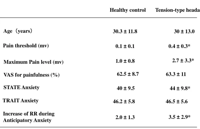
Table 1 shows the pain threshold level, maximum pain level, VAS scores for level of pain, state anxiety, and trait anxiety, and increases in RR (RR during anticipatory anxiety-RR during rest). We found a significant difference in pain thresholds and maximum pain levels between the healthy controls and the TTH group (P<0.05). State anxiety was significantly higher in the TTH group (P<0.001) while we found no
difference in trait anxiety between the two groups. During anticipatory anxiety, the TTH patients showed a greater increase in RR compared with the healthy controls (P<0.05).
We used the MMPI to investigate the relationships between variables related to anxiety
in people with TH. We found no significant differences between the TTH group and healthy controls on the following scales: (L scale, TTH, 44.1 ± 7.1, healthy controls, 43.2 ± 6.7; F scale, TTH, 50.6 ±7.4, healthy controls, 49.2 ± 6.8; K scales, TTH, 50.33
± 8.2, healthy controls, 49.2 ± 7.8; hypochondriasis, TTH, 48.5 ± 9.8, healthy controls, 47 ± 8.8; depression, TTH, 46.8 ± 12, healthy controls, 45.6 ± 9.8; hysteria, TTH, 48.1
± 12, healthy controls, 47.6 ± 11; psychopathic device, TTH, 45.1 ± 9.2, healthy controls, 44.6 ± 8.7; paranoia, TTH, 48.33 ± 6.5, healthy controls, 47.8 ± 7.7;
psychasthenia, TTH, 38.83 ± 14.6, healthy controls, 40.1 ± 13.9; schizophrenia, TTH, 44 ± 9.8, healthy controls, 43.5 ± 8.8; hypomania, 41 ± 15.23, healthy controls, 40.3 ± 14.5; and social introversion, TTH, 52.3 ± 11, healthy controls, 51,2 ± 12) (all P>0.05,
respectively). We did find a significant negative correlation between the MMPI Sc+1K scores (schizophrenia scores) and the state anxiety scores in the TTH patients (r=−0.86, P<0.05) (Figure 2).
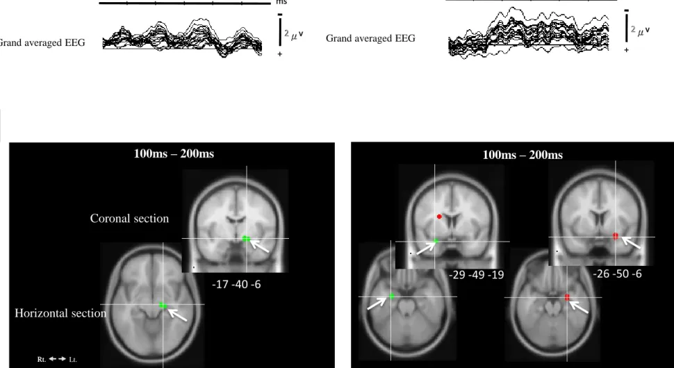
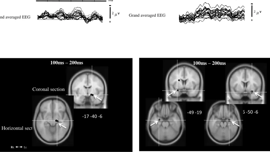
RAP and dipole localizations
Figure 3 shows the grand averaged EEG data from 19 electrodes triggered by the onset of inspiration (top panel indicates as A). The upper panel shows the grand averaged
EEG data from the healthy controls (A-left) and the TTH participants (A-right). We observed RAP with a characteristic 9- to 12-Hz frequency in both groups, as confirmed via a power spectrum analysis. The lower panel (indicates as B) in Figure 3 shows the estimated grand-averaged typical dipole localizations for the normal controls (left) and the TTH participants (right). The dipole locations, which were estimated from individual RAP data during a 400-ms period, are summarized in Table 2 (i.e., the number of dipoles in each anatomical region). Based on the RAP data, the dipoles converged in the right AMG in the normal controls, and in the bilateral AMG in the TTH patients. Figure 3B shows the dipoles localized in the AMG across all healthy controls (left) and TTH patients. The dipoles in the AMG are shown in coronal and horizontal sections and the MNI coordinates are shown at the bottom of each coronal section.
Discussion
In this study, we tested the effect of psychological stress, specifically anticipatory anxiety, on respiratory response and brain activity in healthy controls and individuals with TTH. We also investigated the differences in responses between the two groups,
taking into account personality traits.
We found significantly higher state anxiety scores in the TTH group compared with the healthy controls. Additionally, the TTH individuals exhibited a greater increase in RR during anticipation of an electrical stimulation. A previous study reported that anticipatory anxiety increases the respiratory rate, and that this increase in respiratory rate is directly associated with individual anxiety levels 7). We confirmed the findings of a previous study, i.e., that participants with higher levels of anxiety exhibited greater increases in RR, however, we found this to be the case in a patient population with high levels of anxiety in addition to normal healthy participants 16).
We were surprised to find that there were no differences in trait anxiety scores between TTH and healthy individuals. The state and trait anxiety scales can be defined as follows. The state anxiety scale measures anxiety that is experienced under specific conditions and times, and changes according to external factors. In contrast, the trait anxiety scale measures the general feelings of the individual and reflects their general predisposition to anxiety. In this sense, state anxiety levels in TTH patients might depend on the specific characteristics of stressful situations. Additionally, state anxiety might be easily influenced by anticipation of anxiety, for instance, in the conditions used in this study. Previous reports have indicated that stress and anxiety might trigger
headaches in people with TTH 5, 6). Thus, the tendency of people with TTH to be in a higher state of anxiety might contribute to their increased frequency of headaches. In the TTH patients in our study, a high state of anxiety with an increasing RR was associated with bilateral activation in the AMG.
The AMG plays a role in the reaction element of emotions, especially fear and anxiety 17). Source generators associated with RAP have been observed in the right AMG in healthy controls, indicating that the right AMG is involved in strong emotional reactions 8). The left AMG is thought to be involved in the semantic processing of emotions, as it plays a role in cognitive and resonance processing 8,15). Dolan and Morris 18) suggested that the right AMG might depend on triggers associated with the internal state, and that the left AMG might be related to conscious processing of external stimulation.
However, the bilateral AMG interact, for instance, strong right AMG activity can activate the left AMG 8). Thus, the TTH patients in our study may have been more sensitive to internal anxiety, therefore, have evaluated the external stimuli more consciously compared with the healthy controls. Interestingly, we found a correlation between state anxiety and schizophrenia scores in the TTH patients. The schizophrenia scale measures bizarre thoughts, peculiar perceptions, poor familial relationships, difficulty concentrating, and lack of deep interest 11). Thus, the greater the state of
anxiety, the more abnormal the thought patterns in TTH patients. In terms of brain site, the participants with the highest levels of state anxiety exhibited multiple dipoles in the bilateral AMG.
The psychological factors associated with the above-mentioned brain activation might induce peripheral muscle pressure, thus triggering headaches. Previous reports have described muscle tension and sustained muscle contractions around the neck and scalp of TTH patients 2). In fact, this is the case in many highly anxious individuals, who often exhibit tightness in peripheral muscles. For instance, patients with hyperventilation syndrome often present with chest wall tightness and neck tightness 16). Stress and anxiety affect muscle tone by increasing gamma-motor input at the muscle spindle, as well as increasing respiration-related muscle activity 16).
Although the TTH participants exhibited a high state of anxiety, these individuals had a pain threshold and maximum pain tolerance that was higher than that of healthy controls. However, Bezov et al. 19) found that TTH patients had a decreased electrical pain threshold and pain tolerance threshold, suggesting that TTH is accompanied by non-specific hypersensitivity. This means that general pain sensitivity is altered in TTH patients. A previous imaging study found a decrease in the volume of gray matter brain structures implicated in pain processing in TTH patients. This decrease was associated
with prolonged nociceptive input from pericranial myofascial structures 20). We did not directly evaluate morphological changes in TTH patients; however, this analysis might be worth investigating with respect to the relationship between anxiety-amaygdala responses and pain sensitization.
Recently, behavioral treatments (relaxation training, EEG biofeedback training, and cognitive therapy) had been validated in TTH patients. Relaxation training therapy may reduce headache activities by nearly 50% 2). Cognitive behavioral therapy is an effective treatment for patients with low severity TTH. Future research should test these treatments to investigate associated reductions in state anxiety in individuals with TTH.
Additionally, future studies could assess whether decreases in stress and/or anxiety lead to a reduction in symptoms, potentially inhibiting AMG activation.
Figure legends
Figure 1
Experimental apparatus for measurement of EEG and respiration during anticipation of electrical stimuli.
Figure 2
Correlation between schizophrenia-type MMPI scores and state anxiety scores in TTH patients.
Figure 3
Grand averaged EEGs from 19 electrodes triggered by the onset of inspiration during anticipatory anxiety. The top panel A shows the grand averaged EEG (anxiety-related potentials, RAP) in normal participants and TTH individuals. Dipoles converged in the left AMG in healthy controls and in the bilateral AMG in TTH patients during the interval from 100 ms to 200 ms of RAPs (indicates with white arrows, respectively) (lower panel B). We accepted dipoles with a goodness of fit that was greater than 98%.
References
(1) Stovner L, Hagen K, Jensen R, et al : The global burden of headache:
a documentation of headache prevalence and disability worldwide. Cephalalgia 27(3) : 193-210 (2007)
(2) Shengyuan Yu & Xun Han and Curr Pain : Update of Chronic Tension-Type Headache. Headache Rep 19: 469 (2015)
(3) Schmidt-Hansen PT, Svensson P, Bendtsen L, Graven-Nielsen T and Bach FW : Increased muscle pain sensitivity in patients with tension-type headache. Pain.
129(1-2) : 113-121 (2007)
(4) Bendtsen L : Central sensitization in tension-type headache—possible pathophysiological mechanisms. Cephalalgia. 20(5) : 486-508 (2000)
(5) Yucel B, Kora K, Ozyalcin S, Alcalar N, Ozdemir O and Yucel A : Depression, automatic thoughts, alexithymia, and assertiveness in patients with tension-type headache. Headache. 42(3) : 194-199 (2002)
(6) Cathcart S, Winefield AH, Lushington K and Rolan P : Stress and tension-type headache mechanisms. Cephalalgia. 30(10) : 1250-1267 (2010)
(7) Masaoka Y and Homma I : The effect of anticipatory anxiety on breathing and metabolism in humans. Respir Physiol 128 : 171-177 (2001)
(8) Masaoka Y and Homma I : The source generator of respiratory-related anxiety potential in the human brain. Neurosci Lett 283 : 21-24 (2000)
(9) Headache Classification Committee : The international classification of headache disorders II. Cephalalgia 24 : 21-24 (2000)
(10) Spielberger C., Gorsuch R., Lushene R., Vagg P and Jacobs G. (Eds.) : Manual for
the state-trait anxiety inventory. Consulting Psychologists Press, CA (1983)
(11) Butcher JN, Dahlstrom WG, Graham JR, Tellegen A and Kaemmer B : The Minnesota Multiphasic Personality Inventory-2 (MMPI-2): Manual for administration and scoring. University of Minnesota Press, MN (1989)
(12) Hideyo Kasai, Yuri Masaoka, Takeshi Kuroda, Kanako Satoh, Mitsru Kawamura and Ikuo Homma : Emotional Evaluation of Pain in Migraine Patients. The Showa University Journal of Medical Sciences 24(1) : 43-50 (2012)
(13) Inoue M, Masaoka Y, Kawamura M, Okamoto Y and Homma I : Differences in areas of human frontal medial wall activated by left and right motor execution:
Dipole-tracing analysis of grand-averaged potentials incorporated with MNI three-layer head model. Neuroscience Letters 437 : 82-87 (2008)
(14) Koiwa N, Masaoka Y, Kusumi T and Homma I : Sound localization difficulty affects early and late processing of auditory spatial information: investigation using the dipole tracing method. Clin Neurophysiol 121(9) : 1526-1539(2010)
(15) Masaoka Y, Koiwa N and Homma I : Inspiratory phase-locked alpha oscillation in human olfaction : source generators estimated by a dipole tracing method. J Physiol 566 : 979-997 (2005)
(16) Masaoka Y, Jack S, Warburton C.J. and Homma I : Breathing patterns associated with trait anxiety and breathlessness in humans. Jpn J Physiol 54 : 465-470(2004) (17) Davis M : The role of the amygdala in fear and anxiety. Ann Rev of Neurosci 15:
353-375 (1992)
(18) Dolan RJ and Morris JS : The functional anatomy of innate and acquired fear;
Perspectives from neuroimaging. In R.D. Lane & L. Nadel (Eds). Cognitive neuroscience of emotion. New York:Oxford Press : 225-241 (2000)
(19) Bezov D, Ahina S, Jensen R. and Bendtsen L : Pain perception studies in tension-type headache. Headache 51: 262-270 (2011)
(20) Schmidt-Wilcke T, Leinisch E, Straube A, Kämpfe N, Draganski B, Diener HC, Bogdahn U, May A :Gray matter decrease in patients with chronic
tension type headache. Neurology 65:1483-1486 (2005)
Flow
Volume
EEG monitoring
Experimental apparatus
Flow Transducer
Face Mask
Electrical stimuli
EEG electrodes
Respiratory flow monitor
Figure 1
Increase of RR during
Anticipatory Anxiety 2.0 ± 1.3 3.5 ± 2.9*
Maximum Pain level (mv)
46.2 ± 5.8 46.5 ± 5.6 TRAIT Anxiety
40 ± 9.5 44 ± 9.8*
63.3 ± 11 62.5 ± 8.7
VAS for painfulness (%) STATE Anxiety
1.0 ± 0.8 2.7 ± 3.3*
0.4 ± 0.3*
Pain threshold (mv) 0.1 ± 0.1
30.3 ± 11.8
Healthy control Tension-type headache
Age(years) 30 ± 13.0
Table 1 Comparisons pain perceptions, anxiety and respiratory rate between healthy controls, TH and MG
Table 2 The dipole locations estimated from individual RAP data during a 400-ms period after inspiration onsets.
L=left, R=right
AMG, amygdala; AG, angular gyrus; Cbll, cerebellum; Fusi, fusiform gyrus; Para HC, parahippocampal gyrus; ITG, inferior temporal Gyrus; MTG, middle temporal gyrus; precune, precuneus; retro, retrosplenial cortex; OcG, occipital gurus; STG, superior temporal gyrus
* Significant anatomical regions estimated by one dipole analysis.
Temporal lobe Parietal lobe Others
Fusi ITG MTG STG Para HC AMG OcG AG Precune A
cingulate Retro Cbll
L R L R L R L R L R L R L R L R L R L R L R L R
Healthy Controls 0 2 1 4 2 1 2 3 3 4 8 12 0 2 1 0 2 1 1 0 1 1 0 1 Tension-Type
Headache 0 1 2 2 1 1 2 2 4 4 20 16 1 2 0 0 1 0 4 3 2 1 2 1
20 25 30 35 40 45 50 55 60
30 40 50 60
Correlation between State Anxiety and Schizophrenia scores of MMPI TTH patients
MMPI(Sc+1K)
State Anxiety
r=-0.86
Figure 2
Coronal section
Rt. Lt.
Rt.
2 2 2 μ μ v v + +
2 μ μ v v + + 0 0
- - 100 100 100 100 200 200 300 300 400 400
ms 0 ms
- 0
- 100 100 100 100 200 200 300 300 400 400
ms ms
Grand averaged EEG Grand averaged EEG
Healthy controls TTH patients
Figure 3
100ms – 200ms
Horizontal section
-17 -40 -6
100ms – 200ms
-26 -50 -6 -29 -49 -19
2 2 μ μ v v + +
2 μ μ v v + + 0 0
- - 100 100 100 100 200 200 300 300 400 400
ms 0 ms
- 0
- 100 100 100 100 200 200 300 300 400 400
ms
A
msB
Coronal section
Rt. Lt.
Rt.
2 2 2 μ μ v v + +
2 μ μ v v + + 0 0
- - 100 100 100 100 200 200 300 300 400 400
ms 0 ms
- 0
- 100 100 100 100 200 200 300 300 400 400
ms ms
Grand averaged EEG Grand averaged EEG
Healthy controls TTH patients
Figure 3
100ms – 200ms
Horizontal section
-17 -40 -6 -29 -49 -19 -26 -50 -6
2 2 μ μ v v + +
2 μ μ v v + + 0 0
- - 100 100 100 100 200 200 300 300 400 400
ms 0 ms
- 0
- 100 100 100 100 200 200 300 300 400 400
ms
A
msB
100ms – 200ms