Digeneans Parasitic in Freshwater Fishes (Osteichthyes) of Japan VII. Allocreadiidae: Allocreadium
Takeshi Shimazu
10486–2 Hotaka-Ariake, Azumino, Nagano 399–8301, Japan E-mail: [email protected]
(Received 4 March 2016; accepted 23 March 2016)
Abstract This paper reviews nine identified and four unidentified species of Allocreadium Looss, 1900 (Trematoda, Digenea, Allocreadioidea, Allocreadiidae) and Allocreadium sp. of Kataoka and Momma (1934), incertae sedis, parasitic in freshwater fishes of Japan. Each species is described and figured with a summarized life cycle where known. Morphological characters of adult and cercarial forms of the type species Macrolecithus gotoi Hasegawa and Ozaki, 1926 support the previous hypothesis that Macrolecithus Hasegawa and Ozaki, 1926 is a junior syn- onym of Allocreadium. A neotype is designated for M. gotoi (now Allocreadium gotoi (Hasegawa and Ozaki, 1926)). The type locality becomes Lake Kizaki in Oomachi City, Nagano Prefecture, Japan. Keys to the two genera (Allocreadium and Crepidostomum Braun, 1900) of the family Allocreadiidae in Japan and to the nine identified species of Allocreadium in Japan are presented.
Key words : Digeneans, Allocreadiidae, Allocreadium, Macrolecithus, freshwater fishes, Japan, review.
Introduction
This is the seventh paper of a series that reviews adult digeneans (Trematoda) parasitic in freshwater fishes (Osteichthyes) of Japan (Shimazu, 2013). Many species of the family Allocreadiidae Looss, 1902 sensu Caira and Bogéa (2005) of the Allocreadioidea Looss, 1902 sensu Cribb (2005) have been found in Japanese freshwater fishes. They are classified into two genera, Allocreadium Looss, 1900 and Crepido- stomum Braun, 1900 (see below). This contribu- tion deals with species of Allocreadium and Allo- creadium sp. of Kataoka and Momma (1934), incertae sedis. The next will deal with species of Crepidostomum. The Introduction, Materials, and Methods for the review were given in the first paper (Shimazu, 2013).
Abbreviations used in the figures. c, cercaria;
cbp, cercarial body proper; cp, cirrus pouch; ct, cercarial tail; cvd, common vitelline duct; e, esophagus; ed, ejaculatory duct; egg, eggs in uterus; ep, excretory pore; es, eyespot; esp, eye- spot pigment; ev, excretory vesicle; fc, flame cell;
ga, genital atrium; gp, genital pore; gpr, genital primordium; i, intestine; Lc, Laurerʼs canal; m, metraterm; Mg, Mehlisʼ gland; mr, mother redia;
o, ovary; od, oviduct; os, oral sucker; ot, ootype;
p, pharynx; pc, prostatic cells; pp, pars prostatica;
pr, prepharynx; s, stylet; sd, sperm duct; sr, semi- nal receptacle; sv, seminal vesicle; t, testis; tnc, transverse nerve commissure; u, uterus; vd, vitel- line duct; vf, vitelline follicles; vs, ventral sucker.
Superfamily Allocreadioidea Looss, 1902 Family Allocreadiidae Looss, 1902
Key to genera of the family Allocreadiidae in Japan
1. Oral sucker without muscular papillae ... Allocreadium Looss, 1900
2. Oral sucker with three pairs of muscular papillae ... Crepidostomum Braun, 1900
Genus Allocreadium Looss, 1900 Allocreadium aburahaya Shimazu, 2003
(Figs. 1–3)
Allocreadium aburahaya Shimazu, 2003a: 121–122, figs.
4–6.
Host in Japan. Phoxinus steindachneri Sau- vage, 1883 (Cyprinidae) (type host) (Shimazu, 2003a; this paper).
Site of infection. Intestine.
Geographical distribution. Nagano Prefecture:
Hiroi River (type locality) at Kotobuki, Iiyama
City (Shimazu, 2003a).
Material examined. 18 specimens (NSMT-Pl 5228, type series, holotype and 17 paratypes) of Allocreadium aburahaya, adult, whole-mounted, ex intestine of Phoxinus steindachneri (syn.
Phoxinus lagowskii steindachneri), Hiroi River, 12 June 1999 (Shimazu, 2003a).
Description. After Shimazu (2003a), modified from the present study (Figs. 1–3). Body elon- gate, small, 2.56–2.90 by 0.66–0.80 (holotype 2.73 by 0.78); forebody 0.71–0.79 long, occupy- ing 25–28% of body length. Tegument smooth.
Figs. 1–3. Allocreadium aburahaya, holotype (NSMT-Pl 5228), adult found in intestine of Phoxinus steindach- neri. — 1, entire body, ventral view; 2, terminal genitalia, ventral view; 3, ovarian complex, ventral view.
Scale bars: 1 mm in Fig. 1; 0.2 mm in Figs. 2–3.
Transverse nerve commissure dorsal to prephar- ynx or pharynx. Eyespot pigment present in fore- body, scattered. Small gland cells present in fore- body, with ducts opening at anterior tip of body (not illustrated). Oral sucker globular, 0.23–0.26 by 0.26–0.30, ventrally subterminal. Prepharynx present, very short. Pharynx elliptical, 0.12–0.14 by 0.11–0.12. Esophagus 0.24–0.35 long, sur- rounded by small gland cells, bifurcating dor- sally to ventral sucker. Intestines (or ceca) ending blindly at some distance from posterior extremity of body. Ventral sucker globular, 0.29–0.33 by 0.32–0.39, located at about junction of anterior first and second fourths of body; sucker width ratio 1 : 1.2–1.4. Testes two, slightly indented irregularly, fairly large, 0.27–0.39 by 0.15–0.31, tandem, in middle third of hindbody, slightly separate. Sperm ducts two; common sperm duct absent. Cirrus pouch (or cirrus-sac) broad-ovate, 0.19–0.25 by 0.12–0.16, anterior to ventral sucker, including seminal vesicle, prostatic com- plex, and ejaculatory duct. Seminal vesicle tubu- lar, S-shaped, large, in posterior two thirds of cir- rus pouch. Pars prostatica globular, large, 0.02–0.03 by 0.04–0.05; prostatic cells numer- ous, small. Ejaculatory duct (or cirrus) short, undulating, 0.06–0.09 by 0.01–0.02, surrounded by small gland cells. Genital atrium small. Geni- tal pore median, located at about midlevel of esophagus. Ovary single, globular or somewhat irregular, large, 0.26–0.31 by 0.21–0.27, median, between ventral sucker and anterior testis. Ovar- ian complex postovarian. Seminal receptacle canalicular, flask-shaped, large, 0.18–0.33 by 0.14–0.30, between ovary and anterior testis.
Laurerʼs canal short, surrounded by small gland cells, running backward. Ootype vesicular, large, posterosinistral to ovary. Uterus coiled between anterior testis and cirrus pouch, between intes- tines, overlapping intestines anteriorly, some- times extending posteriorly into intertesticular space; metraterm shorter than cirrus pouch, sur- rounded by small gland cells. Eggs fairly numer- ous, broad-ovate, operculate, light brown, 76–86 by 54–60 μm, unembryonated. Vitellaria follicu- lar; follicles large, usually ventral or rarely dorsal
to intestines, distributed in lateral fields of body from bifurcal level to near posterior extremity of body, separate anteriorly, confluent post-testicu- larly; vitelline ducts two; common vitelline duct serving as vitelline reservoir. Excretory vesicle I-shaped, thick-walled, dorsal to vitelline folli- cles, extending anteriorly to middle of post-tes- ticular region beyond intestinal ends; excretory pore posterodorsal.
Remarks. This species and Allocreadium shinanoense Shimazu, 2003 (see below) were obtained from their respective hosts from the Hiroi River on 12 June 1999. The ootype (Shimazu, 2003a, fig. 6) should have been vesicular.
Life cycle. Not known.
Allocreadium brevivitellatum Shimazu, 1992
(Fig. 4)
Allocreadium sp.: Shimazu, 1988: 18, fig. 15.
Allocreadium brevivitellatum Shimazu, 1992: 213–215, figs. 1–3; Shimazu, 1994: 69, fig. 1.
Host in Japan. Rhynchocypris percnurus (Pal- las, 1814) (Cyprinidae) (type host) (Shimazu, 1988, 1992; this paper).
Site of infection. Intestine.
Geographical distribution. Hokkaido: Saru- runto (or Saruruto-numa, a shallow pond in a marsh) at Toro, Shibecha Town (Shimazu, 1988, 1992; this paper).
Material examined. (1) 1 (NSMT-Pl 3055, Allocreadium sp. of Shimazu (1988), 1 paratype) of A. brevivitellatum, adult, whole-mounted, ex intestine of Rhynchocypris percnurus (syn.
Moroco percnurus sachalinensis Berg, 1907), Sarurunto, 4 July 1984 (Shimazu, 1988, 1992).
(2) 2 (NSMT-Pl 3678–3679, holotype and 1 paratype) of A. brevivitellatum, adult, whole- mounted, ex intestine of R. percnurus, Sarurunto, 3 September 1991 (Shimazu, 1992). One adult paratype and 1 immature voucher (NSMT-Pl 3680–3681) of Shimazu (1992) were not used.
Probably, they had been lost on loan.
Description. After Shimazu (1992), modified
from the present study (Fig. 4). Body elongate
ovate, small, 2.50–3.12 by 0.76–0.88 (holotype 2.68 by 0.84); forebody 0.70–0.96 long, occupy- ing 27–31% of body length. Eyespot pigment dispersed. Oral sucker 0.24–0.26 by 0.29–0.33.
Pharynx 0.12–0.14 by 0.10–0.16. Esophagus bifurcating dorsal to ventral sucker. Intestines extending to near posterior extremity of body.
Ventral sucker 0.33–0.35 by 0.38–0.41; sucker width ratio 1 : 1.2–1.4. Testes smooth or indented irregularly, small, 0.16–0.26 by 0.13–0.20,
almost tandem, separate or contiguous, in middle third of hindbody. Cirrus pouch spherical or elongate-oval, 0.20–0.30 by 0.13–0.21, anterior to ventral sucker. Seminal vesicle sinuous, in posterior third of cirrus pouch. Ejaculatory duct fairly long, slightly everted. Genital atrium small. Genital pore at about midlevel of esopha- gus. Ovary spherical to elliptical, small, 0.16–
0.30 by 0.16–0.21, median. Seminal receptacle clavate, small, 0.18–0.28 by 0.06–0.07. Laurerʼs
Fig. 4. Allocreadium brevivitellatum, holotype (NSMT-Pl 3678), adult found in intestine of Rhynchocypris perc- nurus, entire body, ventral view. Scale bar: 1 mm.
Fig. 5. Allocreadium gotoi, neotype (NSMT-Pl 2929), adult found in intestine of Misgurnus anguillicaudatum, entire body, ventral view. Scale bar: 1 mm.
Fig. 6. Allocreadium hasu, possible paratype (MPM Coll. No. 30006), adult found in intestine of Opsariichthys uncirostris uncirostris, entire body, ventral view. Scale bar: 1 mm.
canal as long as seminal receptacle or slightly shorter than it. Ootype postovarian. Uterus extending posteriorly to anterior testis in 3 speci- mens, but slightly beyond posterior testis in 1 specimen. Eggs 106–130 by 69–80 μm. Vitelline follicles distributed between ovarian level to near posterior extremity of body. Excretory vesicle reaching to middle of posterior testis; excretory pore posterodorsal.
Remarks. This species was found in Sarurunto, but not in Lake Toro nearby.
Life cycle. Not known.
Allocreadium gotoi (Hasegawa and Ozaki, 1926)
(Figs. 5, 7–10)
Macrolecithus gotoi Hasegawa and Ozaki, 1926: 225–227, fig. 1, 1 text table; Yamaguti, 1934: 333–334, fig. 38.
Allocreadium gotoi: Shimazu, 1988: 6–7, figs. 1–3;
Shimazu, 1994: 69, fig. 2; Shimazu, 2002: fig. 7;
Shimazu, Urabe, and Grygier, 2011: 29, figs. 34–35.
Hosts in Japan. Misgurnus anguillicaudatus (Cantor, 1842) (Cobitidae) (type host) (Hasegawa and Ozaki, 1926; Yamaguti, 1934; Shimazu, 1988, 1994, 2002; Shimazu et al., 2011), Gna- thopogon elongatus elongatus (Temminck and Schlegel, 1846) (Cyprinidae) (Shimazu, 1988;
Shimazu et al., 2011), and Gymnogobius opperiens Stevenson, 2002 (Gobiidae) (Shimazu, 1988, 1994; this paper).
Site of infection. Intestine.
Geographical distribution. (1) Hokkaido:
Oono River at Chiyoda, Hokuto City (Shimazu, 1988, 1994; this paper). (2) Miyagi Prefecture:
Shichigo, Sendai City (Shimazu, 1988). (3) Nagano Prefecture: a small river at Midori, Iiyama City; a small river at Okada-machi, Matsumoto City; Furukawa River at Kamiimai, Nakano City; Komi (now Komi Town (?)); Lake Kizaki (type locality) in Oomachi City; and Lake Suwa at Suwa City (Shimazu, 1988, 2002; this paper). (4) Aichi Prefecture: Mikawa (central and eastern district of the prefecture) (Shimazu,
Figs. 7–10. Allocreadium gotoi (continued), life cycle, found in Pisidium nikkoense. — 7, sporocyst; 8, daughter redia; 9, cercaria, lateral view; 10, cercarial body proper, ventral view. Redrawn from Shimazu (2002). Scale bars: 200 μm.
1988). (5) Shiga Prefecture: Lake Biwa basin (Lake Biwa; Terasho, Koka City; and Zeze, Otsu City) (Shimazu, 1988; Shimazu et al., 2011). (6) Kyoto Prefecture: Jonangu, Fushimi-ku, Kyoto City; and Miyakehachiman, Sakyo-ku, Kyoto City (Shimazu, 1988; this paper). (7) Hyogo Pre- fecture: Nishinomiya (Shimazu, 1988).
In China (Okino et al., 2004).
Material examined. (1) 1 specimen (Ozakiʼs Collection, MPM Coll. No. 30009, labeled
“DOJO,” other data not given) of Macrolecithus gotoi, adult, serially sectioned, Misgurnus anguillicaudatus (Shimazu, 1988, 1995). Several sections of the anterior part of the forebody were missing. (2) Yamagutiʼs specimens of M. gotoi, ex intestine of Mi. anguillicaudatus: 2 (MPM Coll. No. 22570), adult, whole-mounted, Komi (Komi Village, now Komi Town, Nagano Prefec- ture (?)), 29 August 1927; 4 (MPM Coll. 22574), adult, whole-mounted, Jonangu, Fushimi-ku, Kyoto City, Kyoto Prefecture, 24 June 1940; 1 (MPM Coll. No. 22288, identified as Allocread- ium by Shunya Kamegai on 19 April 1972), adult, whole-mounted, Miyakehachiman, Sakyo- ku, Kyoto City, 11 August 1941; 9 (MPM Coll.
22575, ex “stomach”), adult, whole-mounted, Mikawa (central and eastern district of Aichi Pre- fecture), 7 July 1937; 1 (MPM Coll. 22573, adult, whole-mounted), Nishinomiya (Nishi- nomiya City, Hyogo Prefecture), 11 June 1940; 7 (MPM Coll. No. 22567, ex “stomach and intes- tine”), adult, serially sectioned, whole-mounted, Shichigo, 3 August 1927; 1 (MPM Coll. No.
22571), adult, whole-mounted, Terasho, 8 July 1927; 1 (MPM Coll. No. 22568), serially sec- tioned, Zeze, 16 July 1928; and 4 (MPM Coll.
No. 22294, locality not given), adult, whole- mounted, 23 June 1932 (Shimazu, 1988;
Shimazu et al., 2011). (3) 4 (NSMT-Pl 2931) of A. gotoi, immature, adult, whole-mounted, ex intestine of Mi. anguillicaudatus, Oono River at Chiyoda, Oono Town, now in Hokuto City, Hok- kaido, 20 August 1983 (Shimazu, 1988). One immature and 2 adult specimens (NSMT-Pl 2932, Oono River, 23 August 1983) of Shimazu (1988) were not used. Probably, they had been
lost on loan. (4) 5 (NSMT-Pl 2929) of A. gotoi, adult, whole-mounted, ex intestine of Mi. anguil- licaudatus, Lake Kizaki, 28 August 1981 (Shimazu, 1988). Five adult specimens (NSMT- Pl 2929, Lake Kizaki, 24 July 1983) of Shimazu (1988) were not used. Probably, they had been lost on loan. (5) Shimazuʼs (2002) specimens of A. gotoi, ex intestine of Mi. anguillicaudatus, Nagano Prefecture: 39 (NSMT-Pl 5216–5219), immature, adult, serially sectioned, whole- mounted, a small river at Midori, 12 July 1987, 3 November 1989, 5 August 1990, 13 August 1999; 5 (NSMT-Pl 5222), immature, adult, a small river at Okada-machi, 3 August 1987; 6 (NSMT-Pl 5220), immature, adult, whole- mounted, Furukawa River in Toyoda Village, now Kamiimai, Nakano City, 17 June 2000; and 1 (NSMT-Pl 5221), immature, whole-mounted, Lake Suwa, 5 October 1991. (6) Yamagutiʼs specimens of M. gotoi, ex intestine of Gnatho- pogon elongatus elongatus [not Gnathopogon elongatus caerulescens of Shimazu (1988)], Lake Biwa: 1 (MPM Coll. No. 22572, labeled
“Macrolecithus gotoi [Moroko]”), adult, whole- mounted, 9 July 1927; and 1 (MPM Coll. No.
22569, labeled “Allocread. [Moroko]”), adult, whole-mounted, 29 July 1927 (Shimazu, 1988;
Shimazu et al., 2011). (7) 1 (NSMT-Pl 2933) of A. gotoi, immature, whole-mounted, ex intestine of Gymnogobius opperiens (Chaenogobius annu- laris (the middle-reaches type) and Chaenogo- bius urotaenia of Shimazu (1994)), Oono River, 20 August 1984 (Shimazu, 1988, 1994). One immature and 2 adult specimens (NSMT-Pl 2932, Oono River, 23 August 1983) of Shimazu (1988) were not used. Probably, they had been lost on loan.
Description. Based on specimens (NSMT-Pl 2929–2930) from Lake Kizaki, after Shimazu (1988), modified from the present study (Fig. 5).
Body elongate-ovate, slightly tapering posteri- orly, fairly small, 2.30–4.54 by 0.80–1.32; fore- body 1.27–1.59 long, occupying 35–42% of body length. Eyespot pigment solid or dispersed.
Oral sucker 0.32–0.47 by 0.30–0.47. Pharynx
0.15–0.25 in diameter. Esophagus winding,
bifurcating midway between two suckers.
Intestines reaching to near posterior extremity of body. Ventral sucker postbifurcal, 0.42–0.61 by 0.40–0.57; sucker width ratio 1 : 1.2–1.3. Testes elliptical, rather small, 0.26–0.47 by 0.24–0.40, almost tandem, usually separated by uterus, in middle third of hindbody. Cirrus pouch clavi- form, small, 0.16–0.34 by 0.08–0.14, between intestinal bifurcation and ventral sucker. Seminal vesicle tubular, sinuous, in posterior two-thirds of cirrus pouch. Ejaculatory duct short. Genital atrium small. Genital pore immediately postbi- furcal. Ovary globular, small, 0.21–0.34 by 0.16–
0.27. Seminal receptacle flask-shaped, 0.16–0.32 by 0.12–0.17. Laurerʼs canal short. Ootype pos- terolateral to ovary; Mehlisʼ gland well devel- oped. Uterus between intestines, entering post- testicular region in fully matured specimens more than 3.60 long; metraterm shorter than cir- rus pouch. Eggs 80–94 by 46–54 μm. Vitelline follicles dorsal to intestines, distributed usually from bifurcal level to posterior extremity of body, rarely extending to pharyngeal level.
Excretory vesicle extending to about midlevel of post-testicular region beyond intestinal ends;
excretory pore posterodorsal.
Remarks. Hasegawa and Ozaki (1926) origi- nally described this species as a new genus and species, Macrolecithus gotoi, on the basis of adult specimens found in the intestine of Misgur- nus anguillicaudatus. They did not specify the locality in Japan. Shimazu (1988) synonymized Macrolecithus with Allocreadium and made a new combination, Allocreadium gotoi (Hasegawa and Ozaki, 1926), for the type species M. gotoi.
Since Tkach (2008) has recently treated Macro- lecithus (syn. Paramacrolecithus Srivastava and Ghosh, 1967) as a genus incertae sedis, the sys- tematic position and validity of Macrolecithus will be discussed later again.
Hasegawa and Ozaki (1926) used several whole-mounted and serially sectioned adult spec- imens for describing M. gotoi. The holotype (No.
P. 270) was lost (see the Materials and Methods in Shimazu (2013)). Shimazu (1988) doubted that the serially sectioned adult specimen (MPM
Coll. No. 30009) belonged to their original speci- mens. However, I now regard it as the only exist- ing original specimen (a paratype (?)). Yamaguti collected several specimens of M. gotoi at Tera- sho, Shichigo, Komi, Zeze, Lake Biwa, and the unknown locality (see Material examined).
When Yamaguti (1934, fig. 38) gave measure- ments of seven whole-mounts and figured the ovarian complex of M. gotoi, he did not mention which ones of them he used at that time.
The present specimens agree well in morphol- ogy and measurements with the original descrip- tion by Hasegawa and Ozaki (1926) for M. gotoi.
The only existing original specimen (MPM Coll.
No. 30009) is serially sectioned. The type local- ity has previously remained unknown. In order to show the adult morphology and type locality, I here designate a neotype for M. gotoi, as follows.
Designation of a neotype for Macrolecithus gotoi Hasegawa and Ozaki, 1926, or now Allo- creadium gotoi (Hasegawa and Ozaki, 1926).
Neotype: an adult specimen (NSMT-Pl 2929, 4.04 mm long by 1.32 mm wide, Fig. 1), slightly flattened, whole-mounted, 28 August 1981.
Type host. Misgurnus anguillicaudatus (Can- tor, 1842) (Cobitidae).
Site of infection. Intestine.
Type locality. Lake Kizaki (36°33′N, 137°50′E) in Oomachi City, Nagano Prefecture, Japan.
Allocreadium gotoi has not as yet been reported from China. However, Okino et al.
(2004) detected adults of A. gotoi in the intestine of Mi. anguillicaudatus that had been commer- cially imported from Zhejiang Province, China, to Japan.
Life cycle. The first intermediate host is Pisid-
ium nikkoense Mori, 1938 (or Pisidium cinereum
nikkoense, P. (P.) cinereum nikkoense) (Bivalvia,
Sphaeriidae) (Japanese name: Nikkou-mameshi-
jimi). Shimazu (2002) found sporocysts, mother
and daughter rediae, and cercariae in Pi. nikkoense
(site of infection not determined) collected in the
small river at Midori, Iiyama City, Nagano Pre-
fecture (see Geographical distribution and Mate-
rial examined; Shimazu, 2007). Cercariae of the
ophthalmoxiphidiocercaria [not ophthalmoxiphi-
diocercous] type (with an unarmed body, one pair of eyespots, a simple tail, a stylet, and an I-shaped epithelial excretory vesicle) were pro- duced in the daughter rediae (NSMT-Pl 5215) (Figs. 7–10). Small protuberances were found arranged in a circle on the periphery of the body around the ventral sucker. The penetration gland cells were not observed clearly. Because the same unusual flame cell formula 2[(2+3+3)
+(3+3+3)]=34 was determined in both adults of A. gotoi and cercariae, Shimazu identified the cercaria as A. gotoi. The second intermediate host is unknown. The final hosts are Misgurnus anguillicaudatus and Gnathopogon elongatus elongatus and possibly Gymnogobius opperiens, in the intestine of which adults live (see Hosts in Japan).
This cercaria was the first to be reported from sphaeriids in Japan (see Ito, 1964). In order to verify the specific identity between the adult and cercaria in molecular data, adults and cercariae collected by me in the small river at Midori in 2012 were submitted to sequencing the cyto- chrome c oxidase I gene of the mitochondrial DNA (COI mtDNA) and first internal transcribed spacer region of the ribosomal DNA (ITS-1 rDNA). The partial COI gene was not sequenced for either the adults or the cercariae, but the entire ITS-1 region was successfully sequenced for adults (Misako Urabe, 2015, unpublished data). Later, the partial COI gene was success- fully sequenced for adults collected by me at the same river in 2015 (Misako Urabe, 2016, unpub- lished data). I attempted to collect cercariae at the same river three times in 2015 without suc- cess.
Allocreadium hasu Ozaki, 1926
(Fig. 6)
Allocreadium hasu Ozaki, 1926: 125, no figure; Yama- guti, 1934: 281–282, no figure; Shimazu, 1988: 9, figs.
4–5 [not 1–2]; Shimazu, Urabe, and Grygier, 2011:
31–33, figs. 36–39.
Allocreadium (Allocreadium) hasu: Yamaguti, 1954: 69;
Yamaguti, 1958: 101; Yamaguti, 1971: 133.
Allocreadium sp.: Shimazu, 1988: 15.
Allocreadium sp. 2: Shimazu, 1999: 71; Shimazu, 2003b:
70.
Hosts in Japan. Opsariichthys uncirostris uncirostris (Temminck and Schlegel, 1846) (Cyprinidae) (type host) (Ozaki, 1926; Yamaguti, 1934; Shimazu, 1988; Shimazu et al., 2011; this paper), Gnathopogon elongatus elongatus (Tem- minck and Schlegel, 1846) (Cyprinidae) (Shimazu, 1988; Shimazu et al., 2011), Nippono- cypris temminckii (Temminck and Schlegel, 1846) (Cyprinidae) (Shimazu, 1988; this paper), and Zacco platypus (Temminck and Schlegel, 1846) (Cyprinidae) (Shimazu, 1988; Shimazu et al., 2011).
Site of infection. Intestine.
Geographical distribution. (1) Shiga Prefec- ture: Lake Biwa basin (Lake Biwa (probably type locality); Imazu-cho, Takashima City; Koma tsu (most likely referring to Kitakomatsu, Otsu City); Moriyama City; Omatsu, Minamikomatsu, Otsu City; and Onoe, Kohoku Town) (Yamaguti, 1934; Shimazu, 1988; Shimazu et al., 2011). (2) Hyogo Prefecture: Asago River (Shimazu, 1988).
(3) Hiroshima Prefecture: Eno River at Yoshida, Yoshida-cho, Akitakata City; and Saijo River at Ooya, Saijo-cho, Shobara City (Shimazu, 1988;
this paper). (4) Oita Prefecture: Chikugo River at Kobuchi Bridge, Miyoshikobuchi-machi, Hita City; and Ooyama River at Seiwa Bridge, Ooyama-machi, Hita City (this paper).
Material examined. (1) 17 specimens (Ozakiʼs Collection, MPM Coll. No. 30006, labeled “A.
hasu HASU [Lake Biwa],” other data not given, paratypes (?)) of Allocreadium hasu, immature, adult, whole-mounted, serially sectioned, ex Opsariichthys uncirostris uncirostris (syn.
Opsariichthys uncirostris), Lake Biwa (Shimazu, 1988; Shimazu et al., 2011). (2) Yamagutiʼs spec- imens of A. hasu, Lake Biwa basin: 6 (MPM Coll. No. 22287, ex “stomach and small intes- tine”), adult, whole-mounted, ex intestine of O.
uncirostris uncirostris, Omatsu, 10 and 17 July
1927; 1 (MPM Coll. No. 22576, labeled “Allo-
cread. hasu”), immature, whole-mounted, ex
intestine of O. uncirostris uncirostris, Lake
Biwa, 10 December 1926; 1 (MPM Coll. No.
22569, labeled “Allocread. [Moroko]”), adult, whole-mounted, ex intestine of Gnathopogon elongatus elongatus, Lake Biwa, 29 July 1927;
and 1 (MPM Coll. No. 22576, labeled “Allo- cread. hasu [Moroko]”), immature, whole- mounted, ex intestine of G. elongatus elongatus, Komatsu (most likely referring to Kitakomatsu), 10 December 1926 (Yamaguti, 1934; Shimazu, 1988; Shimazu et al., 2011). (3) Specimens of A.
hasu, ex intestine of O. uncirostris uncirostris, Lake Biwa basin: 2 (LBM 3-55), adult, whole- mounted, Imazu, 5 May 2000 (Shimazu et al., 2011); 12 (NSMT-Pl 5713), adult, whole- mounted, Moriyama City, 2 May 1992; and 8 (NSMT-Pl 5711–5712), immature, adult, whole- mounted, Omatsu, 30 April 1992, 1 May 1992.
One adult specimen (NSMT-Pl 2934, Onoe, now in Kohoku Town, Shiga Prefecture, 6 June 1980) of Shimazu (1988) was not used. Probably, it had been lost on loan. (4) 1 (NSMT-Pl 5714), adult, whole-mounted, ex intestine of Zacco platypus, Omatsu, 30 April 1992 (Shimazu et al., 2011).
(5) 2 (Yamagutiʼs Collection, MPM Coll. No.
22286), immature, whole-mounted, ex intestine of Nipponocypris temminckii (syn. Zacco tem- minckii), Asago River, 23 March 1932 (Shimazu, 1988). (6) 1 (NSMT-Pl 2936, Allocreadium sp. of Shimazu (1988), Allocreadium sp. 2 of Shimazu (1999, 2003b)), immature, whole-mounted, ex intestine of N. temminckii, Saijo [not Go] River at Hibayama, Saijo Town, now Ooya, Saijo-cho, Shobara City, Hiroshima Prefecture, 31 October 1976 (Shimazu, 1988). (7) Urabeʼs unpublished specimens: 1, immature, whole-mounted, ex intestine of N. temminckii, Ooyama River, 3 November 2003; 1, adult, whole-mounted, ex intestine of O. uncirostris uncirostris, Chikugo River, 25 August 2003; and 3 and 1, adult, whole-mounted, ex intestine of Z. platypus, Chikugo River, 25 August 2003 and 21 October 2003, respectively.
Description. 1) Based on Ozakiʼs specimens (MPM Coll. No. 30006), after Shimazu (1988), modified from the present study (Fig. 6). Body elongate-ovate, rapidly attenuated anteriorly in forebody, fairly small, 4.20–5.80 by 1.10–1.80;
forebody 0.79–1.11 long, occupying 16–26% of body length. Eyespot pigment fine. Oral sucker 0.31–0.37 by 0.28–0.47. Pharynx 0.16–0.23 by 0.15–0.23. Esophagus bifurcating dorsal to ven- tral sucker. Intestines ending some distance ante- rior to posterior extremity of body. Ventral sucker large, 0.43–0.59 by 0.47–0.67; sucker width ratio 1 : 1.4–1.7. Testes deeply indented irregularly, large, 0.55–1.02 by 0.59–1.02, in middle third of hindbody. Cirrus pouch clavate to elliptical, 0.21–0.79 by 0.15–0.47, overlapping ventral sucker (extending to posterior border of ventral sucker in serially sectioned specimens).
Seminal vesicle tubular (possibly constricted once in serially sectioned specimens), convo- luted, in posterior half to two-thirds of cirrus pouch. Ejaculatory duct fairly long [not short], sinuous. Genital atrium small. Genital pore at midlevel of esophagus. Ovary usually 3-lobed but rarely subglobular, fairly large, 0.36–0.47 by 0.31–0.67. Seminal receptacle flask-shaped, 0.15–0.41 by 0.15–0.39, usually submedian.
Laurerʼs canal short. Ootype postovarian. Uterus between anterior testis and intestinal bifurcation, overlapping intestines; metraterm shorter than cirrus pouch. Eggs 64–78 by 46–56 μm. Vitelline follicles mostly dorsal to intestines, distributed from bifurcal level to posterior extremity of body. Excretory vesicle extending to midlevel of post-testicular region beyond intestinal ends;
excretory pore posterodorsal.
Remarks. Ozaki (1926) described this species, in a preliminary note with no figure, based on adult specimens found in the intestine of Opsari- ichthys uncirostris (now O. uncirostris unciros- tris), without indicating the type locality in Japan. The holotype (No. P. 231) (Ozaki, 1926) was lost (see the Materials and Methods in Shimazu (2013)). Ozakiʼs specimens (MPM Coll.
No. 30006) were found in O. uncirostris from Lake Biwa and identified as A. hasu by Ozaki himself. They agree well in morphology and host fish species with Ozakiʼs original description for A. hasu. They are most likely paratypes, which suggests that the type locality is Lake Biwa.
However, since the holotype was lost, there is no
conclusive evidence to prove this (Shimazu, 1988; Shimazu et al., 2011).
Later, Yamaguti (1934) described A. hasu, with no figure either, from his own specimens found in O. uncirostris from Lake Biwa. He seems to have used the six adult specimens (MPM Coll. No. 22287) at least at that time.
Shimazu (1988) described A. hasu from Ozakiʼs, Yamagutiʼs, and his own specimens. Shimazu et al. (2011) described A. hasu from the specimens from the Lake Biwa basin.
With regard to the shape of the ovary, the ovary is 3-lobed (Ozaki, 1926), ovoid (Yamaguti, 1934), 3-lobed or subglobular (Shimazu, 1988), globular to 3-lobed (Shimazu et al., 2011), and usually 3-lobed but rarely subglobular (this paper). It is apparent from the present examina- tion that, as adult worms grow, the ovary changes its shape from globular to deeply 3-lobed.
Shimazu (1988) reported an immature speci- men (NSMT-Pl 2936) as Allocreadium sp. (Allo- creadium sp. 2 of Shimazu (1999, 2003b)) from Nipponocypris temminckii. I identify it as A.
hasu, because the seminal vesicle is weakly con- stricted once and the testes are large and indented irregularly as in Yamagutiʼs and Urabeʼs imma- ture specimens.
One adult specimen (NSMT-Pl 3093) of A.
hasu of Shimazu (1988) (ex intestine of Z. platy- pus, Eno [not Go] River at Yoshida, now in Yoshida-cho, Akitakata City, Hiroshima Prefec- ture, 31 October 1976) was not used. Probably, it had been lost on loan.
Life cycle. Not known.
Allocreadium japonicum Ozaki, 1926
(Fig. 11)
Allocreadum japonicum Ozaki, 1926 [sic, misspelling of Allocreadium]: 124–125, no figure.
Allocreadium japonicum: Yamaguti, 1934: 282, no figure;
Shimazu, 1988: 13–14, figs. 10–11; Shimazu, Urabe, and Grygier, 2011: 35–36, figs. 40–43.
Allocreadium (Allocreadium) japonicum: Yamaguti, 1954:
69; Yamaguti, 1958: 101; Yamaguti, 1971: 133.
Hosts in Japan. Zacco platypus (Temminck
and Schlegel, 1846) (Cyprinidae) (type host) (Ozaki, 1926; Yamaguti, 1934; Shimazu, 1988), Gasterosteus aculeatus leiurus Linnaeus, 1758 (Gasterosteidae) (Shimazu et al., 2011), Nippon- ocypris temminckii (Temminck and Schlegel, 1846) (Cyprinidae) (Yamaguti, 1934; Shimazu, 1988; this paper), Phoxinus oxycephalus (Sau- vage and Dabry de Thiersant, 1874) (Cyprinidae) (Shimazu et al., 2011; this paper), and Tribolo- don brandtii (Dybowski, 1872) (Shimazu, 1988;
this paper).
Site of infection. Intestine.
Geographical distribution. (1) Tokyo: Tama River and Shinkawa River (?) (Shimazu, 1988;
this paper). (2) Shiga Prefecture: Lake Biwa basin (Lake Biwa; Amano River at Samegai, Maibara City; Momose, Momose-gyoko Fishing Port, Chinai, Makino-cho, Takashima City; and Nyuu River at Shiori, Maibara City) (Yamaguti, 1934; Shimazu, 1988; Shimazu et al., 2011).
In China (e.g., Wang, 1984; Wang et al., 1985).
Material examined. (1) 14 specimens (Ozakiʼs Collection, MPM Coll. No. 30007, labeled “Allo- creadium HAYA [Tama River],” other data not given) of Allocreadium japonicum, immature, adult, whole-mounted, serially sectioned, ex Haya (see below), Tama River, Tokyo (?) (Shimazu, 1988). (2) 3 (Ozakiʼs Collection, MPM Coll. No. 30008, labeled “MARUTA [Shinkawa],” other data not given) of A. japoni- cum, adult, whole-mounted, ex Tribolodon brandtii (syn. Tribolodon taczanowskii Stein- dachner, 1881), Shinkawa River (?), Tokyo (?) (Shimazu, 1988). (3) Yamagutiʼs specimens of A.
japonicum, Lake Biwa: 2 (MPM Coll. No.
22285), adult, whole-mounted, ex intestine of Zacco platypus, 7 July 1927; 3 (MPM Coll. No.
22285, labeled “Allocead.”), immature, adult, ex intestine of Z. platypus, 10 July 1927; and 1 (MPM Coll. No. 22577), adult, whole-mounted, ex intestine of Nipponocypris temminckii (syn.
Zacco temminckii), 7 July 1927 (Yamaguti, 1934;
Shimazu, 1988; Shimazu et al., 2011). (4) 1
(LBM 8-18) of A. japonicum, adult, whole-
mounted, ex intestine of Gasterosteus aculeatus
leiurus, Amano River, 10 May 1998 (Shimazu et al., 2011). (5) 5 (LBM 8-25) of A. japonicum, adult, whole-mounted, ex intestine of Phoxinus oxycephalus (syn. Rhynchocypris oxycephalus), Nyuu River, 26 April 2001 (Shimazu et al., 2011). (6) 1 (LBM 8-54) of A. japonicum, adult, whole-mounted, ex intestine of Z. platypus, Momose, 24 November 2007 (Shimazu et al., 2011).
Description. Based on Ozakiʼs specimens (MPM Coll. No. 30007), after Shimazu (1988), modified from the present study (Fig. 11). Body
elongate-oval, fairly small, 4.00–5.40 by 0.86–
1.35; forebody 1.03–1.27 long, occupying 24–30% of body length. Eyespot pigment solid or dispersed. Oral sucker 0.31–0.39 by 0.33–0.41 (missing in 2 of 7 specimens). Pharynx 0.15–
0.22 by 0.15–0.23. Esophagus bifurcating dor- sally to anterior half of ventral sucker. Intestines ending at near posterior extremity of body. Ven- tral sucker 0.39–0.57 by 0.39–0.55; sucker width ratio 1 : 1.3–1.4. Testes weakly to deeply indented irregularly, large, 0.51–0.86 by 0.35–
0.79, almost contiguous, in middle third of hind-
Fig. 11. Allocreadium japonicum, adult (MPM Coll. No. 30007) found in intestine of Haya (Tribolodon hakonensis [?]), entire body, ventral view. Scale bar: 1 mm.
Fig. 12. Allocreadium shinanoense, holotype (NSMT-Pl 5227), adult found in intestine of Phoxinus steindachneri, entire body, ventral view. Scale bar: 1 mm.
Fig. 13. Allocreadium tamoroko, holotype (NSMT-Pl 5858), adult found in intestine of Gnathopogon elongatus elongatus, entire body, ventral view. Scale bar: 1 mm.
body. Cirrus pouch clavate, 0.27–0.63 by 0.17–
0.30, anterior to or overlapping ventral sucker.
Seminal vesicle S-shaped, in posterior two-thirds of [not filling] cirrus pouch. Ejaculatory short, duct slightly undulating. Genital atrium fairly large. Genital pore at midlevel of esophagus.
Ovary elliptical to weakly 3-lobed, small, 0.39–
0.59 by 0.23–0.47, median. Laurerʼs canal short.
Seminal receptacle flask-shaped, 0.19–0.39 by 0.19–0.31. Ootype postovarian. Uterus usually between anterior testis and ventral sucker, rarely extending to posterior testis, slightly overlapping intestines; metraterm shorter than cirrus pouch.
Eggs 80–84 by 44–50 μm. Vitelline follicles dis- tributed from about bifurcal level to posterior extremity of body, mostly dorsal to intestines.
Excretory vesicle extending to anterior third of post-testicular region beyond intestinal ends;
excretory pore posterodorsal.
Remarks. Ozaki (1926) described this species, in a preliminary note with no figure, based on adult specimens found in the intestine of Zacco platypus, without indicating the type locality in Japan. The holotype (No. P. 226) was lost (see the Materials and Methods in Shimazu (2013)).
Because Ozaki reexamined his specimens at his disposal for Peters (Peters, 1957), he must have still possessed some specimens of A. japon- icum then. Ozakiʼs Collection today includes the specimens of Allocreadium (MPM Coll. Nos.
30006, 30007, and 30008) found in the Hasu (see A. hasu above), Haya, and Maruta, respectively.
The checklist (Shimazu, 1995) does not include the last specimens.
Ozakiʼs specimens (MPM Coll. No. 30007) were found in the Haya from the Tama River in [Tokyo (?)]. The question is whether the current scientific name of the Haya is Z. platypus. It is either Z. platypus or Tribolodon hakonensis (Shimazu, 1988). However, local people have traditionally called T. hakonensis Haya around the Tama River (Fisheries Cooperative Associa- tion of Tamagawa in Fuchu, personal communi- cation, 2011). Ozaki (1926) described the excre- tory vesicle as reaching anteriorly to the posterior testis in A. japonicum. However, the
excretory vesicle ends at the anterior third of the post-testicular region in the specimens. As men- tioned above, the holotype was lost, the labels are incomplete, the scientific name of the Haya is unknown, and the specimens are different from Ozakiʼs original description of A. japonicum in the anterior extent of the excretory vesicle. In conclusion, it remains unsolved as to whether the specimens (MPM Coll. Nos. 30007) are part of Ozakiʼs (1926) original specimens of A. japoni- cum from Z. platypus, and so the identification of them as A. japonicum is provisional. The excre- tory vesicle does not reach the posterior testis in the others of the present specimens, either (Shimazu, 1988; Shimazu et al., 2011). Accord- ingly, the identification of them as A. japonicum is also provisional. The records of A. japonicum by Wang (1984) and Wang et al. (1985) from Fujian Province, China, also need validation. I was unsuccessful in obtaining any specimens of Allocreadium from 36 individuals of Z. platypus and 4 of T. hakonensis collected in the Tama River at Fuchu City, Tokyo, on 13 August 2011.
Ozaki found no eyespot pigment in the speci- mens of A. japonicum reexamined except in one
“doubtful” specimen (Peters, 1957). Peters (1957) considered that Ozakiʼs species A. japoni- cum consisted of two genera. One of them was an opecoelid genus owing to the absence of the eyespot pigment and the presence of the 3-lobed ovary. Shimazu (1988) did not agree with him.
Eyespot pigment was solid or dispersed in the present specimens.
What are morphological differences between
A. japonicum and A. hasu? Ozaki (1926) seems
to have separated A. japonicum from A. hasu by
the testes being smaller and more weakly
notched, the vitelline follicles being larger and of
irregular shape, and the excretory vesicle being
longer and reaching to the posterior testis (see
also Shimazu, 1988). Yamaguti (1934) merely
stated that A. japonicum differed from A. hasu in
the size of the body, characters of the testes, and
extent of the excretory vesicle. The above-men-
tioned differences are slight, and future studies
may demonstrate that A. japonicum and A. hasu
are conspecific (Shimazu, 1988).
Life cycle. Not known.
Allocreadium shinanoense Shimazu, 2003
(Fig. 12)
Allocreadium shinanoense Shimazu, 2003a: 119–120, figs. 1–3.
Host in Japan. Phoxinus steindachneri Sau- vage, 1883 (Cyprinidae) (type host) (Shimazu, 2003a; this paper).
Site of infection. Intestine.
Geographical distribution. Nagano Prefecture:
Hiroi River (type locality) at Kotobuki, Iiyama City (Shimazu, 2003a).
Material examined. 2 specimens (NSMT-Pl 5227, holotype and 1 paratype) of Allocreadium shinanoense, adult, whole-mounted, ex intestine of Phoxinus steindachneri (syn. Rhynchocypris lagowskii steindachneri), Hiroi River, 12 June 1999 (Shimazu, 2003a).
Description. After Shimazu (2003a), modified from the present study (Fig. 12). Body elongate, fairly small, 3.74–3.80 by 1.16–1.20 (holotype 3.74 by 1.16); forebody 1.27–1.30 long, occupy- ing 33–34% of body length. Eyespot pigment solid. Oral sucker 0.38–0.41 by 0.41–0.42. Phar- ynx 0.17–0.19 by 0.18–0.19. Esophagus bifurcat- ing immediately anterior to ventral sucker. Intes- tines ending some distance anterior to posterior extremity of body. Ventral sucker 0.55–0.57 by 0.53–0.57; sucker width ratio 1 : 1.3. Testes ellip- tical, somewhat irregular in outline, fairly small, 0.35–0.55 by 0.26–0.44, slightly oblique, slightly separated by uterus, in middle third of hindbody.
Cirrus pouch claviform, 0.39–0.51 by 0.13–0.14, anterior to ventral sucker, slightly overlapping it posteriorly. Seminal vesicle claviform, 0.34–0.48 by 0.11, in posterior two-thirds of cirrus pouch.
Ejaculatory duct fairly long, everted. Genital atrium small. Genital pore immediately anterior to intestinal bifurcation. Ovary globular, some- what irregular in outline, fairly small, 0.27–0.33 by 0.33–0.39 [not 0.39–0.33]. Seminal receptacle ovate, 0.23–0.27 by 0.13–0.16, sinistrally subme-
dian. Laurerʼs canal short. Ootype postovarian.
Uterus between midlevel of posterior testis and cirrus pouch, overlapping intestines; metraterm shorter than cirrus pouch. Eggs 88–96 by 58–64 μm. Vitelline follicles distributed from level of pharynx to posterior extremity of body.
Excretory vesicle reaching to posterior testis;
excretory pore posterodorsal.
Remarks. This species and A. aburahaya (see above) were obtained from their respective indi- viduals of Phoxinus steindachneri from the Hiroi River on 12 June 1999 (Shimazu, 2003a). These two species are considered to be specific to the same fish species, P. steindachneri, but very rare in the Hiroi River. The ootype (Shimazu, 2003a, fig. 3) should have been vesicular.
Allocreadium aburahaya and A. shinanoense are somewhat similar to Allocreadium elongatum (Park, 1939) (syn. Macrolecithus elongatus Park, 1939) and Allocreadium phoxinusi (Park, 1939) (syn. Macrolecithus phoxinusi [sic] Park, 1939), respectively, both parasitic in Rhynchocypris lagowskii (Dybowski, 1869) (syn. Phoxinus lagowskii) from Sensen, North Korea (Park, 1939; Shimazu, 2003a). It is desirable that these four species be morphologically and molecularly compared closely.
Life cycle. Not known.
Allocreadium tamoroko Shimazu and Urabe, 2013
(Fig. 13)
Allocreadium tamoroko Shimazu and Urabe, 2013: 70, figs. 1–2.
Host in Japan. Gnathopogon elongatus elon- gatus (Temminck and Schlegel, 1846) (Cyprini- dae) (type host) (Shimazu and Urabe, 2013).
Site of infection. Intestine (Shimazu and Urabe, 2013).
Geographical distribution. Shiga Prefecture:
Kayao River at Nakano, Otsu City (Shimazu and Urabe, 2013).
Material examined. 2 specimens (NSMT-Pl
5858, holotype and 1 paratype) of A. tamoroko,
adult, whole-mounted, ex intestine of G.
elongatus elongatus, Kayao River, 1 May 2009 (Shimazu and Urabe, 2013).
Description. After Shimazu and Urabe (2013), modified from the present study (Fig. 13). Body elongate, fairly small, 4.71–4.73 by 1.59–1.63 (holotype 4.73 by 1.59); forebody rapidly taper- ing anteriorly, 1.11–1.19 long, occupying 23–25% of body length. Eyespot pigment fine.
Position of transverse nerve commissure not con- firmed. Oral sucker 0.36–0.42 by 0.46–0.47.
Pharynx 0.20–0.23 by 0.15–0.16. Esophagus bifurcating at posterior border of ventral sucker.
Intestines ending near posterior extremity of body. Ventral sucker large, 0.78–0.80 by 0.82–
0.84; sucker width ratio 1 : 1.8. Testes weakly indented irregularly, large, 0.35–0.51 by 0.57–
0.84, slightly oblique, separated by uterus, in middle third of hindbody. Cirrus pouch clavate, large, 0.84–1.27 by 0.17–0.22, extending back- ward to posterior border of ventral sucker. Semi- nal vesicle sinuous, in posterior half of cirrus pouch. Ejaculatory duct long, slightly everted into metraterm in the holotype. Genital atrium small. Genital pore shifted a little to right or left of median line of body, opening near pharynx in the holotype, not clearly observed (possibly in contact with anterior border of ventral sucker) in the paratype. Ovary elliptical to globular, fairly small, 0.35–0.38 by 0.44–0.47, median. Laurerʼs canal short. Seminal receptacle flask-shaped, 0.32 by 0.23–0.25. Ootype between ovary and seminal receptacle. Uterus between posterior tes- tis and ventral sucker, overlapping intestines;
metraterm clavate, large, almost as long as cirrus pouch, internally warty. Eggs 71–83 by 44–59 μm. Vitelline follicles distributed from midlevel of ventral sucker (or slightly anterior to intestinal bifurcation) to posterior extremity of body, dorsal to intestines. Excretory vesicle extending to midlevel of post-testicular region beyond intestinal ends; excretory pore pos- terodorsal.
Remarks. This species is characterized by a long esophagus reaching to the posterior border of the ventral sucker, a large ventral sucker with
the sucker width ratio of 1 : 1.8, a large cirrus pouch extending posteriorly to the posterior bor- der of the ventral sucker, and a large internally warty metraterm reaching to the posterior border of the ventral sucker (Shimazu and Urabe, 2013).
Allocreadium tosai Shimazu, 1988
(Fig. 14) Allocreadium sp.: Seki, 1975b: 13, table 1.
Allocreadium transversale (not of (Rudolphi, 1802) Odh- ner, 1901): Shimazu, 1981: 17, 19, figs. 8–10.
Allocreadium tosai Shimazu, 1988: 15–16, figs. 12–14;
Shimazu, 1994; 70, fig. 4.
Hosts in Japan. Tribolodon hakonensis (Gün- ther, 1877) (Cyprinidae) (type host) (Shimazu, 1981, 1988, 1994; this paper), Oncorhynchus mykiss (Walbaum, 1792) (Salmonidae) (Shimazu, 1988, 1994; this paper), Rhynchocypris percnurus (Pallas, 1814) (Cyprinidae) (Shimazu, 1981, 1988, 1994; this paper), Salvelinus leucomaenis leucomaenis (Pallas, 1814) (Salmonidae) (Seki, 1975a, b; Shimazu, 1981, 1988, 1994; this paper), and Tribolodon sachalinensis (Nikolskii, 1889) (Shimazu, 1981, 1988, 1994; this paper).
Site of infection. Intestine.
Geographical distribution. Hokkaido: Lake Toro (type locality), Lake Shirarutoro, and Kush- iro River at Toro, Shibecha Town (Shimazu, 1981, 1988, 1994; this paper); Lake Panketo at Akan-cho, Kushiro City (Seki, 1975a, b;
Shimazu, 1981, 1988, 1994; this paper); and Lake Shikaribetsu at Shikaoi Town (Shimazu, 1988, 1994).
Material examined. (1) 32 specimens (NSMT-
Pl 3051, holotype and 31 paratypes) of Allo-
creadium tosai, adult, whole-mounted, ex intes-
tine of Tribolodon hakonensis, Lake Toro, 22
July 1984 (Shimazu, 1988). (2) 2 (NSMT-Pl
3053, 2 paratypes, broken) of A. tosai, immature,
whole-mounted, ex intestine of T. sachalinensis
(syn. Tribolodon ezoe Okada and Ikeda, 1937),
Lake Toro, 28 June 1984 (Shimazu, 1988). One
immature and 20 adult specimens (NSMT-Pl
3052, 21 paratypes) of Shimazu (1988) were not
used. Probably, they had been lost on loan. (3) 4
(NSMT-Pl 1843–1844, A. transversale of Shimazu (1981), 4 paratypes) of A. tosai, adult, whole-mounted, ex intestine of T. sachalinensis, Kushiro River, 10 May 1977 (Shimazu, 1981, 1988). (4) 2 (NSMT-Pl 3054, 2 paratypes) of A.
tosai, adult, whole-mounted, ex intestine of Rhynchocypris percnurus (syn. Moroco percnu- rus sachalinensis Berg, 1907), Lake Shirarutoro, 19 September 1981 (Shimazu, 1988).
Description. Based on specimens (NSMT-Pl 3051), after Shimazu (1988), modified from the present study (Fig. 14). Body elongate-ovate, small, 1.80–2.90 by 0.62–0.88 (holotype 2.62 by
0.76); forebody 0.52–0.79 long, occupying 26–32% of body length. Eyespot pigment dis- persed. Oral sucker 0.17–0.24 by 0.24–0.27.
Pharynx 0.09–0.13 by 0.08–0.12. Esophagus bifurcating just anterior or anterodorsal to ventral sucker. Intestines ending at about midlevel of post-testicular region. Ventral sucker 0.23–0.35 by 0.27–0.34; sucker width ratio 1 : 1.1–1.3. Tes- tes globular to elliptical, small, 0.15–0.27 by 0.15–0.21, oblique, separate or contiguous, in middle third of hindbody. Cirrus pouch ovate, small, 0.12–0.19 by 0.08–0.12, anterior or anterolateral to ventral sucker. Seminal vesicle
Fig. 14. Allocreadium tosai, holotype (NSMT-Pl 3051), adult found in intestine of Tribolodon hakonensis, entire body, ventral view. Scale bar: 1 mm.
Fig. 15. Allocreadium tribolodontis, holotype (NSMT-Pl 1942), adult found in intestine of Tribolodon hakonen- sis, entire body, ventral view. Scale bar: 1 mm.
Fig. 16. Allocreadium sp. of Shimazu (1988), adult (NSMT-Pl 2935) found in intestine of Gymnogobius opperiens, entire body, ventral view. Scale bar: 1 mm.
sinuous, in posterior half of cirrus pouch.
Ejaculatory duct fairly long [not short]. Genital atrium small. Genital pore prebifurcal, usually just anterior to ventral sucker. Ovary elliptical, fairly small, 0.17–0.25 by 0.13–0.19, median or submedian. Seminal receptacle flask-shaped, 0.13–0.19 by 0.05–0.06, submedian. Laurerʼs canal short. Ootype postovarian. Uterus between posterior testis and ventral sucker, overlapping intestines; metraterm slightly shorter than cirrus pouch. Eggs 92–102 by 60–70 μm. Vitelline folli- cles distributed from midlevel of esophagus to near posterior extremity of body. Excretory vesi- cle extending to middle of posterior testis; excre- tory pore posterodorsal.
Remarks. Shimazu (1981) found four adult specimens (NSMT-Pl 1843–1844) in the intestine of Tribolodon sachalinensis and erroneously referred them to a European species, Allocread- ium transversale (Rudolphi, 1802). Later, Shimazu (1988) obtained additional specimens and proposed a new species, A. tosai, for the specimens. This species is distinct from A. trans- versale in that the sucker width ratio is much lower, less than 1 : 1.3 instead of more than 1 : 1.5;
the cirrus pouch and genital pore are located pre- bifurcal instead of postbifurcal; the vitelline folli- cles extend into the forebody instead of to the bifurcal level; and the excretory vesicle is longer, reaching to the posterior testis instead of saccular and very small (Shimazu, 1988).
Allocreadium tosai most closely resembles A.
brevivitellatum (see above) in having small testes and a long excretory vesicle extending to the midlevel of the posterior testis, but it differs from the latter in having the vitelline follicles distrib- uted anteriorly to the midlevel of the esophagus instead of to the ovarian level and smaller eggs measuring 84–102 by 60–70 μm instead of 106–
130 by 69–80 μm (Shimazu, 1992). The latter has been recorded from only Rhynchocypris percnu- rus of Sarurunto near Lake Toro (Shimazu, 1992).
Seki (1975a, b) reported Allocreadium sp.
(1975a, pp. 13–14, pl. 1, fig. 6) from the intestine of Salvelinus leucomaenis leucomaenis (syn. S.
leucomaenis) collected in Lake Panketo at Akan,
now Akan-cho, Kushiro City, Hokkaido. Reex- amining part of Sekiʼs specimens, which were then deposited in the Department of Parasitology, Faculty of Veterinary Medicine, Hokkaido Uni- versity, Sapporo, Shimazu (1981, 1988) identi- fied the following specimens as A. tosai: 2 (No.
379) of Allocreadium sp. of Seki (1975a), adult, whole-mounted, ex intestine of S. leucomaenis leucomaenis, Lake Panketo; and 13 (No. 374), immature, adult, whole-mounted, ex intestine of Oncorhynchus mykiss (syn. Salmo gairdnei iri- deus Gibbons, 1855), Lake Shikaribetsu at Shikaoi Town, Hokkaido, 20 June 1974.
Life cycle. Not known.
Allocreadium tribolodontis Shimazu and Hashimoto, 1999
(Figs. 15)
Allocreadium isoporum (not of (Looss, 1894) Looss, 1900): Shimazu, 1981: 16–17, figs. 5–7; Shimazu, 1988: 10–12, figs. 6–8; Shimazu, 1994: 70, fig. 3.
Allocreadium tribolodontis Shimazu and Hashimoto, 1999: 28–30, figs. 1–4.
Hosts in Japan. Tribolodon hakonensis (Gün- ther, 1877) (Cyprinidae) (Hashimoto, 1998;
Shimazu and Hashimoto, 1999) and Tribolodon sachalinensis (Nikolskii, 1889) (Cyprinidae) (type host) (Shimazu, 1981, 1988, 1994;
Shimazu and Hashimoto, 1999; this paper).
Site of infection. Intestine.
Geographical distribution. (1) Hokkaido:
Kushiro River (type locality) at Toro, Shibecha Town (Shimazu, 1981, 1988, 1994; Shimazu and Hashimoto, 1999). (2) Iwate Prefecture: Hei River at Kawai, Miyako City (Hashimoto, 1998;
Shimazu and Hashimoto, 1999; this paper).
Material examined. (1) 20 specimens (NSMT-
Pl 1840–1844, Allocreadium isoporum of
Shimazu, 1981, 1988, 1994, holotype and 19
paratypes) of A. tribolodontis, immature, adult,
whole-mounted, ex intestine of Tribolodon
sachalinensis (syn. Tribolodon ezoe Okada and
Ikeda, 1937), Kushiro River, 10 May 1977
(Shimazu, 1981, 1988, 1994; Shimazu and
Hashimoto, 1999). One immature and 1 adult
specimens (NSMT-Pl 1838–1839, 2 paratypes) of Shimazu (1981, 1988) were not used. Proba- bly, they had been lost on loan. (2) 23 (NSMT-Pl 4576, Allocreadium sp. of Hashimoto (1998), 23 paratypes) of A. tribolodontis, immature, adult, whole-mounted, ex intestine of T. hakonensis, Hei River at Kanioka, Kawai Village, now Kawai, Miyako City, 16 April 1997 (Hashimoto, 1998; Shimazu and Hashimoto, 1999).
Description. Based on specimens (NSMT-Pl 1838–1844) from Hokkaido, after Shimazu (1981, 1988) and Shimazu and Hashimoto (1999), modified from the present study (Fig.
15). Body elongate, small, 2.34–3.10 by 0.80–
1.20 (holotype 3.00 by 0.90); forebody 0.63–0.90 long, occupying 25–36% of body length. Eyespot pigment dispersed or not seen. Oral sucker 0.25–
0.32 by 0.26–0.32. Pharynx 0.12–0.14 in diame- ter. Esophagus bifurcating immediately posterior or posterodorsal to ventral sucker. Intestines end- ing in anterior half of post-testicular region. Ven- tral sucker 0.31–0.39 by 0.32–0.46; sucker width ratio 1 : 1.1–1.5. Testes round or elliptical, large, 0.24–0.47 by 0.31–0.58, tandem, contiguous, in middle third of hindbody. Cirrus pouch clavi- form, 0.24–0.35 by 0.08–0.13, anterolateral to ventral sucker, extending posteriorly to middle of ventral sucker. Seminal vesicle bipartite [not tri- partite], 0.14–0.25 by 0.06–0.12, in posterior half to two-thirds of cirrus pouch; anterior portion thick-walled, globular, small; posterior one thin- walled, large, usually constricted once weakly.
Ejaculatory duct short. Genital atrium small.
Genital pore at about midlevel of esophagus, anterior to ventral sucker. Ovary globular, reni- form or weakly 3-lobed, fairly small, 0.18–0.31 by 0.24–0.35, median or submedian, immedi- ately posterior to ventral sucker, immediately postbifurcal. Seminal receptacle retort-shaped, 0.12–0.16 by 0.06–0.08, posterolateral to ovary.
Laurerʼs canal short. Ootype posterolateral to ovary. Uterus usually between anterior testis and ventral sucker, occasionally extending posteri- orly to posterior testis, overlapping intestines;
metraterm much shorter than cirrus pouch [not as long as cirrus pouch]. Eggs 60–80 by 50–60 μm.
Vitelline follicles distributed from bifurcal level to posterior extremity of body, ventral and dorsal to intestines. Excretory vesicle extending to ante- rior half of post-testicular region beyond intesti- nal ends; excretory pore terminal.
Remarks. Shimazu (1981, 1988) erroneously identified the specimens found in Tribolodon sachalinensis (syn. T. ezoe) from the Kushiro River as a European species, Allocreadium iso- porum (Looss, 1894) Looss, 1900 [not Odhner, 1901]. Later, Hashimoto (1998, p. 31, tables 7 and 9) found unidentified specimens of Allo- creadium in T. hakonensis from the Hei River.
Shimazu and Hashimoto (1999) described their specimens as a new species, A. tribolodontis.
This species differs from A. isoporum in having a higher sucker width ratio, a larger ovary, and smaller eggs (Shimazu and Hashimoto, 1999).
The seminal vesicle is bipartite: the anterior por- tion is small and thick-walled, and the posterior one is large and thin-walled (Fig. 14). This shape of the seminal vesicle also separates the species from A. isoporum.
This freshwater species A. tribolodontis has been recorded from eastern Hokkaido and Tohoku (Iwate Prefecture) across the Tsugaru Straits. It is desirable that this record be con- firmed from further morphological and molecular studies.
Life cycle. Not known.
Allocreadium sp. of Shimazu (1988)
(Fig. 16)
Allocreadium sp.: Shimazu, 1988: 13, fig. 9; Shimazu, 1994: 70, fig. 5.
Allocreadium sp. 3: Shimazu, 1999: 71; Shimazu, 2003b:
70.
Host in Japan. Gymnogobius opperiens Ste- venson, 2002 (Gobiidae) (Shimazu, 1988; this paper).
Site of infection. Intestine.
Geographical distribution. Hokkaido: Shu- monbetsu River in Mashike Town (Shimazu, 1988; this paper).
Material examined. 1 specimen (NSMT-Pl
2935) of Allocreadium sp., adult, whole-mounted, ex intestine of Gymnogobius opperiens (Chaeno- gobius annularis Gill, 1859 (the middle-reaches type) of Shimazu, 1988, Chaenogobius urotaenia of Shimazu, 1994), Shumonbetsu [not Shubun- betsu] River, 30 July 1984 (Shimazu, 1988).
Description. After Shimazu (1988), modified from the present study (Fig. 16). Body 2.44 by 0.78; forebody 0.68 long, occupying 28% of body length. Eyespot pigment fine. Oral sucker 0.21 by 0.26. Pharynx 0.11 by 0.15. Esophagus bifurcating dorsally to ventral sucker. Intestines ending near posterior extremity of body. Ventral sucker 0.31 by 0.32; sucker width ratio 1 : 1.2.
Testes large, 0.26–0.39 by 0.31–0.35. Cirrus pouch globular, 0.22 by 0.19, anterior to ventral sucker. Seminal vesicle convoluted, in posterior two thirds of cirrus pouch. Ejaculatory duct short. Genital atrium small. Genital pore at about midlevel of esophagus. Ovary fairly small, 0.26 in diameter. Seminal receptacle very small, 0.12 by 0.03. Laurerʼs canal short. Ootype posterodor- sal to ovary. Uterus between anterior testis and ventral sucker; metraterm shorter than cirrus pouch. Eggs 74–80 by 50–54 μm. Vitelline folli- cles distributed from bifurcal level to posterior extremity of body. Excretory vesicle not clearly observed (possibly ending near posterior testis).
Remarks. This specimen is similar to A. isopo- rum of Shimazu, 1988 (or now A. tribolodontis) (see above) but different from it in that the cirrus pouch is larger and the seminal vesicle is E-shaped and unipartite.
Life cycle. Not known.
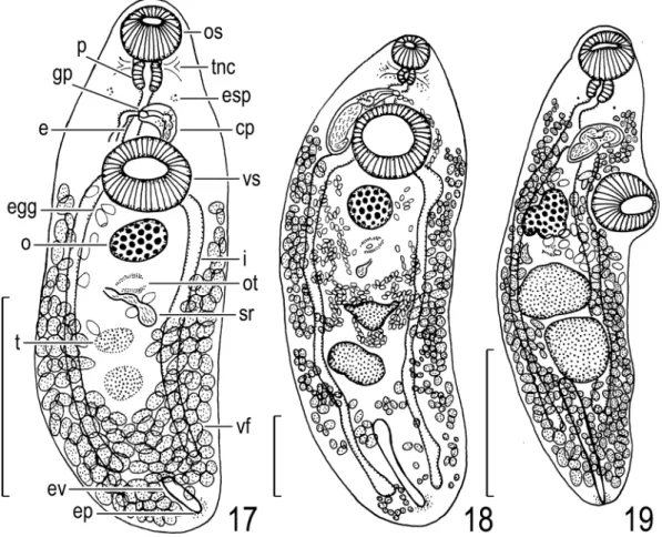
Allocreadium sp. of Shimazu (2005)
(Fig. 17)
Allocreadium sp.: Shimazu, 2005: 140, fig. 4.
Host in Japan. Tribolodon hakonensis (Gün- ther, 1877) (Cyprinidae) (Shimazu, 2005).
Site of infection. Intestine.
Geographical distribution. Aomori Prefecture:
Lake Ogawara at Kamikita-kita, Tohoku Town (Shimazu, 2005; this paper).
Material examined. 1 specimen (NSMT-Pl 5246) of Allocreadium sp., adult, whole- mounted, ex intestine of Tribolodon hakonensis, Lake Ogawara at Kamikita Town, now Kamikita- kita, Tohoku Town, 4 September 1997 (Shimazu, 2005).
Description. After Shimazu (2005), modified from the present study (Fig. 17). Body 2.69 by 0.88; forebody 0.83 long, occupying 31% of body length. Eyespot pigment scattered. Oral sucker 0.28 by 0.31. Pharynx 0.15 in diameter. Esopha- gus bifurcating dorsally to ventral sucker. Intes- tines ending some distance from posterior extremity of body. Ventral sucker 0.39 by 0.44;
sucker width ratio 1 : 1.4. Testes slightly degener- ated, small, 0.15–0.29 by 0.15–0.20. Cirrus pouch clavate, 0.21 by 0.10, anterior to ventral sucker.
Seminal vesicle bipartite, in posterior half of cir- rus pouch; posterior portion larger than anterior one. Ejaculatory duct short. Genital atrium small.
Genital pore located at midlevel of esophagus.
Ovary fairly small, 0.21 by 0.29. Seminal recepta- cle retort-shaped, 0.43 by 0.15. Laurerʼs canal short. Uterus between anterior testis and ventral sucker. Eggs 77–87 by 50–60 μm. Vitelline folli- cles distributed from bifurcal level to posterior extremity of body. Excretory vesicle extending to midlevel of post-testicular region beyond intesti- nal ends; excretory pore posterodorsal.
Remarks. This specimen is similar to A. tri- bolodontis (this paper) in having a bipartite semi- nal vesicle and short excretory vesicle and in the same host fish species (Shimazu, 2005). Addi- tional better-prepared specimens are necessary for definitive species identification.
Life cycle. Not known.
Allocreadium sp. of Shimazu (2008)
(Fig. 18)
Allocreadium sp.: Shimazu, 2008: 48, figs. 3–5.
Host in Japan. Nipponocypris temminckii (Temminck and Schlegel, 1846) (Cyprinidae) (Shimazu, 2008; this paper).
Site of infection. Intestine.
Geographical distribution. Tokushima Prefecture:
Kaifu River at Yoshino, Kaiyo Town (Shimazu, 2008).
Material examined. 1 specimen (NSMT-Pl 5525) of Allocreadium sp., adult, whole- mounted, ex intestine of Nipponocypris tem- minckii (syn. Zacco temminckii (Temminck and Schlegel, 1846)), Kaifu River, 11 September 1998 (Shimazu, 2008).
Description. After Shimazu (2008), modified from the present study (Fig. 18). Body elongate, rapidly tapering anterirly, fairly small, 6.40 by 2.19; forebody 1.28 long, occupying 20% of body length. Eyespot pigment present. Oral sucker
small, 0.40 by 0.41. Pharynx 0.20 in diameter.
Esophagus bifurcating dorsally to ventral sucker.
Intestines ending at near posterior extremity of body. Ventral sucker large, 0.94 by 0.98; sucker width ratio 1 : 2.4. Testes apparently atrophied, small, 0.38–0.69 by 0.44–0.55. Cirrus pouch claviform, long, 1.16 by 0.31, extending posteri- orly slightly beyond midlevel of ventral sucker.
Seminal vesicle sinuous, large, in posterior two- thirds of cirrus pouch, constricted to form small anterior and large posterior portions. Pars prostat- ica globular, 0.05 by 0.03 [not oblong, 0.02 by 0.01]. Ejaculatory duct long, undulating, slightly everted. Genital atrium small. Genital pore at
Fig. 17. Allocreadium sp. of Shimazu (2005), adult (NSMT-Pl 5246) found in intestine of Tribolodon hakonen- sis, entire body, ventral view. Scale bar: 1 mm.
Fig. 18. Allocreadium sp. of Shimazu (2008), adult (NSMT-Pl 5525) found in intestine of Nipponocypris tem- minckii, entire body, ventral view. Scale bar: 1 mm.
Fig. 19. Allocreadium sp. of Shimazu et al. (2011), adult (LBM 8-26) found in intestine of Tanakia lanceolata, entire body, ventral view. Redrawn from Shimazu et al. (2011). Scale bar: 1 mm.
about midlevel of esophagus. Ovary fairly small, 0.57 in diameter. Seminal receptacle retort- shaped, 0.16 [not 0.63] by 0.14. Laurerʼs canal short. Ootype postovarian. Uterus between poste- rior tests and ventral sucker; metraterm short.
Eggs 80–86 by 51–56 μm. Vitelline follicles dis- tributed from bifurcal level to posterior extremity of body. Excretory vesicle extending to about midlevel of post-testicular region; excretory pore posterodorsal.
Remarks. This specimen is somewhat similar to A. tamoroko (see above) in having a high sucker width ratio, long esophagus, long cirrus pouch, and short excretory vesicle, and in the anterior extent of the vitelline follicles. However, it is dif- ferent from the latter in that the sucker width ratio is higher, 1 : 2.4 instead of 1 : 1.8; the metraterm is small and internally smooth instead of large and internally warty; and eggs are larger, 80–86 by 51–56 μm instead of 71–83 by 44–59 μm.
Life cycle. Not known.
Allocreadium sp. of Shimazu, Urabe, and Grygier (2011)
(Fig. 19)
Allocreadium sp.: Shimazu, Urabe, and Grygier, 2011: 36, figs. 44–46.
Host in Japan. Tanakia lanceolata (Temminck and Schlegel, 1846) (Cyprinidae) (Shimazu et al., 2011).
Site of infection. Intestine.
Geographical distribution. Shiga Prefecture:
Irrigation canal closely connected to Yogo River at Nishiyama, Kinomoto Town (Shimazu et al., 2011).
Material examined. 2 specimens (LBM 8-26) of Allocreadium sp., adult, whole-mounted, ex intestine of Tanakia lanceolata, Nishiyama, 26 April 2001 (Shimazu et al., 2011).
Description. After Shimazu et al. (2011), mod- ified from the present study (Fig. 19). Body 3.20–3.56 by 0.88–0.94; forebody 1.17–1.20 long, occupying 33–36% of body length. Eyespot pigment solid. Position of transverse nerve com-
missure not confirmed. Oral sucker 0.32–0.34 by 0.36. Pharynx 0.15–0.17 by 0.19–0.20. Esopha- gus bifurcating anteriorly to ventral sucker. Intes- tines ending at near midlevel of post-testicular region. Ventral sucker postbifurcal, 0.41 by 0.43;
sucker width ratio 1 : 1.2. Testes elliptical, large, 0.37–0.61 by 0.41–0.50. Cirrus pouch claviform, 0.42–0.43 by 0.15–0.17, anterior to ventral sucker. Seminal vesicle S-shaped, in posterior half of cirrus pouch. Ejaculatory duct fairly long.
Genital atrium small. Genital pore shifted to left, about bifurcal. Ovary weakly 3-lobed, fairly large, 0.35–0.36 by 0.28–0.30. Seminal recepta- cle small, 0.14–0.50 by 0.05. Laurerʼs canal short. Uterus between posterior margin of ante- rior testis and cirrus pouch; metraterm as long as cirrus pouch. Eggs 80–89 by 57–62 μm. Vitelline follicles distributed from near posterior margin of pharynx to posterior extremity of body. Excre- tory vesicle reaching to posterior testis; excretory pore posteroterminal.
Remarks. These specimens are similar to A.
shinanoense (see above) and A. japonicum as described by Ozaki (1926) especially in having a long excretory vesicle reaching to the posterior testis. However, they are different from A. shina- noense in having a smaller body, smaller oral and ventral suckers, a lower sucker width ratio, larger testes, a weakly 3-lobed ovary, the uterus extend- ing to the posterior margin of the anterior testis, and smaller eggs; and from A. japonicum in hav- ing a more posterior ventral sucker, elliptical tes- tes, and a more posterior genital pore (see also Shimazu et al., 2011).
Life cycle. Not known.
Allocreadium sp. of Kataoka and Momma (1934), incertae sedis
Allocreadium sp.: Kataoka and Momma, 1934: 59–60, no figure; Shimazu, 1988: 18–19; Shimazu, Urabe, and Grygier, 2011: 89.
Allocreadium sp. 1: Shimazu, 1999: 71; Shimazu, 2003b:
70.

![Fig. 11. Allocreadium japonicum, adult (MPM Coll. No. 30007) found in intestine of Haya (Tribolodon hakonensis [?]), entire body, ventral view](https://thumb-ap.123doks.com/thumbv2/123deta/7183863.2372406/11.773.95.685.108.638/allocreadium-japonicum-adult-intestine-tribolodon-hakonensis-entire-ventral.webp)