Title: Utility of liquid biopsy using urine in patients with pancreatic ductal adenocarcinoma
Hiroyuki Terasawa, Hideaki Kinugasa, Soichiro Ako, Mami Hirai, Hiroshi Matsushita, Daisuke Uchida, Takeshi Tomoda, Kazuyuki Matsumoto, Shigeru Horiguchi, Hironari Kato, Kazuhiro Nouso, Hiroyuki Okada
Department of Gastroenterology and Hepatology, Okayama University Graduate School of Medicine, Dentistry and Pharmaceutical Sciences
Corresponding Author: Hideaki Kinugasa; (Postal address) 2-5-1 Shikata-Cho Kita-Ku, Okayama, Japan, 7008558; (E-mail address) [email protected]; (Phone)
E-mail: Hiroyuki Terasawa: [email protected], Hideaki Kinugasa:
[email protected], Soichiro Ako: [email protected], Mami Hirai:
[email protected], Hiroshi Matsushita: [email protected], Daisuke Uchida:
[email protected], Takeshi Tomoda: [email protected], Kazuyuki Matsumoto: [email protected], Shigeru Horiguchi:
[email protected], Hironari Kato: [email protected], Kazuhiro Nouso:
[email protected], Hiroyuki Okada: [email protected]
Authors’ contributions: Dr. HK designed and Dr. HT drafted the manuscript. Dr. SA, Dr.
MH, Dr. DU, Dr. TT, Dr. KM, Dr. SH and Dr.HK was responsible for diagnosis. Dr. HK, Dr. KN and Dr. HO supervised the manuscript preparation. All authors approved the final manuscript.
Disclosure of Potential Conflicts of Interest: None.
Grant support: None.
Abstract
In recent years, liquid biopsy for blood and body fluid in cancer patients has attracted attention. However, there have been few reports of liquid biopsy focusing on urine of pancreatic ductal adenocarcinoma (PDAC).
In 56 patients with PDAC, DNA was extracted from urine and plasma prior to treatment, and KRAS mutations were analyzed with droplet digital PCR to
examine the mutation detection rate. Our study showed that KRAS mutations were found in 27 cases (48%) in urine and 27 cases (48%) in plasma. The detection rate of urine KRAS mutations varied by renal functions. The rates were 70% (14/20) and 36% (13/36) in the creatinine clearance rate (CCr) < 70 mL/min group and in the CCr ≥ 70 mL/min group, respectively (P=0.024). Whereas, no influence of the CCr was observed in the detection rates of plasma KRAS mutations. The rates were 50% (10/20) and 47% (17/36) in cases with the CCr < 70 mL/min group and the CCr ≥ 70 mL/min group, respectively.
Although the sample size was small, this study clearly indicated a new possibility of less invasive urine liquid biopsy in PDAC patient.
Introduction
Liquid biopsy, for detecting molecules such as circulating tumor cells (CTC), circulating cell-free DNA (cfDNA), circulating tumor DNA (ctDNA), and exosomes in peripheral blood and other body fluids from cancer patients, has been developed recently
123. Detecting these molecules enables not only early diagnosis and improved prognosis
but also monitoring and prediction of treatment response 4. A blood sample is widely used for liquid biopsy, whereas other body fluid samples such as saliva, cerebrospinal fluid (CSF), bile, urine, and stools have been reported for liquid biopsy 4. Of these samples, urine may have a remarkable advantage as an ultra-non-invasive, simple sample source over tissue and blood.
Urinary cell-free DNA (ucfDNA) originates either from the shedding of cells into urine from the genitourinary tract or from cfDNA and ctDNA in circulation passing through glomerular filtration, known as transrenal DNA. Next generation sequencing (NGS) and digital PCR could make it possible to detect rare mutations and methylation for ucfDNA. UcfDNA has been applied for not only urological cancer but also
non-urological cancers such as colon cancer, lung cancer, hepatocellular cancer, and
gastric cancer. However, to our knowledge, ucfDNA for pancreatic ductal adenocarcinoma (PDAC) with NGS or digital PCR has not been reported.
The KRAS oncogene is a driver mutation that is presented in greater than 90%
of PDAC. The concordance rate between tissue DNA and blood ctDNA with a KRAS mutation was reported to be 60–80% 56. Detection of a KRAS mutation in blood could be used to improve prognosis and monitor treatment and disease progression. If we could gather the same information from a urinary liquid biopsy, we could have an ideal, non-invasive option for cancer treatment.
In this study, we analyzed KRAS mutations in urine and investigated the potential of urine liquid biopsy in patients with PDAC.
Materials and Methods Patients
We enrolled 56 patients with PDAC who were admitted to Okayama University Hospital between May 2015 and April 2018. All patients were diagnosed histopathologically. We investigated the diurnal variation of cfDNA concentration in
urine for 9 patients as a preliminary step. Then, we investigated the detection rate of KRAS mutations (G12D, G12V) in urine and plasma for 56 patients. In 40 of the 56 patients, we also examined the KRAS mutations in tissue using a needle washing solution after fine needle aspiration (FNA). All patients provided written informed consent prior to enrolment. This study was approved by the ethics committee of Okayama University Hospital, conducted in accordance with the Declaration of Helsinki.
DNA extraction
All samples were collected prior to initial treatment. DNA was extracted from each source according to the manufacturer’s instructions. Urine samples were collected and stored in tubes with ethylenediaminetetraacetic acid (EDTA) at -80°C for later DNA extraction. After removal of the sediment by centrifugation (16000×g, 10 min, room temperature), ucfDNA was extracted from 3 mL of urine with a QIAamp Circulating Nucleic Acid Kit (Qiagen, Hilden, Germany). Plasma samples were separated by
centrifugation (3000 rpm, 10 min, 4°C) within 3 h of blood collection. The samples were then stored at -30°C for later DNA extraction. CfDNA was extracted from 1 mL of
plasma with the same kit. Needle washing solutions after FNA for diagnosis were stored at -30°C for later DNA extraction. CfDNA was extracted from 200 μL of the solutions by a QIAamp DNA Mini Kit (Qiagen, Hilden, Germany). All DNAs were eluted in a final volume of 50 μL and stored at -30°C. DNAs extracted from plasma and urine were quantified using a Qubit fluorometer (Thermo Fisher Scientific, Waltham, MA, USA).
Droplet digital PCR
The presence of KRAS mutations was detected via droplet digital PCR (QX200 system; Bio-Rad Laboratories, Hercules, CA, USA) using the two probes (G12D and G12V), as follows: DNA eluent (5 μL) from urine, plasma, and needle washing solution was mixed with Droplet PCR Supermix (10 μL; Bio-Rad Laboratories, Hercules, CA, USA), primer/probe mixture (2 μL), and sterile DNase- and RNase-free water (5 μL). The mixture (22 μL) was added to droplet generation oil (70 μL; Bio-Rad Laboratories, Hercules, CA, USA) to produce a droplet. The emulsion was thermal cycled as follows:
an initial temperature setting of 95°C for 10 min, 40 cycles at 94°C for 30 s, and 55°C for 1 min. A final step of 98°C for 10 min for enzyme deactivation was performed, and the
reaction mixes were analyzed using a droplet reader (Bio-Rad Laboratories, Hercules, CA, USA). For quantification, a fluorescence signal readout was obtained using QuantaSoft software (Bio-Rad Laboratories, Hercules, CA, USA). In this study, we set the threshold of fluorescence intensity as 2000 using a positive control and a negative control, which were cancerous and non-cancerous lesion tissues of surgical resection samples in patients with PDAC. All experiments were conducted in duplicate to diminish the effect of a false negative result, although its frequency was low in these materials. A positive result was accepted if it was positive even once.
Statistical analysis
We used the software JMP v14.0 (SAS Institute, Cary, NC, USA) and set the significance at P<0.05. The Wilcoxon test (non-parametric) was used for the analysis of continuous data, and the Chi-square test was used for the analysis of categorical data.
Survival curves were generated by the Kaplan–Meier method and compared by log rank test.
DNA fragment analysis
We analyzed the DNA fragments of ucfDNA by Qsep100 DNA Fragment Analyzer (BiOptic inc, Taiwan).
Results
Diurnal variation of cell-free DNA concentration in urine
Prior to the study of KRAS mutations in urine, we investigated diurnal
variation of ucfDNA concentration because no data about the variation had been reported.
We collected urine at 7:00 am, 11:00 am, 3:00 pm, and 7:00 pm from the first 9 PDAC patients, and measured the DNA concentration (Figure 1). Median concentrations were 8.5 ng/mL, 5.6 ng/mL, 11.2 ng/mL, and 14.5 ng/mL, respectively, but the variation was not significant (P=0.321). Therefore, we decided not to define the time of collecting urine samples in subsequent analyses.
Patient characteristics
The characteristics of 56 patients at baseline are shown in Table 1. Median patient age was 66.5 years, and 30 patients (54%) were male. Creatinine clearance rate
(CCr) ranged from 23.9 mL/min to 164.5 mL/min, with a median of 74.1 mL/min.
Median tumor size was 35 mm. About half of the cases were stage IV, and none were stage I. Seven patients underwent surgical resection after neo-adjuvant chemotherapy, and 48 patients received chemotherapy (modified FOLFIRINOX for 17 cases,
gemcitabine plus nab-paclitaxel for 28 cases, and other drugs for 3 cases). For the stage IV patients, only one patient had renal metastasis.
Detection of KRAS mutations
In urine, G12D and G12V were detected in 25 of 56 patients (45%) and 4 of 56 patients (7%), respectively. The detection rate of KRAS mutations (G12D and/or G12V) in urine was 48% (27 of 56 patients). In plasma, G12D and G12V were detected in 17 of 56 patients (30%) and 11 of 56 patients (20%), respectively. The detection rate of KRAS mutations (G12D and/or G12V) in plasma was 48% (27 of 56 patients), which was equal to that in urine (Table 2).
The median value of cfDNA concentration was 16.5 ng/mL in plasma and 5.7 ng/mL in urine (Table 2). CfDNA concentration in plasma and urine was significantly different (P<0.001).
Effect of renal function
One patient did not receive treatment. Patient age and existence of diabetes mellitus were statistically different between the CCr < 70 mL/min group and the CCr ≥ 70 mL/min group, but no other factor was statistically different between the two groups (Table 3). For stage IV patients, there was a significant difference only in age (P<0.001, median age for the CCr < 70 mL/min group and the CCr ≥ 70 mL/min group of 75 and 58, respectively), but no significant difference was observed in other factors including tumor marker and tumor size.
DNA concentration in plasma and urine, divided into two groups by CCr, is shown in Table 4. In the CCr < 70 mL/min group, cfDNA concentration in plasma and urine was not significantly different (P=0.098); however, it was significantly different (P<0.001) in the CCr ≥ 70 mL/min group. The mean concentration of urinary cfDNA in
the CCr < 70 mL/min group was higher than that in the CCr ≥ 70 mL/min group (Table 4;
P=0.038). In plasma, there was no influence of CCr for DNA concentration (Table 4;
P=0.993). These results may suggest that cfDNA tends to translocate the kidney barrier with renal function degeneracy.
Interestingly, the detection rate in urine was remarkably different in the
reexamination by renal function, because the detection rate was 70% (14/20) in the CCr <
70 mL/min group, in contrast to 36% (13/36) in the CCr ≥ 70 mL/min group. The detection rate was significantly higher in the group with the lower CCr (P=0.024). In plasma, the detection rate was 50% (10/20) in the CCr < 70 mL/min group and 47%
(17/36) in the CCr ≥ 70 mL/min group, but the influence of CCr for plasma mutation was not clear (P=1.00; Figure 2).
In tissue, G12D and G12V were detected in 28 of 40 patients (70%) and 12 of 40 patients (30%), respectively. The detection rate of KRAS mutations (G12D and/or G12V) in tissue was 83% (33 of 40 patients). The sensitivity of the urine liquid biopsy was 42% (14/33). After separation of the low and high CCr groups, the sensitivity was 80% (8/10 cases) in the CCr < 70 mL/min group and 26% (6/23 cases) in the CCr ≥ 70
mL/min group (Figure 3). The sensitivity was significantly higher in the group with the lower CCr (P=0.007). On the other hand, the sensitivity of plasma liquid biopsy was 70%
(7/10 cases) in the CCr < 70 mL/min group and 48% (11/23 cases) in the CCr ≥ 70 mL/min group (Figure 3), but the influence of CCr for sensitivity was not clear (P=0.282).
Liquid biopsy on urine showed the influence of renal function on detection rate and sensitivity, unlike plasma.
DNA fragment size
The size of ucfDNA was a bimodal distribution of about 200 bp and 1000bp (Figure 4).
Discussion
The detection of KRAS mutations in blood from PDAC patients is possible, and we have reported its utility for diagnosis, treatment, and prognosis 57. However, there have been no reports for liquid biopsy using urine in PDAC patients. In this study, we showed that detection of KRAS mutations in urine from PDAC patients was possible, and the rate was comparable to that with plasma. The detection rate of urine KRAS mutations
was high especially in patients with poor renal function and was higher than that of plasma in these patients.
KRAS mutations present in urine from cancer patients were reported to be detectable, and the usefulness of analyzing mutations was reported. In PDAC patients, KRAS mutations have been detected previously by PCR 8. However, the utility of analyzing these mutations is unknown. Transrenal DNA has been identified, but the mechanism of transfer is unclear. The non-globular shape or the deformability of the DNA complexes may enable them to pass through the glomerular filtration. Another explanation is that cfDNA may be included in liposomes; therefore, the cfDNA could pass through the glomerular filtration 910.
In this study, we found that ucfDNA concentration varied by renal function, and this fact suggested that the renal barrier becomes more permeable to cfDNA due to renal function degeneracy. An increase of urine protein with renal function degeneracy is well known. It is considered that the size barrier of glomerular filtration collapses with renal function degeneracy because podocytes, which are responsible for the size barrier, tend to disappear 11. With renal function degeneracy, the charge barrier function may
decrease too. Therefore, albumin and other proteins pass the barrier. Considering this mechanism, dysfunction of the barrier may affect the diffusibility of cfDNA. The size of cfDNA in blood is reported to be about 180 to 200 bp 12. UcfDNA can be classified generally by its size into high-molecular-weight and low-molecular-weight.
Low-molecular-weight DNA is about 250 bp 13 or shorter 14, and these DNAs originate either from circulation or apoptotic cells in contact with urine 131516. We analyzed the DNA fragments of ucfDNA and the result was about 200 bp. We speculate that
high-molecular-weight DNA cannot pass easily through the glomerular filtration barrier, but low-molecular-weight DNA can. Targeting of ucfDNA from circulation passing through the kidney barrier might further increase the detection rate and sensitivity of the urine liquid biopsy.
On the other hand, we tried to address stage specific confounders leading to changes in renal function. However, no significant difference in characteristics but age was observed between the CCr < 70 mL/min group and the CCr ≥ 70 mL/min group.
In non-urological cancers with urine liquid biopsy, mutation rate rather than methylation and integrity was mainly studied. EGFR and KRAS mutations in NSCLC 17
181920, KRAS and TP53 mutations in colorectal cancer 212213, EGFR mutations in gastric
cancer 23, and in other cancers have been reported, but not for PDAC. Chen et al. reported that the concordance rate of EGFR mutations in NSCLC patients between tissue and ucfDNA was 88% 17. Fajiu et al. reported that the concordance rate of EGFR mutations in NSCLC patients between tissue and ucfDNA was 42% in the early stage, and 93% in the advanced stage 18. In this study, the concordance rate of KRAS mutations between tissue and ucfDNA was 47.5% (19/40). In 56 patients, seventeen patients with positive KRAS mutation in the plasma also had the same KRAS mutation in the urine. On the other hand, ten patients with KRAS mutation in urine had no KRAS mutation in plasma. The false positive and false negative rate of using this urine testing method were 37% (10/27) and 34.4% (10/29), respectively. Concordance rate between urine KRAS mutation and plasma KRAS mutation was 64.2% (36/56). It is difficult to compare these results because the type of cancer was different, and the results might have been affected by sample volume. We used 1 ml of plasma and 3 ml of urine, but a greater volume (10 ml) was used in the previous studies. In terms of urine liquid biopsy, a larger amount of sample is available, and it is less invasive, which is the greatest advantage. In NSCLC
patients, the sensitivity of EGFR mutations analyzed by next-generation sequencing assays was higher in a larger volume of urine sample 19. If we had used a larger volume of urine sample, the results might have been even more significant.
Urine liquid biopsy, a less invasive technique, is also feasible. This study suggested the potential of urine liquid biopsy compared with plasma liquid biopsy, but the clinical effect is still unknown. We conducted a sub-analysis about progression free survival (PFS) and overall survival (OS) for patients who received systemic
chemotherapy. For plasma liquid biopsy, there was no significant difference in PFS between the group with KRAS mutations and that without mutations (P=0.451, median PFS for KRAS-positive and KRAS-negative patients of 138 and 201 days, respectively).
The same result was obtained by urine liquid biopsy (P=0.935, median PFS for
KRAS-positive and KRAS-negative patients of 160 and 154 days, respectively). There was no significant difference in OS between the group with KRAS mutations and that without mutations for plasma liquid biopsy (P=0.261, median survival times for
KRAS-positive and KRAS-negative patients of 406 and 449 days, respectively) or urine liquid biopsy (P=0.882, median survival times for KRAS-positive and KRAS-negative
patients of 470 and 449 days, respectively). Further study will reveal the clinical effect of urine liquid biopsy in the context of renal function.
The limitations of the current study are shown below. This was a single center study. The patient characteristics were also biased, and there was no early disease stage.
Also, the number of patients enrolled in this study was small. Therefore, all stages and a larger number of patients should be enrolled in further studies. Further, we substituted the needle washing solutions after FNA for tissue samples. More than 90% of cases of pancreatic intraepithelial neoplasia of all grades have KRAS mutations 24, and the detection rate of FNA samples was 83% in our study. Finally, we analyzed KRAS mutations of G12D and G12V, but other mutations such as G12R, G13D, and Q61L were not investigated. G12D and G12V are the most common KRAS mutations 25, but other mutations could have been analyzed.
In conclusion, urine liquid biopsy in PDAC patients is a simple and ultra-non-invasive technique. Moreover, a sufficient amount of urine sample can be easily secured, unlike for other samples. The detection rate and the sensitivity are
comparable to plasma liquid biopsy and are higher for PDAC patients with renal function
degeneracy. Further investigation might allow the technique to substitute for liquid biopsy using a blood sample.
Figure legends
Figure 1. Urine at 7:00 am, 11:00 am, 3:00 pm, and 7:00 pm from the first 9 PDAC patients was collected, and the DNA concentration measured. Median concentrations were 8.5 ng/mL, 5.6 ng/mL, 11.2 ng/mL, and 14.5 ng/mL, respectively, but the variation was not significant (P=0.321).
Figure 2. In urine, the detection rate of KRAS mutations was 70% (14/20) with the CCr <
70 mL/min group, in contrast to 36% (13/36) with the CCr ≥ 70 mL/min group. The detection rate was significantly higher in the group with the lower CCr (P=0.024). In plasma, the detection rate was 50% (10/20) in the CCr < 70 mL/min group and 47%
(17/36) in the CCr ≥ 70 mL/min group, but the influence of CCr for plasma mutation was not clear (P=1.00).
Figure 3. The sensitivity of the urine liquid biopsy was 42% (14/33). The sensitivity was 80% (8/10 cases) in the CCr < 70 mL/min group and 26% (6/23 cases) in the CCr ≥ 70 mL/min group. The sensitivity was significantly higher in the group with the lower CCr (P=0.007). In contrast, the sensitivity of plasma liquid biopsy was 70% (7/10 cases) in the
CCr < 70 mL/min group and 48% (11/23 cases) in the 70 mL/min ≤ CCr group, but the influence of CCr for sensitivity was not clear (P=0.282).
Figure 4. Size distribution of ucfDNA samples from patients with PDAC was analyzed.
All samples showed a bimodal distribution of about 200 bp and 1000bp, indicating that ucfDNA originates either from cfDNA and ctDNA in circulation passing through
glomerular filtration or from the shedding of cells into urine from the genitourinary tract.
References
[1] Murtaza M, Dawson SJ, Tsui DW, Gale D, Forshew T, Piskorz AM, Parkinson C, Chin SF, Kingsbury Z, Wong AS, et al. Non-invasive analysis of acquired resistance to cancer therapy by sequencing of plasma DNA. Nature. 2013; 497: 108-12.
[2] Dawson SJ, Tsui DW, Murtaza M, Biggs H, Rueda OM, Chin SF, Dunning MJ, Gale D, Forshew T, Mahler-Araujo B, et al. Analysis of Circulating Tumor DNA to Monitor Metastatic Breast Cancer. N Engl J Med. 2013; 368: 1199-209.
[3] Lu T, Li J. Clinical applications of urinary cell-free DNA in cancer: current insights and promising future. Am J Cancer Res. 2017; 7: 2318-32.
[4] Peng M, Chen C, Hulbert A, Brock MV, Yu F. Non-blood circulating tumor DNA detection in cancer. Oncotarget. 2017; 8: 69162-73.
[5] Kinugasa H, Nouso K, Miyahara K, Morimoto Y, Dohi C, Tsutsumi K, Kato H, Matsubara T, Okada H, Yamamoto K. Detection of K-ras gene mutation by liquid biopsy in patients with pancreatic cancer. Cancer. 2015; 121: 2271-80.
[6] Heitzer E, Ulz P, Geigl JB. Circulating Tumor DNA as a Liquid Biopsy for Cancer. Clin Chem. 2014; 61: 112-23.
[7] Ako S, Nouso K, Kinugasa H, Dohi C, Matushita H, Mizukawa S, Muro S, Akimoto Y, Uchida D, Tomoda T, et al. Utility of serum DNA as a marker for KRAS mutations in pancreatic cancer tissue. Pancreatology. 2017; 17: 285-90.
[8] Botezatu I, Serdyuk O, Potapova G, Shelepov V, Alechina R, Molyaka Y, Ananév V, Bazin I, Garin A, Narimanov M, et al. Genetic analysis of DNA excreted in urine: a new approach for detecting specific genomic DNA sequences from cells dying in an organism. Clin Chem. 2000; 46: 1078-84.
[9] Foglieni C, Bragonzi A, Cortese M, Cantù L, Boletta A, Chiossone I, Soria MR, Monaco L. Glomerular filtration is required for transfection of proximal tubular cells in the rat kidney following injection of DNA complexes into the renal artery. Gene Ther.
2000; 7: 279-85.
[10] Lichtenstein AV, Melkonyan HS, Tomei LD, Umansky SR. Circulating nucleic acids and apoptosis. Ann N Y Acad Sci. 2001; 945: 239-49.
[11] Garg P. A Review of Podocyte Biology. Am J Nephrol. 2018; 47 Suppl 1:
3-13.
[12] Diaz LA, Jr., Bardelli A. Liquid biopsies: genotyping circulating tumor DNA. J Clin Oncol. 2014; 32: 579-86.
[13] Su YH, Wang M, Brenner DE, Ng A, Melkonyan H, Umansky S, Syngal S, Block TM. Human urine contains small, 150 to 250 nucleotide-sized, soluble DNA derived from the circulation and may be useful in the detection of colorectal cancer. J Mol Diagn. 2004; 6: 101-7.
[14] Bryzgunova OE, Skvortsova TE, Kolesnikova EV, Starikov AV, Rykova EY, Vlassov VV, Laktionov PP. Isolation and comparative study of cell-free nucleic acids from human urine. Ann N Y Acad Sci. 2006; 1075: 334-40.
[15] Salvi S, Martignano F, Molinari C, Gurioli G, Calistri D, De Giorgi U,
Conteduca V, Casadio V. The potential use of urine cell free DNA as a marker for cancer.
Expert Rev Mol Diagn. 2016; 16: 1283-90.
[16] Bryzgunova OE, Laktionov PP. Extracellular Nucleic Acids in Urine: Sources, Structure, Diagnostic Potential. Acta Naturae. 2015; 7: 48-54.
[17] Chen S, Zhao J, Cui L, Liu Y. Urinary circulating DNA detection for dynamic tracking of EGFR mutations for NSCLC patients treated with EGFR-TKIs. Clin Transl Oncol. 2017; 19: 332-40.
[18] Li F, Huang J, Ji D, Meng Q, Wang C, Chen S, Wang X, Zhu Z, Jiang C, Shi Y, et al. Utility of urinary circulating tumor DNA for EGFR mutation detection in different stages of non-small cell lung cancer patients. Clin Transl Oncol. 2017; 19: 1283-91.
[19] Reckamp KL, Melnikova VO, Karlovich C, Sequist LV, Camidge DR, Wakelee H, Perol M, Oxnard GR, Kosco K, Croucher P, et al. A Highly Sensitive and Quantitative Test Platform for Detection of NSCLC EGFR Mutations in Urine and Plasma. J Thorac Oncol. 2016; 11: 1690-700.
[20] Wang X, Meng Q, Wang C, Li F, Zhu Z, Liu S, Shi Y, Huang J, Chen S, Li C.
Investigation of transrenal KRAS mutation in late stage NSCLC patients correlates to disease progression. Biomarkers. 2017; 22: 654-60.
[21] Su YH, Wang M, Aiamkitsumrit B, Brenner DE, Block TM. Detection of a K-ras mutation in urine of patients with colorectal cancer. Cancer Biomark. 2005; 1:
177-82.
[22] Su YH, Wang M, Brenner DE, Norton PA, Block TM. Detection of mutated K-ras DNA in urine, plasma, and serum of patients with colorectal carcinoma or adenomatous polyps. Ann N Y Acad Sci. 2008; 1137: 197-206.
[23] Shi XQ, Xue WH, Zhao SF, Zhang XJ, Sun W. Dynamic tracing for epidermal growth factor receptor mutations in urinary circulating DNA in gastric cancer patients.
Tumour Biol. 2017; 39: 1010428317691681.
[24] Kanda M, Matthaei H, Wu J, Hong SM, Yu J, Borges M, Hruban RH, Maitra A, Kinzler K, Vogelstein B, et al. Presence of somatic mutations in most early-stage
pancreatic intraepithelial neoplasia. Gastroenterology. 2012; 142: 730-3 e9.
[25] Schultheis B, Reuter D, Ebert MP, Siveke J, Kerkhoff A, Berdel WE, Hofheinz R, Behringer DM, Schmidt WE, Goker E, et al. Gemcitabine combined with the
monoclonal antibody nimotuzumab is an active first-line regimen in KRAS wildtype patients with locally advanced or metastatic pancreatic cancer: a multicenter, randomized phase IIb study. Ann Oncol. 2017; 28: 2429-35.
Table 1. Patient Characteristics
N = 56
Age, years (range) 66.5 (38–91)
Sex, male (%) 30 (54%)
Diabetes mellitus, no. (%) 19 (34%)
Creatinine, mg/dL (range) 0.6 (0.4–1.4)
CEA, ng/mL (range) 3.9 (0.7–274)
CA19-9, IU/mL (range) 233.9 (0.9–129164)
SPAN1, U/mL (range) 96.8 (10–28000)
DUPAN2, U/mL (range) 348 (25–16000)
Tumor location (head of pancreas), no. (%) 25 (45%)
Tumor size, mm (range) 35 (18–80)
Stage of disease (UICC), no. (%) I 0 (0%)
II 12 (21.5%)
III 18 (32%)
IV 26 (46.5%)
Initial therapy, no. (%) Chemotherapy 48 (86%)
NAC + surgical resection 7 (12%)
BSC 1 (2%)
CEA: carcinoembryonic antigen; CA19-9: carbohydrate antigen 19-9; NAC: neoadjuvant chemotherapy
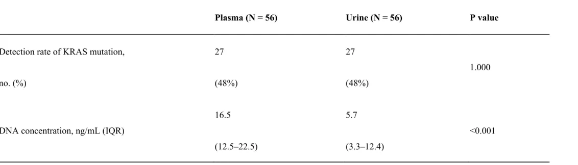
Table 2. Comparison between plasma and urine samples in patients with PADC
Plasma (N = 56) Urine (N = 56) P value
Detection rate of KRAS mutation,
no. (%)
27
(48%)
27
(48%)
1.000
DNA concentration, ng/mL (IQR)
16.5
(12.5–22.5)
5.7
(3.3–12.4)
<0.001
Table 3. Patient Characteristics by CCr
CCr < 70 (N = 20) CCr ≥ 70 (N = 36) P value
Age, years (range) 75 (63–91) 61.5 (38–71) <0.001
Sex, male (%) 10 (50%) 20 (56%) 0.782
Diabetes mellitus, no. (%) 3 (15%) 16 (44%) 0.038
CEA, ng/mL (range) 5.0 (1.0–115.8) 3.9 (0.7–274) 0.521
CA19-9, IU/mL (range) 280.5 (0.9–129164) 194.4 (1.7–50600) 0.918
SPAN1, U/mL (range) 84.4 (10–28000) 110.7 (10–11000) 0.681
DUPAN2, U/mL (range) 348 (25–16000) 294 (25–16000) 0.632
Tumor location (head of pancreas), no. (%) 8 (40%) 17 (47%) 0.779
Tumor size, mm (range) 35.5 (20–60) 35 (18–80) 0.803
Stage of disease (UICC), no. (%) I 0 (0%) 0 (0%) 0.286
II 3 (15%) 9 (25%)
III 6 (30%) 12 (33%)
IV 11 (55%) 15 (42%)
Initial therapy, no. (%) Chemotherapy 16 (80%) 32 (89%) 0.912
NAC + surgical resection 3 (15%) 4 (11%)
BSC 1 (5%) 0 (0%)
Table 4. Comparison between CCr < 70 and CCr ≥ 70
Plasma (N = 56) Urine (N = 56)
CCr < 70 (N = 20)
CCr ≥ 70 (N = 36)
P value
CCr < 70 (N = 20)
CCR ≥ 70 (N = 36)
P value
Detection rate of KRAS mutation,
no. (%)
10
(50%)
17
(47%)
1.000
14
(70%)
13
(36%)
0.024
DNA concentration, ng/mL (IQR) 16.9 (12.2–24.4) 16.2 (13.0–21.7) 0.993 8.0 (3.5–40.3) 5.5 (2.9–9.1) 0.038
Figure 1
Figure 2
Figure 3
Figure 4