Study of novel function of vitamin K2 in mice
with humanized pregnane X receptor
著者
Sultana Halima
学位授与機関
Tohoku University
学位授与番号
11301甲第18369号
Study of novel function of vitamin K
2in mice with
humanized pregnane X receptor
ヒト pregnane X 受容体導入マウスを用いたビタミン K
2の新規作用に関する研究
Ph.D. Dissertation
Study of novel function of vitamin K
2in mice with humanized
pregnane X receptor
ヒト pregnane X 受容体導入マウスを用いたビタミン K
2の新規作用に関する研究
Laboratory of Nutrition
Department of Science of Food Function and Health
Division of Bioscience and Biotechnology for Future Bioindustries
Tohoku University
Supervisor
Associate Professor
Acknowledgement
At first I would like to express my especial indebted to Associate Professor Hitoshi Shirakawa, laboratory of Nutrition, Department of Science of Food Function and Health, Division of Bioscience and Biotechnology for Future Bioindustries, Tohoku University under whose supervision the dissertation was materialized. I am very grateful to him for his expert supervision, encouragement and research facilities without which it would not be possible to conduct this study. I could not have imagined having a better supervisor for my Ph.D. study.
I would like to gratefully acknowledge Professor Michio Komai for his valuable suggestions throughout the study. My sincere thanks also go to Isono Kunio sensei and all the members of laboratory of Nutrition for their candid help and support during my study. I would like to express my sincere gratitude to my former mentor Professor Yearul Kabir, Department of Biochemistry and Molecular Biology, University of Dhaka for his precious guidance and motivation. In addition, I would like to express my warmest thanks to the Ministry of Education, Science, and Culture for the scholarship during my study in Japan.
Last but not the least; I would like to express my highest regards to all the members of my family for supporting me spiritually throughout the study. I am grateful for their continuous, encouragement and unlimited love which is my actual strength.
LIST OF CONTENTS
CHAPTER 1: Introduction 1
PXR 1
Vitamin K 2
Humanized PXR 3
PXR and drug metabolism 3
PXR and bile acids synthesis 4
PXR and energy homeostasis 5
Purpose of the study 6
CHAPTER 2: Validation of the diet and animal 10
Section 1: Effects of F2 diet and CRF-1 diet on basal mRNA levels of genes encoding
drug metabolizing proteins
10
Section 2: Effects of PCN and rifampicin on mRNA levels of genes encoding drug
metabolizing proteins in WT and hPXR mice
16
CHAPTER 3: Effects of single dose of MK-4 treatment on the expression of genes involved in drug metabolism, bile acids synthesis and energy homeostasis in mice
22
Section 1: MK-4 content in the liver at different time interval after treatment in mice 21
Section 2: MK-4 content in the liver after treatment of mice with different dose of MK-4 25
Section 3: Investigation of effects of MK-4 treatment on typical PXR target genes
involved in drug metabolism by quantitative RT-PCR
27
Section 4: Investigation of effects of MK-4 treatment in mice by DNA microarray
analysis and evaluation of the obtained data by quantitative RT-PCR
30
Section 5: Effects of lower dose of MK-4 treatment on genes encoding proteins involved
in bile acids synthesis and energy homeostasis in mice
38
Section 6: Effects of different doses of MK-4 on Cyp7a1 protein levels in mice 41
CHAPTER 4: Effects of chronic pharmacological dose of MK-4 treatment on the expression of genes involved in drug metabolism, bile acids synthesis and energy homeostasis in hPXR mice
44
CHAPTER 5: Effects of MK-4 treatment on the expression of genes involved in bile acids synthesis and drug metabolism in human hepatocellular carcinoma cells
49
CHAPTER 6: General discussion and conclusions 52
1
Chapter 1: Introduction
PXR
The pregnane X receptor (PXR, also known as SXR or NR1I2) was identified by three different research groups in 1998 as a member of the nuclear receptor superfamily of ligand activated transcription factors. PXR is mainly expressed in the liver and intestine ‒ the major organs where detoxification occurs [1–3]. This receptor is generally known to be activated by xenobiotics and pharmacological compounds; it plays an important role in the activation of many drug metabolizing enzymes and membrane-bound drug transporters. Among these enzymes, the most important are those of the 3A (CYP3A) subfamily of the cytochrome P450 superfamily because they are involved in the metabolism of approximately 50% of prescribed drugs [4, 5].
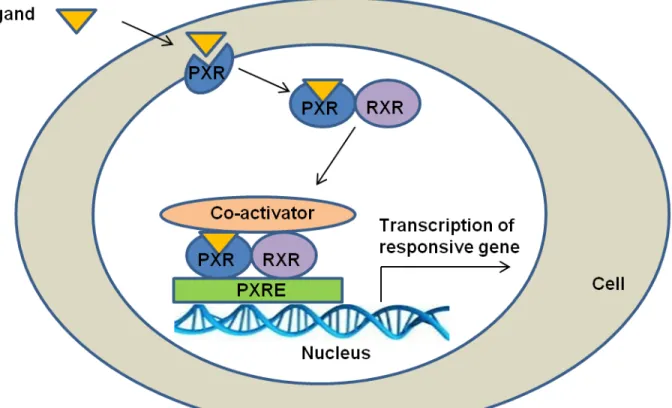
Nuclear receptors have common structural features: an N-terminal domain, a DNA-binding domain (DBD), and a ligand DNA-binding domain (LBD). PXR has attracted a particular attention because of its enlarged, flexible, hydrophobic LBD that allows PXR to be activated by a large variety of substances. Because of its structural features, PXR LBD can change its shape, thereby fitting various ligands, depending on their nature [6]. PXR can be activated by xenobiotic compounds, pharmacological agents, dietary substances, and endobiotics like lithocholic acid [6–10]. Moreover, recent studies have revealed the involvement of PXR in many physiological functions other than drug metabolism, such as inflammation, bile acids synthesis, bone homeostasis, vitamin D metabolism, lipid homeostasis, energy homeostasis, and cancer development [9]. PXR activation is ligand dependent; following activation by ligand binding, PXR forms a heterodimer with the retinoid X receptor. This heterodimer complex binds to PXR
response element which is located in the 5′-flanking regions of PXR target genes, resulting in
2
Vitamin K
Vitamin K (VK) is a well-known nutrient that acts as a co-factor of γ-glutamyl
carboxylase and plays an important role in blood coagulation and bone formation. VK is also involved in many physiological and pathological conditions, such as inflammation, testosterone production, cancer progression, and type 2 diabetes [11–15]. Inadequate intake of VK may cause osteoporosis, oseteoarthritis, Alzheimer’s disease, and coronary artery disease [16–18]. Vitamin
K naturally occurs in 2 forms- phylloquinone (vitamin K1), which is synthesized in plants, and
menaquinone (vitamin K2, or MK-n), which is produced mainly by microorganisms, including
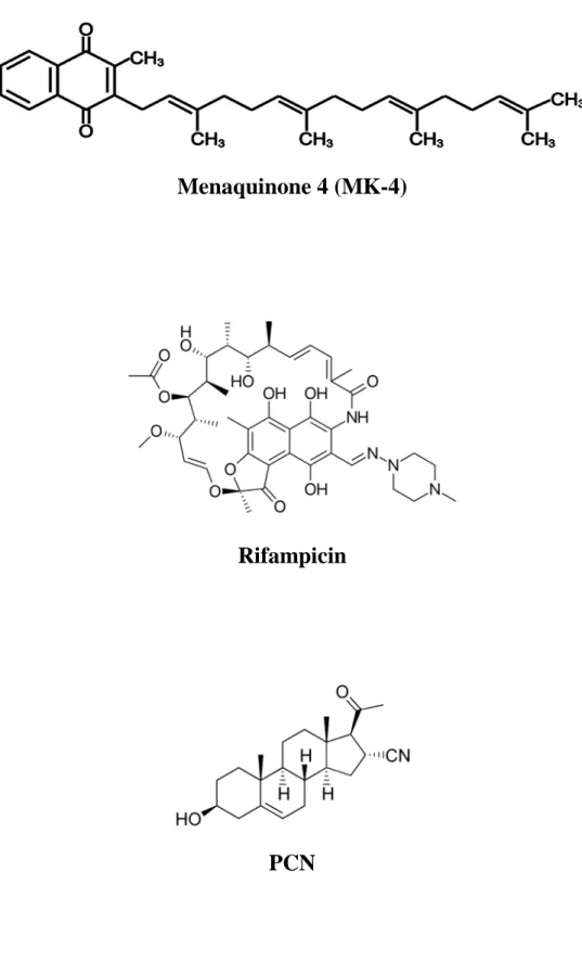
intestinal bacteria (Figure 2). Menaquinone-4 (MK-4) is a vitamin K2 analog, which has been
used as a therapeutic agent in osteoporosis in many East and South East Asia countries [19]. In 2003, another mode of action of MK-4 has been revealed by Tabb et al. [14]. It was demonstrated that MK-4 directly binds to and transcriptionally activates PXR as well as promotes the association between PXR and coactivators. Activated PXR, in turn, plays an important role in bone homeostasis by modulating gene expression [14]. The function of PXR activation by MK-4 in bone homeostasis was further evaluated by analyzing gene expression changes caused by both rifampicin (Rif) and MK-4 [20]. Later, PXR-mediated effect of VK was also shown in human hepatocellular carcinoma [21]. However, the research related to the PXR-mediated effects of MK-4 has concentrated mostly on bone physiology and cell-based experiments. Therefore, potential effects of MK-4 mediated by the activation of PXR should be investigated in other organs. This may shed light on many mechanisms related to diseases in which VK can be used as a preventive or therapeutic agent.
3
Humanized PXR
Species-specific ligand preference by PXR constitutes a significant challenge for the studies of PXR function. For example, pregnane 16α-carbonitrile (PCN) is a much stronger activator of rat or mouse PXR than of human or rabbit PXR. On the other hand, Rif is a strong activator of human or rabbit PXR but a very weak activator of mouse or rat PXR [22]. These species-specific preferences show that evaluation of toxicity and functionality of PXR ligands in rodents has an inherent limitation as it may not be readily translated into human physiology. To overcome this issue, several groups of researchers have developed a mouse model with humanized PXR (hPXR) by using different strategies [4, 23–25].
PXR and drug metabolism
Drugs and xenobiotic substances are mostly lipophilic in nature, so can easily be absorbed. After absorption from the gastrointestinal lumen, drugs and other xenobiotics are transported via the portal vein into the liver by uptake-transporters such as members of the solute carrier family, where drug metabolism mainly takes place. Within hepatocytes, phase I enzymes, for example, cytochrome P450 monooxygenases, and alcohol and aldehyde dehydrogenases, facilitate the oxidation of hydrophobic drugs to increase their water solubility. Phase II conjugating enzymes like UDP-glucuronosyltransferases, glutathione S-transferases and sulfotransferases further increase hydrophilicity by adding polar molecules. The products of phase I and II reactions are finally exported via efflux transporters (phase III), for example, members of the ATP-binding cassette family into blood or bile, and undergo renal or biliary excretion [26, 27]. PXR, being a master regulator, protects body by facilitating the clearance of toxic substances via regulating many of the above mentioned enzymes and transporters.
4
PXR and bile acids synthesis
Bile acids are physiological detergents required for the intestinal digestion and absorption of lipids, including fat-soluble vitamins [28]. These amphipathic sterols are synthesized from cholesterol in the liver and secreted into the intestine where they function to emulsify dietary lipids. There are two pathways for the synthesis of bile acids: classic or neutral pathway and alternative or acidic pathway. The classic pathway is the major pathway, which is initiated by CYP7A1 enzyme. Cholesterol is converted into two primary bile acids in human liver — chenodeoxycholic acid (CDCA) and cholic acid (CA). The distribution of these two bile acids is determined by the activity of CYP8B1 enzyme. Most of bile acids are conjugated to glycine or taurine to decrease their toxicity and increase solubility for the secretion into bile. In the intestine, glyco- and tauro-conjugated CA and CDCA are deconjugated and eventually transformed into secondary bile acids, deoxycholic acid (DCA) and lithocholic acid (LCA), respectively, by intestinal flora. CA, CDCA, DCA and a small amount of LCA are transported back to the liver to inhibit bile acid synthesis in the entero-hepatic circulation system [28].
Kliewer et al. has shown that both mouse PXR and human PXR can be activated by LCA and its metabolite 3-keto-LCA [10]. They have also shown that PCN-mediated PXR activation regulates the expression of genes involved in the biosynthesis, transport, and metabolism of bile acids, including Cyp7a1 in mice [10]. It has been reported that PXR with bound Rif interacted
with hepatic nuclear factor 4α (HNF4α) and enhanced the dissociation of peroxisome
proliferator-activated receptor γ coactivator α (PGC1α) from HNF4α to inhibit the transcription of CYP7A1 in human primary hepatocytes [29]. Another report suggested that Rif treatment did
5
rather seemed to promote the dissociation of PGC1α from HNF4α in HepG2 cells, because
ligand-activated PXR interacts with PGC1α [30].
PXR and energy homeostasis
PXR plays an important role in energy metabolism by modulating the metabolism of fatty acids, lipids and glucose through direct transcriptional regulation or through crosstalk with other transcriptional regulators. Being a complex regulator, PXR promotes lipogenesis whereas suppress gluconeogenesis and β-oxidation of fatty acid [31, 32]. Different groups of researchers have provided strong evidences that establish PXR as an important regulator of glucose and lipid metabolism thus affecting the overall energy homeostasis of the body [33]. However, there are several contradictory results regarding the significance of PXR in energy homeostasis since varying results have been observed in mice models. PXR induces lipogenesis in a SREBP-1 (sterol regulatory element binding protein) independent manner unlike other nuclear receptor. Lipid accumulation is associated with increased expression of the fatty acid translocase CD36 and several lipogenic enzymes, such as stearoyl-CoA desaturase-1 and long-chain free fatty acid
elongase [32]. Another study demonstrated that PXR represses β-oxidation by suppressing the
expression of related genes, such as carnitine palmitoyltransferase 1a and mitochondrial 3- hydroxy-3-methyl-glutarayl-CoA synthase 2, through crosstalk with forkhead box protein A2 [34]. Furthermore, this study also has shown that PXR interacts with forkhead box protein O1 which is the main transcription factor of gluconeogenesis and suppresses gluconeogenic genes [34]. However, in human and mouse hepatocytes PXR activation may affects gluconeogenesis differently. In the mouse liver PEPCK and G6Pase are repressed by PXR activation whereas both up- and down-regulation was observed in human primary hepatocytes [35, 36].
6
Purpose of the study
The purpose of the present study was to investigate the novel function of MK-4 in the humanized mouse line (hPXR) in which the LBD region of the human PXR gene was homologously knocked-in to the mouse Pxr gene, replacing the sequence that encoded endogenous LBD (Figure 3).
7
Figure 1: PXR pathway (RXR: Retinoid X receptor PXRE: PXR responsive element)
8
Menaquinone 4 (MK-4)
Rifampicin
PCN
9
Figure 3: Hinge region & LBD of human PXR are knocked-in to mouse PXR, resulting in
10
Chapter 2: Validation of the diet and animal
PXR can be activated by a large variety of substances including prescribed drugs, herbal medicines, dietary supplements, environmental pollutants, and xenobiotics. In this study, WT mice and hPXR mice were used. Therefore, it was necessary to establish a condition where environment has no effect on PXR target gene expression in both types of mice. For this purpose, the effects of F2 and CRF-1- two common commercially available non-purified diets were investigated. Moreover, the response of mouse PXR in WT mice and human PXR in hPXR mice to their specific ligands were investigated by the treatment of pregnane 16α-carbonitrile (PCN) which is a much stronger activator of rat or mouse PXR than of human or rabbit PXR and rifampicin (Rif) which is a strong activator of human or rabbit PXR but a very weak activator of mouse or rat PXR [22]. PXR is generally known to be activated by xenobiotics and pharmacological compounds which plays an important role in the activation of many drug metabolizing enzymes and membrane-bound drug transporters. Cyp3a11, Ces2a and Mdr1 are very common target genes of PXR involved in drug metabolism. Therefore, first the effects of diet and PCN as well as Rif were investigated on the expression of these common genes.
Section 1: Effects of F2 diet and CRF-1 diet on basal mRNA levels of genes encoding drug metabolizing proteins.
First the effects of F2 diet and CRF-1 diet on basal mRNA levels of genes encoding drug metabolizing proteins were investigated. F2 and CRF-1 are commercially available non- purified diets for rodents.
11
Table 1: Composition of F2 and CRF-1 diet.
Content F2 diet (%) CRF-1 diet (%)
Water 8 8.2 Protein 20.8 21.9 Fat 4.8 5.4 Crude fiber 3.2 2.9 Ash 5 6.3 Other 58.2 55.3 Experimental procedure Animal
hPXR mice on the C57BL/6NCrSlc background, originally established by Igarashi et al. [4], were obtained from RIKEN BioResource Center (Tsukuba, Japan). Homozygous hPXR and WT female mice (12–14 weeks of age) were maintained in plastic cages (3–4 mice per cage) with free access to commercial diet (F2, Funabashi Farms Co., Funabashi, Japan or CRF-1, Oriental Yeast Co. Ltd., Tokyo, Japan) and tap water under controlled temperature (23 ± 2 °C), humidity (50 ± 10%) and a 12:12 light:dark cycle (lights on at 8:00).
Treatment
The mice were sacrificed without any treatment and a part of the liver and intestine were preserved immediately in RNAlater (Thermo Fishier Scientific, Waltham, MA, USA). The remaining part was snap-frozen in liquid nitrogen and then preserved at −80 °C for further analysis.
RNA extraction and mRNA quantification
Total RNA was isolated from the liver and HepG2 cells using the Isogen reagent (Nippon Gene, Tokyo, Japan) according to the manufacturer’s instructions. The quantity and quality of RNA were determined spectrophotometrically, by measuring the absorbance at 260 nm in
12
relation to that at 280 nm, and subsequent agarose gel electrophoresis. Four microgram of total RNA was denatured at 65 °C for 5 min with 2.5 μM oligo-dT primer (Hokkaido System Science Co., Sapporo, Japan) and 0.5 mM dNTP (GE Healthcare, Tokyo, Japan) for cDNA synthesis. RNA was incubated in 20 μL of RT buffer [50 mM Tris–HCl (pH 8.3), 75 mM KCl, 3 mM
MgCl2, 5 mM dithiothreitol] containing 50 U SuperScript III reverse transcriptase (Invitrogen,
Carlsbad, CA, USA) and 20 U RNaseOUT RNase inhibitor (Invitrogen) for 60 min at 50 °C. An aliquot of synthesized cDNA was used as template for quantitative RT-PCR (qRT-PCR) using an Applied Biosystems 7300 Real-Time PCR System (Foster City, CA, USA). Target cDNAs were amplified using gene-specific primers (Table 1) and SYBR Premix Ex Taq (TaKaRa Bio, Otsu, Japan) solution. The relative mRNA levels were normalized to the amount of eukaryotic translation elongation factor 1α1 (Eef1a1) mRNA [37].
Statistical analysis
Data are presented as the mean ± SEM (standard error of the mean). Statistical analysis was performed by the Student’s t-test using SigmaPlot version 12.5 (San Jose, CA). P<0.05 was considered statistically significant.
Results and discussion:
To investigate whether diet has any effect on the basal expression of genes encoding drug metabolizing proteins which are commonly regulated by PXR, mice were maintained by two commercially available diets; F2 or CRF-1. Without any treatment or fasting condition, mice were sacrificed.
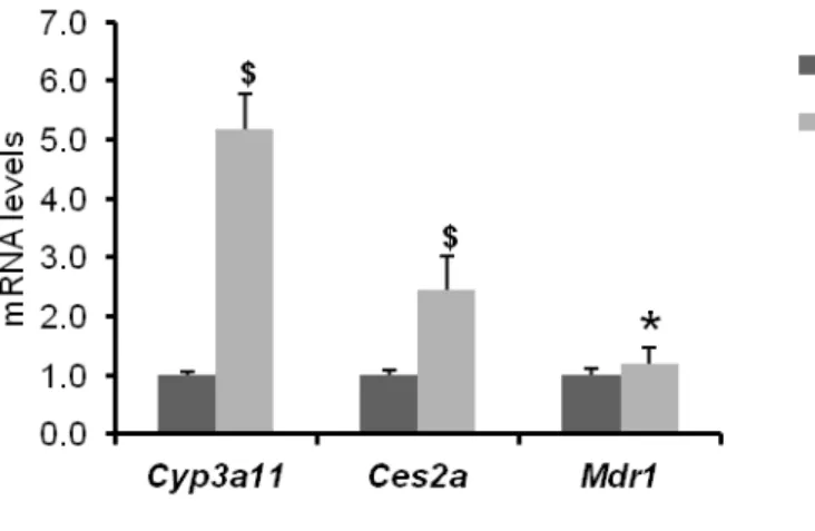
It was found that the basal mRNA levels of Cyp3a11, Ces2a and Mdr1 were significantly high in liver of hPXR mice compared to WT mice when the mice were maintained on F2 diet
13
(Figure 4). In contrast, there was no significant difference in the basal mRNA levels of Cyp3a11, Ces2a and Mdr1 in liver (Figure 5A) or intestine (Figure 5B) of WT and hPXR mice. Therefore, mice were maintained on CRF-1 diet throughout the study.
14
Figure 4: Effect of F2 diet on basal expression of genes encoding drug metabolizing proteins in
liver of WT and hPXR mice. mRNA levels of genes that encode drug metabolizing proteins were measured by quantitative RT-PCR. The relative mRNA levels were normalized to the amount of eukaryotic translation elongation factor 1α1 mRNA. Data are expressed as the mean ± SEM, n = 4. $P < 0.01, *P < 0.05, compared to WT group.
15
(A)
(B)
Figure 5: Effect of CRF-1 diet on basal expression of genes encoding drug metabolizing
proteins in liver (A) and intestine (B) of WT and hPXR mice. mRNA levels of genes that encode drug metabolizing proteins were measured by quantitative RT-PCR. The relative mRNA levels were normalized to the amount of eukaryotic translation elongation factor 1α1 mRNA. Data are expressed as the mean ± SEM, n =3-4.
0.0 0.5 1.0 1.5 2.0 Cyp3a11 Ces2a Mdr1 m R N A lev el s WT hPXR 0.0 0.5 1.0 1.5 2.0 Cyp3a11 Ces2a Mdr1 m R N A l ev el s WT hPXR
16
Section 2: Effects of PCN and rifampicin on mRNA levels of genes encoding drug metabolizing proteins in WT and hPXR mice.
Pregnane 16α-carbonitrile (PCN) is a much stronger activator of rat or mouse PXR than of human or rabbit PXR and rifampicin (Rif) is a strong activator of human or rabbit PXR but a very weak activator of mouse or rat PXR. Here, the responses of mouse PXR in WT mice and human PXR in hPXR mice to the treatment of PCN and Rif were investigated.
Experimental procedure
Animal
As shown in section 1, chapter 2. Treatment
After 14–16 h of fasting, PCN at a concentration of 100 mg/kg body weight (BW) dissolved in corn oil containing 0.1% dimethyl sulfoxide (DMSO) was given to mice by intraperitoneally and Rif at a concentration of 50 mg/kg BW dissolved in corn oil containing 0.1% DMSO was given to mice by orally. In each case, control mice received only the vehicle. In 6 h after treatment, the mice were sacrificed and a part of the liver and intestine were preserved immediately in RNAlater. The remaining part was snap-frozen in liquid nitrogen and then preserved at −80 °C for further analysis.
RNA extraction and mRNA quantification As shown in section 1, chapter 2. Statistical analysis
17
Results and discussion:
To confirm the functional activity of human PXR in hPXR mice, it was sought to compare the responses of mouse PXR in WT mice and human PXR in hPXR mice to the treatment of PCN and Rif.
It was found that mRNA levels of all of the tested genes (Cyp3a11, Ces2a and Mdr1) encoding the common drug metabolizing proteins were significantly increased by the treatment of PCN in liver of WT mice (Figure 6A). In contrast, no significant change was observed in case of hPXR mice (Figure 6B). Similarly, no significant change in the mRNA levels of Cyp3a11, Ces2a or Mdr1 was observed in both liver (Figure 7A) and intestine (Figure 7B) of WT mice when treated by Rif - the ligand of human PXR. In contrast, the mRNA levels of all the genes were significantly increased by the treatment of Rif in both liver (Figure 8A) and intestine (Figure 8A) of hPXR mice. These results demonstrated that WT mice were responsive to only PCN (strong ligand of rodent PXR) treatment, not to Rif treatment. In contrast, hPXR mice were responsive to only Rif (strong ligand of human PXR) treatment, not to PCN treatment.
Overall these data suggest that mice and the diet condition are in favorable condition for further experiments.
18
(A)
(B)
Figure 6: Effect of PCN on mRNA levels of genes encoding drug metabolizing proteins in liver
of WT (A) and hPXR (B) mice. mRNA levels of genes that encode drug metabolizing proteins were measured by quantitative RT-PCR. The relative mRNA levels were normalized to the amount of eukaryotic translation elongation factor 1α1 mRNA. Data are expressed as the mean ±
19
(A)
(B)
Figure 7: Effect of rifampicin on mRNA levels of genes encoding drug metabolizing proteins in
liver (A) and intestine (B) of WT mice. mRNA levels of genes that encode drug metabolizing proteins were measured by quantitative RT-PCR. The relative mRNA levels were normalized to
the amount of eukaryotic translation elongation factor 1α1 mRNA. Data are expressed as the
20
(A)
(B)
Figure 8: Effect of rifampicin on mRNA levels of genes encoding drug metabolizing proteins in
liver (A) and intestine (B) of hPXR mice. mRNA levels of genes that encode drug metabolizing proteins were measured by quantitative RT-PCR. The relative mRNA levels were normalized to the amount of eukaryotic translation elongation factor 1α1 mRNA. Data are expressed as the
21
Table 2: Nucleotide sequences of primers used in quantitative RT-PCR.
Gene symbol Forward primer Reverse primer
Abca3 TCCTTTGCCTTCATAGCACAGTT GACCAAGCCAGGGATGCTAA
Aldh1b1 TCAGCCTGAGTTCCCAGTGA GGTTGACACTTCTTGTAACACTTCA
Aldoc TCATCTCACATCTCACATGCCTT GGTCTCGGGTACAGCAATGT
Ces2a GTATCGCTTGGGGGTCCTTG GCCACTTGGTCCAGGTATCC
Cyp1a1 TGCATCAGCTCTTGGTCTCTC CAAATGCATAAGCAAAATACAGTCCAT
Cyp2s1 AGCTGACCTCTTTGTGACTTGA TTTATTGCAAAACGGGAACCTTCA
Cyp39a1 GAAGGTGGGGAACGGAAACT CCTGGCACAAAACCAGATGC
Cyp3a11 CCTGGGTGCTCCTAGCAATC GGCCCAGGAATTCCCTGTTT
Cyp7a1 AAAACAAGTTTTATGACTCCCTGAAC GGAAGGTATGTGGATACATTCAGTT
Cyp8b1 AAACCTGGAAAAAGACGGCATAAG ACTGAAGCATGTAGCCTAACCAA
Eef1al GATGGCCCCAAATTCTTGAAG GGACCATGTCAATGGCAG
Gstp1 CAGGGCCAGAGCTGGAAG AGCCTTGCATCCAGGTATCTATG
Lipin1 TGGGTACCATACATTTCAAAGTTGC CTATGGAGCAAGTCGCTCATTTC
Mdr1 ATGCTGAGACAGGATGTGAGC AGACCCTGTAGCCCCTTTCA
Nceh1 TTTTTCATATGTGTCCATGTCTGGG ACCAGTACTCATATGCACATACCC
Pdk4 ATGAACCCATGGGAGACTTTAA GCCTGGGCATTTAGCATCTAT
Slc2a5 CCATTTGCGAAGACACACTGAG CGCTTACAGTTAATAATCCACGCTC
Sult1b1 GACCCACAATCTGATACAACCTC AAATTAGTCTAGGTCACAGATGCTT
hCYP7A1* AAACGGGTGAACCACCTCTAGA AACTCAAGAGGATTGGCACCA
hCYP8B1 CTGTTCCGCCATGGAGACAT TGTGCACTGAGAGGTAGGGA
hCYP3A4 TGGTGATGATTCCAAGCTATGCT AATGCAGTTTCTGGGTCCACTTC
hMDR1 CCCATCATTGCAATAGCAGG TGTTCAAACTTCTGCTCCTGA
22
Chapter 3: Effects of single dose of MK-4 treatment on the expression of genes
involved in drug metabolism, bile acids synthesis and energy homeostasis in
mice.
It has been reported by other research group that menaquinone-4 (MK-4) is a type 2 vitamin K which acts as a ligand of PXR. However, species specific effect of PXR activation by MK-4 has not been yet revealed. Here, the effects of MK-4 treatment on mRNA levels of genes encoding proteins involved in drug metabolism, bile acids synthesis and energy homeostasis in both WT and hPXR mice were investigated.
Section 1: MK-4 content in the liver at different time interval after treatment in mice.
To find out proper time for the investigation of the effects of MK-4 on the expression of genes, a time-course experiment was conducted in WT mice. MK-4 content in the liver was measured by high performance liquid chromatography (HPLC) at different time interval after treatment of mice.
Experimental procedure
Animal
As shown in section 1, chapter 2. Treatment
After 14–16 h of fasting, MK-4 at a concentration of 10 mg/kg body weight (BW) dissolved in corn oil containing 0.1% DMSO was given to mice by oral gavage. Control mice received only the vehicle. The mice were sacrificed at 1, 3, 6, and 9 h after the treatment. The liver was snap-frozen in liquid nitrogen and then preserved at −80 °C for further analysis.
23 High-performance Liquid Chromatography (HPLC)
Liver samples (0.1 g) were homogenized in five volumes of 66% 2-propanol. VK was extracted from the homogenate using six volumes of n-hexane as described previously [40] and measured using a fluorescent HPLC system (Agilent 1260 infinity, Agilent Technologies, Santa Clara, CA, USA; Puresil C18, 5 µm, 4.6 × 150 mm column; Waters Co., Milford, MA, USA; RC-10, 4.0 × 50 mm, Shiseido-IRICA, Kyoto, Japan; fluorescence detection, excitation at 240 nm, emission at 430 nm). The concentration of MK-4 was calculated using the relative fluorescent intensities of menaquinone-3 (Eisai Co., Ltd., Tokyo, Japan) as internal standard [39].
Results and discussion:
Because there is a very limited data regarding the bioavailability of MK-4 in animal, it was important to find out proper time for the investigation of the effects of MK-4 on the expression of genes. It was found that MK-4 was rapidly absorbed by the liver. Moreover, within 9 h of the treatment, MK-4 content was drastically reduced in the liver (Figure 9). Igarashi et al [4] who originally developed these hPXR mice, showed the effect of Rif and PCN on the gene expression involved in drug metabolism. In their study mice were sacrificed after 8 h of the treatment. Therefore, in the present study, 6 h duration of treatment was selected for further experiments.
24
Figure 9: Menaquinone-4 concentration in the liver of wild type mice at different time interval
25
Section 2: MK-4 content in the liver after treatment of mice with different dose of MK-4.
To confirm the availability of MK-4 in the liver, MK-4 content was measured after the treatment by different doses of MK-4 in mice.
Experimental procedure
Animal
As shown in section 1, chapter 2. Treatment
After 14–16 h of fasting, MK-4 at a concentration of 10, 50, and 100 mg/kg body weight (BW) dissolved in corn oil containing 0.1% DMSO was given to mice by oral gavage. Control mice received only the vehicle. The mice were sacrificed at 6 h after the treatment. The liver was
snap-frozen in liquid nitrogen and then preserved at −80 °C for further analysis.
High-performance Liquid Chromatography (HPLC) As shown in section 1, chapter 3.
Statistical analysis
As shown in section 1, chapter 2.
Result and discussion:
It was found that MK-4 concentration was elevated in a dose-dependent manner, but no significant difference was observed between MK-4 levels in hPXR and WT mice except for when the highest dose of MK-4 was used (Figure 10).
26
Figure 10: Menaquinone-4 concentration in the liver of wild type and hPXR mice after
treatment of menaquinone-4. The data are expressed as the mean ± SEM, n = 4-9. *P < 0.05, compared to wild type groups.
27
Section 3: Investigation of effects of MK-4 treatment on typical PXR target genes involved in drug metabolism by quantitative RT-PCR.
It was hypothesized that MK-4 may affect the expression of genes encoding the proteins involved in drug metabolism. Therefore, first the effects of MK-4 in the alteration of the expression of common PXR target genes involved in drug metabolism were investigated by quantitative RT-PCR.
Experimental procedure
Animal
As shown in section 1, chapter 2. Treatment
After 14–16 h of fasting, MK-4 at a concentration of 100 mg/kg body weight (BW) dissolved in corn oil containing 0.1% DMSO was given to mice by oral gavage. Control mice received only the vehicle. At 6 h after gavage, the mice were sacrificed and a part of the liver was preserved immediately in RNAlater. The remaining part was snap-frozen in liquid nitrogen and then preserved at −80 °C for further analysis.
RNA extraction and mRNA quantification As shown in section 1, chapter 2. Statistical analysis
28
Results and discussion:
The effects of MK-4 in the alteration of the expression of common PXR target genes involved in drug metabolism were investigated by qRT-PCR. It was found that in both WT and hPXR mice, mRNA levels of none of the analyzed genes (Cyp3a11, Ces2a and Mdr1) in liver were affected by MK-4 treatment (Figure 11). Similar phenomenon was also observed in intestine (Figure 12).
29
Figure 11: Effects of menaquinone-4 (MK-4) treatment on mRNA levels of common drug
metabolizing genes in liver of WT (A) and hPXR mice (B). mRNA levels of genes that encode drug metabolizing proteins were measured by quantitative RT-PCR. The relative mRNA levels were normalized to the amount of eukaryotic translation elongation factor 1α1 mRNA. Data are expressed as the mean ± SEM, n = 4.
Figure 12: Effects of menaquinone-4 (MK-4) on mRNA levels of common drug metabolizing
genes in intestine of WT (A) and hPXR mice (B). mRNA levels of genes that encode drug metabolizing proteins were measured by quantitative RT-PCR. The relative mRNA levels were normalized to the amount of eukaryotic translation elongation factor 1α1 mRNA. Data are expressed as the mean ± SEM, n = 4.
30
Section 4: Investigation of effects of MK-4 treatment in mice by DNA microarray analysis and evaluation of the obtained data by quantitative RT-PCR.
To establish whether genes other than those involved in drug metabolism are affected by the treatment with MK-4, the DNA microarray assay was performed. The data obtained from DNA microarray was evaluated by quantitative RT-PCR.
Experimental procedure
Animal
As shown in section 1, chapter 2. Treatment
As shown in section 3, chapter 3. DNA Microarray assay
Pooled RNA of each group (100 µg) was submitted to TaKaRa Bio, where whole gene expression was analyzed by using Agilent mouse DNA array (SurePrint G3 Mouse GE v2 8x60K). Expression levels of genes up-regulated (more than twice) or down-regulated (to below 0.5-fold) by MK-4 and Rif treatments were compared to those in control group. Selected genes were analyzed by the web-based software David (https://david.ncifcrf.gov/) and Enricher (http://amp.pharm.mssm.edu/Enrichr/).
RNA extraction and mRNA quantification As shown in section 1, chapter 2. Statistical analysis
31
Results and discussion:
DNA microarray assay was done to clarify if genes other than those involved in drug metabolism are affected by the treatment with MK-4. Venn diagram shows the differentially expressed genes between rifampicin/menaquinone-4 treated and control mice in DNA microarray analysis (Figure 13). Genes that were (A) up-regulated (more than twice), (B) down-regulated (to below 0.5-fold), or (C) overall differentially expressed after the treatment with Rif or MK-4 in comparison with the levels in control (Cont) hPXR mice are shown. Genes that were (D) up-regulated (more than twice), (E) down-up-regulated (to below 0.5-fold), or (F) overall differentially expressed in comparison with the levels in control WT and hPXR mice are also shown in Figure 13.
Selected genes, whose expression was up-regulated (to more than twice) or down-regulated (to below 0.5-fold) in MK-4 and Rif treated groups in comparison with that in control, were analyzed by web-based software programs, David and Enricher. It was found that genes involved in drug metabolism as well as in bile acids synthesis and energy homeostasis were affected by MK-4 treatment in hPXR mice. Expression levels of these genes in MK-4-treated hPXR and WT mice as well as in Rif-treated hPXR mice are compared in Table 2. The ratios were deduced by comparisons with respective control groups. Furthermore, it was found that the effects of MK-4 on PXR target genes in hPXR mice were different from those of Rif in hPXR mice or MK-4 in WT mice.
The data regarding some genes of interest obtained from DNA microarray was also evaluated by qRT-PCR. From qRT-PCR data it was found that Rif significantly affected expression of some genes (Figure 14A). Moreover, though MK-4 treatment had no effect on the expression of typical PXR target genes, in hPXR mice, MK-4 treatment significantly affected
32
mRNA levels of ATP-binding cassette, sub-family A, member 3 (Abca3), cytochrome P450, family 2, subfamily s, polypeptide 1 (Cyp2s1), and sulfotransferase family 1B, member 1 (Sult1b1) genes in liver (Figure 14B), which are not known as common targets of PXR. In contrast, WT mice were almost unaffected by MK-4 treatment (Figure 14C). In intestine, mRNA level of only Abca3 was affected by Rif treatment in hPXR mice. Other genes were affected neither by the MK-4 nor Rif treatment in both hPXR and WT mice (Figure 15).
In accordance with other studies, Rif had a tendency to suppress Cyp7a1 (cytochrome P450, family 7, subfamily a, polypeptide 1) expression in hPXR mice although the effect did not reach statistical significance. Cyp8b1 mRNA level (cytochrome P450, family 8, subfamily b, polypeptide 1) was not significantly altered by Rif treatment either (Figure 16A, upper). However, mRNA levels of these genes were significantly reduced by MK-4 treatment in hPXR mice (Figure 16A, middle), whereas in WT mice, expression levels of these genes were not significantly altered (Figure 16A, lower). Furthermore, although none of the energy homeostasis genes was affected by Rif treatment, expression levels of Aldoc (aldolase C) and Slc2a5 (solute carrier family 2 member 5) were significantly suppressed by MK-4 treatment in hPXR mice (Figure 16B, upper and middle). Interestingly, MK-4 treatment increased mRNA level of Aldoc in WT mice. However, expression levels of other energy homeostasis genes were not affected in WT mice (Figure 16B, lower).
33
Up-regulated Down-regulated Overall
Figure 13: Venn diagram of differentially expressed genes between rifampicin/ menaquinone-4
treated and control mice. (A) Up-regulated (more than twice), (B) down-regulated (less than 0.5 fold), and (C) overall differentially expressed genes of rifampicin (Rif) or menaquinone-4 (MK-4) treatment in comparison with control (Cont) in hPXR mice. Overall, there are 29 shared differentially expressed genes between Rif and MK-4 treated groups (8 up-regulated and 21 down-regulated). 502 (135 up-regulated and 367 down-regulated) and 590 (299 up-regulated and 291 down-regulated) differentially expressed genes were unique for Rif and MK-4 treated mice, respectively. (D) Up-regulated (more than twice), (E) down-regulated (less than 0.5 fold), and (F) overall differentially expressed genes of menaquinone-4 (MK-4) treatment in comparison with control (Cont) in hPXR and wild type (WT) mice. Overall, there are 26 shared differentially expressed genes between MK-4 treated hPXR and WT groups (9 up-regulated and 17 down-regulated). 593 (298 up-regulated and 295 down-regulated) and 437 (257 up-regulated and 180 down-regulated) differentially expressed genes were unique for MK-4 treated hPXR and WT mice, respectively.
34
Table 3: Comparison of fold changes of gene expression (compared to corresponding control) in WT (treated by MK-4) and hPXR (treated by MK-4 or rifampicin) mice.
Function Gene symbol WT MK-4 hPXR MK-4 hPXR Rif Description Drug metabolism Cyp1a1 1.05 2.56 1.42
Mus musculus cytochrome P450, family 1, subfamily a, polypeptide 1 (Cyp1a1), transcript variant 1, mRNA
Abca3 0.84 2.31 0.60 ATP-binding cassette, sub-family A (ABC1), member 3
[Source:MGI
Cyp2s1 1.41 2.06 1.00 Mus musculus cytochrome P450, family 2, subfamily s,
polypeptide 1 (Cyp2s1), mRNA
Gstp1 0.87 2.02 1.28 Mus musculus glutathione S-transferase, pi 1 (Gstp1), mRNA
Aldh1b1 0.60 0.48 0.66 Mus musculus aldehyde dehydrogenase 1 family, member B1
(Aldh1b1), mRNA
Sult1b1 1.18 0.29 0.85 Mus musculus sulfotransferase family 1B, member 1
(Sult1b1), mRNA
Cyp3a11 1.45 1.43 1.72 Mus musculus cytochrome P450, family 3, subfamily a,
polypeptide 11 (Cyp3a11), mRNA
Ces2a 1.01 1.30 2.23 Mus musculus carboxylesterase 2A (Ces2a), transcript variant
1, mRNA
Abcb1a 0.97 0.92 1.66 Mus musculus ATP-binding cassette, sub-family B
(MDR/TAP), member 1A (Abcb1a), mRNA
Bile acids homeostasis
Cyp7a1 2.24 0.07 0.39 Cytochrome P450, family 7, subfamily a, polypeptide 1
(Cyp7a1)
Cyp8b1 1.11 0.45 0.99 Cytochrome P450, family 8, subfamily b, polypeptide 1
(Cyp8b1)
Cyp39a1 0.93 1.82 1.12 Cytochrome P450, family 39, subfamily a, polypeptide 1
(Cyp39a1), transcript variant 1
Nceh1 0.97 1.83 1.91 Neutral cholesterol ester hydrolase 1
Energy homeostasis
Aldoc 1.44 0.44 0.73 Aldolase C, fructose-bisphosphate (Aldoc), transcript variant 1
Lipin1 1.03 2.00 0.75 Lipin 1 (Lipin1), transcript variant 3
Pdk4 0.61 2.12 0.96 Pyruvate dehydrogenase kinase, isoenzyme 4 (Pdk4)
Slc2a5 0.99 0.51 0.71 Solute carrier family 2 (facilitated glucose transporter),
35
Figure 14: Effects of rifampicin and menaquinone-4 on the expression of drug metabolizing
genes in the liver of wild type and hPXR mice. The data are expressed as the mean ± SEM, n =
36
Figure 15: Effects of rifampicin and menaquinone-4 on the expression of drug metabolizing
genes in the intestine of wild type and hPXR mice. The data are expressed as the mean ± SEM, n = 4-5. *P < 0.05, compared to control groups.
37
Figure 16: Effects of menaquinone-4 and rifampicin on mRNA levels of genes involved in bile
acids synthesis and energy homeostasis in the liver of wild-type and hPXR mice.The data are
expressed as the mean ± SEM, n = 4-5. $P < 0.01, *P < 0.05, compared to control group
38
Section 5: Effects of lower dose of MK-4 treatment on genes encoding proteins involved in bile acids synthesis and energy homeostasis in mice.
Here we have investigated the effects of lower doses of MK-4 (10 or 50 mg/kg BW) on the mRNA levels of bile acids synthesis and energy homeostasis genes by qRT-PCR.
Experimental procedure
Animal
As shown in section 1, chapter 2. Treatment
After 14–16 h of fasting, MK-4 at a concentration of 10 and 50 mg/kg body weight (BW) dissolved in corn oil containing 0.1% DMSO was given to mice by oral gavage. Control mice received only the vehicle. At 6 h after gavage, the mice were sacrificed and a part of the liver was preserved immediately in RNAlater. The remaining part was snap-frozen in liquid nitrogen and then preserved at −80 °C for further analysis.
RNA extraction and mRNA quantification As shown in section 1, chapter 2. Statistical analysis
Data are presented as the mean ± SEM (standard error of mean). Statistical analysis was performed by Dunnet’s test using SigmaPlot version 12.5 (San Jose, CA). P<0.05 was considered statistically significant.
39
Results and discussion:
Because the administered MK-4 dose (100 mg/kg BW) was relatively higher than usual pharmacological doses, the effects of lower doses of MK-4 (10 or 50 mg/kg BW) was investigated on the mRNA levels of bile acids synthesis and energy homeostasis genes by qRT-PCR. In accordance with previous findings, expression levels of Cyp7a1 and Cyp8b1 were significantly suppressed also by the treatment with lower doses of MK-4 in hPXR mice, but not in WT mice (Figure 17A). Energy homeostasis gene Slc2a5 mRNA level was suppressed by MK-4 at lower dose in hPXR mice (Figure 17B). However, in WT mice, Pdk4 was significantly affected by the treatment with the lowest dose of MK-4 (Figure 17B).
40
Figure 17: Effects of lower doses of menaquinone-4 on the expression of genes involved in bile
acids synthesis and energy homeostasis. Mean ± SEM, n=4. $P < 0.01, *P < 0.05, compared to
41
Section 6: Effects of different doses of MK-4 on Cyp7a1 protein levels in mice.
Here the effects of different doses of menaquinone-4 on Cyp7a1 protein levels was investigated by western blot analysis in both WT and hPXR mice.
Experimental procedure
Animal
As shown in section 1, chapter 2. Treatment
As shown in section 2, chapter 3. Western blot analysis
Frozen liver samples were homogenized in ice-cold extraction buffer containing inhibitors for proteinase (Complete Mini proteinase inhibitor cocktail tablet, Roche Applied Science, Mannheim, Germany) and phosphatase (PhosSTOP phosphatase inhibitor cocktail tablet, Roche Applied Science). Tissue homogenates were centrifuged at 130,000 x g for 20 min at 4° C, and the supernatant was collected. The protein concentration was determined using a protein assay kit (Bio-Rad Laboratories, Tokyo, Japan). Twenty micrograms of protein were mixed with SDS gel loading buffer and resolved by 10–20 % SDS-polyacrylamide gel electrophoresis (Wako Pure Chemical Industries). The proteins were then transferred onto polyvinylidene fluoride membranes (Millipore, Billerica, MA, USA). Membranes were subjected to blocking for 1 h with TBS-T (10 mM Tris–HCl, pH 7.4, 150 mM NaCl, and 0.1 % Tween-20) containing 5 % dried milk, incubated with antibodies against Cyp7a1 (bs-2399R, Bioss, USA) and detected by Immobilon Western Detection Reagent (Millipore) using a luminescent image
42
analyzer LAS-4000mini (Fujifilm, Tokyo, Japan). The relative expression level of each protein was normalized according to the amount of α-tubulin (Sigma) detected in each sample [40]. Statistical analysis
As shown in section 1, chapter 2
Result and discussion:
It was found that single dose of 50 and 100 mg/kg BW MK-4 treatment significantly suppressed the mRNA levels of Cyp7a1 which encode the rate limiting enzyme of bile acids synthesis. Therefore, it was sought to investigate the MK-4 treatment on the protein level of hepatic Cyp7a1 by western blot analysis. It was found that though mRNA levels of Cyp7a1 were suppressed after 6 h of the MK-4 treatment, protein levels were not changed significantly (Figure 18). Possibly, it may take longer time for MK-4 to affect the protein levels.
43
Figure 18: Effects of different doses of menaquinone-4 on Cyp7a1 protein levels in WT and
hPXR mice. Representative image of gel is showing the protein levels in the liver of hPXR mice. The data are expressed as the mean ± SEM.
44
Chapter 4: Effects of chronic pharmacological dose of MK-4 treatment on the
expression of genes involved in drug metabolism, bile acids synthesis and
energy homeostasis in hPXR mice.
To investigate whether the effect of MK-4 is also consistently observed when treated by pharmacological dose of MK-4, a chronic dose study was conducted by treating the hPXR mice for 30 days.
Experimental procedure
Animal
As shown in section 1, chapter 2. Treatment
75 mg/kg MK-4 supplemented AIN93M diet was given to 12–14 week old hPXR female mice for 30 days. Only AIN93M diet was given to the control group. After treatment, the mice were sacrificed and a part of the liver was preserved immediately in RNAlater. The remaining
part was snap-frozen in liquid nitrogen and then preserved at −80 °C for further analysis.
High-performance Liquid Chromatography (HPLC) As shown in section 1, chapter 3.
RNA extraction and mRNA quantification As shown in section 1, chapter 2. Western blot analysis
As shown in section 6, chapter 3. Statistical analysis
45
Results and discussion:
It was sought to investigate the chronic pharmacological dose of MK-4 treatment on the expression of genes in hPXR mice. After completing 30 days MK-4 treatment no significant difference was found in average body weight gain (Figure 19) or average food intake (Figure 20) between the groups. However, MK-4 content was found to be significantly high in the liver of treatment group after the treatment (Figure 21).
Drug metabolizing genes - Abca3 and Gstp1 were significantly reduced by the chronic treatment (Figure 22A). None of the genes involved in bile acids synthesis (Figure 22B) and energy homeostasis (Figure 22C) was significantly affected by the chronic MK-4 treatment. However, no significant change was observed in Cyp7a1 protein level either (Figure 23).
From the above data, it can be concluded that though pharmacological dose of MK-4 can modulate a few genes expression involved in drug metabolism, this dose is likely to be not enough to affect the expression of genes involved in bile acids synthesis or energy homeostasis.
46
Figure 19: Average weight gain of each group. The data are expressed as the mean ± SEM, n=6.
Figure 20: Average food intake by each group. The data are expressed as the mean ± SEM, n=6.
Figure 21: Menaquinone-4 concentration in the liver of hPXR mice after chronic dose treatment
47
Figure 22: Effects of chronic pharmacological dose of MK-4 on genes expression involved in
drug metabolism (A), bile acids synthesis (B) and energy homeostasis (c) in hPXR mice. The data are expressed as the mean ± SEM, n = 6. *P < 0.05, compared to control groups.
48
Figure 23: Effect of chronic pharmacological dose of MK-4 on Cyp7a1 protein levels in hPXR
49
Chapter 5: Effects of MK-4 treatment on the expression of genes involved in
bile acids synthesis and drug metabolism in human hepatocellular carcinoma
cells.
Finally it was sought to evaluate the effect of MK-4 on the expression levels of CYP7A1 and CYP8B1 in human hepatocarcinoma HepG2 cells.
Experimental procedure
Cell culture and treatment
Human hepatocarcinoma HepG2 cells were maintained in Dulbecco's modified Eagle's medium (DMEM; Sigma-Aldrich Co., St. Louis, MI, USA) supplemented with 10% fetal bovine serum and antibiotics (100 U/mL penicillin and 100 μg/mL streptomycin; Gibco, Thermo Fisher
Scientific) at 37 °C in a humidified atmosphere of 95% air and 5% CO2. Cells were seeded into
35 mm plates at a density of 2.5 × 10⁵ cells per plate and incubated overnight. The culture
medium was then replaced with fresh medium, and MK-4 was added to the cells at a final concentration of 0 (control), 10, 20, or 30 μM. Then cells were harvested after 24 h of treatment and RNA was extracted.
RNA extraction and mRNA quantification As shown in section 1, chapter 2. Statistical analysis
50
Results and discussion:
It was found that MK-4 treatment significantly suppress the expression of genes involved in bile acids synthesis in mice. Because CYP7A1, which encodes cholesterol 7α-hydroxylase, the
rate limiting enzyme in bile acids synthesis, and of CYP8B1, which encodes sterol
12α-hydroxylase, another important enzyme in bile acids synthesis, it was sought to confirm this effects of MK-4 in human hepatocarcinoma HepG2 cells. After 24 h of the treatment with 30 µM MK-4, the expression levels of both genes involved in bile acids synthesis were markedly suppressed (Figure 24A) even though mRNA levels of CYP3A4 and MDR1 were not changed (Figure 24B).
51
Figure 24: Effects of menaquinone-4 on the expression of genes encoding bile acids synthesis
(A) and drug metabolism (B) in human hepatocarcinoma HepG2 cells. The data are expressed as the mean ± SEM, n=3. *P < 0.05, compared to control.
52
Chapter 6: General discussion and conclusions
The present study focused on the investigation of the novel function of MK-4 in mice with humanized PXR. In the present study, it has been shown that genes involved in drug metabolism as well as in bile acids synthesis and energy homeostasis are affected significantly by high dose of MK-4 treatment in hPXR mice. In WT mice, however, the effect of MK-4 on the expression of these genes was likely to be weak. Moreover, it has been found that the MK-4 has an inhibitory effect on bile acids synthesis.
It has been reported that MK-4binds to and activates PXR, affecting bone homeostasis
[14]. Moreover, the tumor-suppressive effect of MK-4 is also mediated by PXR [21]. However, the effect of VK on the genes involved in bile acids synthesis in hPXR mice has not been reported so far. In the present study, PXR humanized mouse line was used which was generated by replacing the mouse PXR LBD with the human PXR LBD. There are some advantages of this mouse line over other PXR humanized mice: the expression of PXR in different tissues is comparable to that in WT PXR and given that only LBD is replaced, the binding affinity of DBD of PXR to target genes in mice is not altered [4]. In the present study, before starting the main experiment, diet and mice conditions were investigated.
Masuyama et al. showed that PXR interacts with co-activators in ligand-species-dependent manner [41]. This characteristic of PXR indicates that different ligands are able to specifically change the conformation of PXR-LBD, resulting in different interactions with co-activators. This mechanism allows PXR to alter the expression of many genes involved in drug metabolism and homeostasis [41, 42]. In the present study, it was also found that Rif and MK-4 specifically affected different sets of PXR target genes, even though both are able to bind to PXR.
53
PXR is a master regulator of drug metabolizing enzymes and transporters; it protects the body by facilitating the clearance of toxic substances. Therefore, initially, the effects of MK-4 on common PXR target genes, including some other genes involved in drug metabolism were analyzed, and then compared it with the effects of Rif on the expression of these genes in hPXR mice. Ces2a, Cyp3a11, Gstp1, and Mdr1are well-known PXR target genes [4, 43]. In the present study, it was found that expression levels of all these common PXR target genes were increased significantly by Rif treatment, as expected. However, although some genes (Abca3, Cyp2s1 and Sult1b1) encoding drug metabolizing proteins were affected by MK-4 treatment in hPXR mice, the expression levels of many common PXR target genes were not altered. These results indicate that MK-4 has no or little potency to modulate the expression levels of drug metabolism-related, endogenous PXR target genes in mice liver.
Abca3, Cyp2s1, and Sult1b1, whose expression was modulated by MK-4, are not typical PXR target genes. From the in silico analysis of ChIP-Seq database, it was found that Abca3 has a PXR binding site in its promoter region [44]. Cyp2s1 is known to be regulated mainly by aryl hydrocarbon receptor (AHR). However, the regulation of Cyp2s1 is species-dependent, and it was found that Cyp2s1 is not regulated by the PXR ligand dexamethasone in male Sprague Dawley rats [45]. The contradictory finding of present study may be due to the difference in species used. Cyp1a1 and Sult1b1, which also encode drug metabolizing proteins, are known AHR target genes regulated by PXR activation [43, 46].
DNA microarray assay was conducted to clarify whether any genes unrelated to drug metabolism was affected by MK-4 treatment. It was found that some genes involved in bile acids homeostasis, including Cyp7a1, the gene encoding rate limiting enzyme of bile acids synthesis, were affected by single dose MK-4 treatment in hPXR mice. Furthermore, some energy
54
homeostasis genes were also affected. These findings were confirmed by qRT-PCR. To the best of our knowledge, these were the first observations of MK-4 treatment effects on the expression of bile acids synthesis genes.
Bile acids are physiological detergents required for the intestinal transport and absorption of lipids, including fat-soluble vitamins [28]. Between two pathways for the synthesis of bile acids the classic pathway is the major pathway, which is initiated by CYP7A1 enzyme. The distribution of the bile acids is determined by the activity of CYP8B1 enzyme. Staudinger et al. has shown that PCN-mediated PXR activation regulates the expression of genes involved in the biosynthesis, transport, and metabolism of bile acids, including Cyp7a1 in mice [10]. Hepatic nuclear factor 4α (HNF4α) and its coactivator, peroxisome proliferator-activated receptor γ coactivator α (PGC1α) are crucial transcription factors for the expression of CYP7A1 and CYP8B1. It has been reported that liganded PXR interacts with HNF4α to inhibit the transcription of CYP7A1 in human primary hepatocytes [29]. Another report suggested that Rif
treatment seems to promote the dissociation of PGC1α from the promoters of CYP7A1 and
CYP8B1 in HepG2 cells, because ligand-activated PXR interacts with PGC1α [30]. In present study, it was found that mRNA levels of Cyp7a1 and Cyp8b1 were significantly suppressed by MK-4 treatment in hPXR mice, but not in WT mice. Moreover, it was found that MK-4 treatment for 24 h resulted in a significant suppression of both CYP7A1 and CYP8B1 mRNA in HepG2 cells. The biological significance of VK for the regulation of bile acids synthesis is not clear, but recently, Maldonado et al. reported that VK deficiency is associated with intrahepatic cholestasis of pregnancy in certain patients. Low levels of VK may cause dysregulation of bile acids synthesis with concomitant up-regulation of CYP7A1 and CYP8B1 expression levels, which occurs during the progression of this disease [47].
55
It has already been revealed that PXR plays an important role in energy homeostasis by modulating the metabolism of lipids and glucose through direct gene regulation or through crosstalk with other transcriptional regulators. However, there are also several contradictory outcomes regarding the significance of PXR in energy metabolism. Variation in the results may be due to the use of different mouse models or methods that critically affected the results. Moreover, species-specific and even gender-specific differences in response to PXR activation may partly explain those variable results [33]. Notwithstanding these discrepancies, VK has been shown to have a beneficial effect on glucose homeostasis as well as on the sensitivity to insulin, although the mechanism of these actions has not been revealed so far [48].
Aldolase is a glycolytic enzyme that catalyzes the cleavage of fructose-1,6-bisphosphate into dihydroxyacetone phosphate and glyceraldehyde-3-phosphate in the glycolytic pathway. In mammalian tissues, there are three primary forms of aldolase: aldolase A is found predominantly in the muscles, aldolase B — in the liver and kidney, and aldolase C — in the brain [49]. In present study, no significant effect of MK-4 was found on aldolase B mRNA level by DNA microarray analysis (data not shown). However, MK-4 treatment significantly upregulated the expression of Aldoc in WT mice, whereas in hPXR mice, Aldoc mRNA was significantly downregulated. Further research is necessary to explain this result.
Fructose absorption is mediated by the fructose transporter GLUT5 encoded by Slc2a5 gene. Slc2a5 is mainly expressed in the intestine [50]. A previous study revealed that ligand-mediated PXR activation induced significant hepatic Slc2a5 expression in rats [51]. On the contrary, in the present study, it was found that Slc2a5 expression was significantly suppressed by MK-4 treatment in hPXR mice.
56
These data showed that though chronic pharmacological dose of MK-4 treatment had no significant effects on the expression of the genes involved in drug metabolism, bile acids synthesis and energy homeostasis in hPXR mice, high dose of MK-4 may affects these genes expression significantly. Because MK-4 is used as a nutraceutical, the results suggest that there is a risk of undesired interactions between high dose of MK-4 and prescribed drugs. Therefore, awareness of this potential nutraceutical-drug interaction is critical. Furthermore, because MK-4 may inhibit bile synthesis, MK-4 should have a protective effect in cholestasis. To our knowledge, this is the first study showing the effect of MK-4 in hPXR mice. Further study should be conducted to clarify the mechanism of how MK-4 modulates the gene expression through regulation of PXR.
In conclusion the present study demonstrates-
1. Single administration of MK-4 altered mRNA levels of genes involved in drug metabolism, bile acids synthesis, and energy homeostasis possibly through the interaction with hPXR. 2. In WT mice, the effect of MK-4 on the expression of these genes seemed to be weaker.
These data indicate that MK-4 is likely a strong ligand of human PXR and a weak modulator of murine PXR in the regulation of metabolism-related genes.
3. The effects of MK-4 on PXR target genes in hPXR mice were different from those of Rif in hPXR mice or MK-4 in WT mice.
4. Similar mRNA changes of CYP7A1 and CYP8B1 were observed in human hepatocarcinoma HepG2 cells treated with MK-4. These results suggest that MK-4 may modulate bile acids synthesis in human.
57
References
1. Kliewer, S.A.; Moore, J.T.; Wade, L.; Staudinger, J.L.; Watson, M.A.; Jones, S.A.; McKee, D.D.; Oliver, B.B.; Willson, T.M.; Zetterstrom, R.H.; Perlmann, T.; Lehmann, J.M. An Orphan Nuclear Receptor Activated by Pregnanes Defines a Novel Steroid Signaling Pathway. Cell 1998, 92, 73-82, doi:10.1016/S0092-8674(00)80900-9.
2. Bertilsson, G.; Heidrich, J.; Svensson, K.; Asman, M.; Jendeberg, L.; Sydow-Backman, M.; Ohlsson, R.; Postlind, H.; Blomquist, P.; Berkenstam, A. Identification of a human nuclear receptor defines a new signaling pathway for CYP3A induction. Proc. Natl. Acad. Sci., USA,
1998, 95, 12208-12213, doi:10.1073/pnas.95.21.12208
3. Blumberg, B.; Sabbagh, W., Jr.; Juguilon, H.; Bolado, J., Jr, van Meter, C.M.; Ong, E.S.; Evans, R.M. SXR, a novel steroid and xenobiotic-sensing nuclear receptor. Genes Dev. 1998, 12, 3195–3205, doi:10.1101/gad.12.20.3195.
4. Igarashi, K.; Kitajima, S.; Aisaki, K.; Tanemura, K.; Taquahashi, Y.; Moriyama, N.; Ikeno, E.; Matsuda, N.; Saga, Y.; Blumberg, B.; Kanno, J. Development of humanized steroid and xenobiotic receptor mouse by homologous knock-in of the human steroid and xenobiotic receptor ligand binding domain sequence. J. Toxicol. Sci. 2012, 37, 373-380.
5. Guengerich, F.P. Cytochrome P-450 3A4: regulation and role in drug metabolism. Ann. Rev. Pharmacol. Toxicol. 1999, 39, 1-17.
6. Watkins, R.E.; Davis-Searles, P.R.; Lambert, M.H.; Redinbo, M.R. Coactivator Binding Promotes the Specific Interaction Between Ligand and the Pregnane X Receptor. J. Mol. Biol.
58
7. Watkins, R.E.; Wisely, G.B.; Moore, L.B.; Collins, J.L.; Lambert, M.H.; Williams, S.P.; Willson, T.M.; Kliewer, S.A.; Redinbo, M.R. The Human Nuclear Xenobiotic Receptor PXR: Structural Determinants of Directed Promiscuity. Science 2001, 292, 2329-2333.
8. Watkins, R.E.; Maglich, J.M.; Moore, L.B.; Wisely, G.B.; Noble, S.M.; Davis-Searles, P.R.; Lambert, M.H.; Kliewer, S.A.; Redinbo, M.R. 2.1 Å Crystal Structure of Human PXR in Complex with the St. John's Wort Compound Hyperforin. Biochemistry 2003, 42, 1430-1438. 9. Zhou, C.; Verma, S.; Blumberg, B. The steroid and xenobiotic receptor (SXR), beyond
xenobiotic metabolism. Nucl. Recept. Signal. 2009, 7, e001.
10. Staudinger, J.L.; Goodwin, B.; Jones, S.A.; Hawkins-Brown, D.; MacKenzie, K.I.; LaTour, A.; Liu, Y.; Klaassen, C.D.; Brown, K.K.; Reinhard, J.; Willson, T.M.; Koller, B.H.; Kliewer, S.A. The nuclear receptor PXR is a lithocholic acid sensor that protects against liver toxicity. Proc. Natl. Acad. Sci. USA. 2001, 98, 3369–3374, doi:10.1073/pnas.051551698.
11. Ohsaki, Y.; Shirakawa, H.; Miura, A.; Giriwono, P.E.; Sato, S.; Ohashi, A.; Iribe, M.; Goto T.; Komai, M. Vitamin K suppresses the lipopolysaccharide-induced expression of inflammatory cytokines in cultured macrophage-like cells via the inhibition of the activation of nuclear factor κB through the repression of IKKα/β phosphorylation. J. Nutr. Biochem.
2010, 21, 1120-1126, doi:10.1016/j.jnutbio.2009.09.011.
12. Ito, A.; Shirakawa, H.; Takumi, N.; Minegishi, Y.; Ohashi, A.; Howlader, Z.H.; Ohsaki, Y.; Sato, T.; Goto, T.; Komai, M. Menaquinone-4 enhances testosterone production in rats and testis-derived tumor cells. Lipids Health Dis. 2011, 10, 158, doi:org/10.1186/1476-511X-10-158.
13. 23. Lamson, D.W.; Plaza, S.M. The Anticancer Effects of Vitamin K. Altern. Med. Rev. 2003, 8, 303-318.
59
14. Tabb, M.M.; Sun, A.; Zhou, C.; Grun, F.; Errandi, J.; Romero, K.; Pham, H.; Inoue, S.; Mallick, S.; Lin, M.; Forman, B.M.; Blumberg, B. Vitamin K2 Regulation of Bone Homeostasis Is Mediated by the Steroid and Xenobiotic Receptor SXR. J. Bio.l Chem. 2003, 278, 43919–43927, doi:10.1074/jbc.M303136200.
15. Beulens, J.W.; van der A, D.L.; Grobbee, D.E.; Sluijs, I.; Spijkerman, A.M.; van der Schouw, Y.T. Dietary Phylloquinone and Menaquinones Intakes and Risk of Type 2 Diabetes. Diabetes Care 2010, 33, 1699–1705.
16. Neogi, T.; Booth, S.L.; Zhang, Y.Q.; Jacques, P.F.; Terkeltaub, R.; Aliabadi, P.; Felson, D.T. Low Vitamin K Status Is Associated With Osteoarthritis in the Hand and Knee. Arthritis. Rheum. 2006, 54, 1255–1261, doi:10.1002/art.21735.
17. Presse, N.; Shatenstein, B.; Kergoat, M.J.; Ferland, G. Low Vitamin K Intakes in Community-Dwelling Elders at an Early Stage of Alzheimer’s Disease. J. Am. Diet. Assoc.
2008, 108, 2095–2099, doi:10.1016/j.jada.2008.09.013.
18. Geleijnse, J.M.; Vermeer, C.; Grobbee, D.E.; Schurgers, L.J.; Knapen, M.H.; van der Meer, I.M.; Hofman, A.; Witteman, J.C. Dietary Intake of Menaquinone Is Associated with a Reduced Risk of Coronary Heart Disease: The Rotterdam Study. J. Nutr. 2004, 134, 3100– 3105.
19. Azuma, K.; Inoue, S. Vitamin K, SXR, and GGCX. In Vitamin K2 - Vital for Health and Wellbeing, InTechOpen: London, United Kingdom, 2017; pp. 21-32; ISBN: 978-953-51-3020-8.
20. Ichikawa, T.; Horie-Inoue, K; Ikeda, K; Blumberg, B; and Inoue, S. Steroid and Xenobiotic Receptor SXR Mediates Vitamin K2-activated Transcription of Extracellular Matrix-related
60
Genes and Collagen Accumulation in Osteoblastic Cells. J. Biol. Chem. 2006, 281, 16927-16934, doi:10.1074/jbc.M600896200.
21. Azuma, K.; Urano, T.; Ouchi, Y.; Inoue, S. Vitamin K2 Suppresses Proliferation and Motility of Hepatocellular Carcinoma Cells by Activating Steroid and Xenobiotic Receptor. Endocr. J. 2009, 56, 843-849.
22. Jones, S.A.; Moore, L.B.; Shenk, J.L.; Wisely, G.B.; Hamilton, G.A.; McKee, D.D.; Tomkinson, N.C.; LeCluyse, E.L.; Lambert, M.H.; Willson, T.M.; Kliewer, S.A.; Moore, J.T. The Pregnane X Receptor: A Promiscuous Xenobiotic Receptor That Has Diverged during Evolution. Mol. Endocrinol. 2000, 1, 27-39.
23. Xie, W.; Barwick, J.L.; Downes, M.; Blumberg, B.; Simon, C.M.; Nelson, M.C.; Neuschwander-Tetri, B.A.; Brunt, E.M.; Guzelian, P.S.; Evans, R.M. Humanized xenobiotic response in mice expressing nuclear receptor SXR. Nature, 2000, 406, 435-439.
24. Ma, X.; Shah, Y.; Cheung, C.; Guo, G.L.; Feigenbaum, L.; Krausz, K.W.; Idle, J.R.; Gonzalez, F.J. The Pregnane X Receptor Gene-Humanized mouse: A Model for Investigating drug-drug Interactions Mediated by Cytochromes P450 3A. Drug. Metab. Dispos. 2007, 35, 194-200, doi:10.1124/dmd.106.012831.
25. Scheer, N.; Ross, J.; Rode, A.; Zevnik, B.; Niehaves, S.; Faust, N.; Wolf, C.R. A novel panel of mouse models to evaluate the role of human pregnane X receptor and constitutive androstane receptor in drug response. J. Clin. Invest. 2008, 118, 3228-3239, doi:10.1172/JCI35483.
26. Anzenbacher, P.; Zanger, U.M. Metabolism of Drugs and Other Xenobiotics . In Wiley-VCH: Germany, 2012; ISBN: 978-3-527-32903-8.
![Figure 3: Hinge region & LBD of human PXR are knocked-in to mouse PXR, resulting in chimeric protein having murine N-terminal domain & DBD [4]](https://thumb-ap.123doks.com/thumbv2/123deta/5899534.1049159/14.918.234.714.107.358/figure-hinge-region-knocked-resulting-chimeric-protein-terminal.webp)