IRUCAA@TDC : Tooth inclination in elderly with many remaining teeth observed by 3-D imaging.
9
0
0
全文
(2) 69. Bull Tokyo Dent Coll (2010) 51(2): 69–76. Original Article. Tooth Inclination in Elderly with Many Remaining Teeth Observed by 3-D Imaging Hiroko Fukagawa, Etsuko Motegi*, Asuka Fuma*, Mayumi Nomura*, Masataka Kano*, Kenji Sueishi* and Shigeru Okano** Division of Orthodontics, Department of Oral Health Clinical Science, Tokyo Dental College, 2-9-18 Misaki-cho, Chiyoda-ku, Tokyo 101-0061, Japan * Department of Orthodontics, Tokyo Dental College, 1-2-2 Masago, Mihama-ku, Chiba 261-8502, Japan ** Department of Oral Science Center, Tokyo Dental College, 1-2-2 Masago, Mihama-ku, Chiba 261-8502, Japan. Received 31 October, 2009/Accepted for Publication 3 February, 2010. Abstract Tooth inclination has been discussed many times in terms of esthetics and functionality, but reports related to aging are extremely rare. The purpose of this study was to evaluate tooth inclination in the elderly from the orthodontic point of view. The dental casts of twenty elderly persons with many remaining teeth were digitized with a 3-D laser scanner (VMS-100F,UNISN INC., Osaka, Japan) for reconstruction into 3-D images. Inclination of each tooth was then measured with an analytical software (SURFLACER, UNISN INC. and IMAGEWARE 12, UGS PLM Solutions, MO, USA). The occlusal plane formed by the incisal edge of the central incisor and distal buccal cusp tip of the first molar on either side was used as a reference plane to measure tooth inclination, and the complementary angle as tooth inclination was measured. The average tooth inclinations (degrees) of the maxillary teeth were 8.08 for central incisors, 8.10 for lateral incisors, 4.85 for canines, ⳮ6.68 for first premolars, ⳮ5.58 for second premolars, ⳮ5.15 for first molars, and ⳮ5.41 for second molars. The corresponding values for the mandibular teeth were 6.78 for central incisors, 4.87 for lateral incisors, ⳮ5.73 for canines, ⳮ13.74 for first premolars, ⳮ19.21 for second premolars, ⳮ23.76 for first molars, and ⳮ28.63 for second molars. There was no statistical difference between men and women, except for in the maxillary lateral incisors (p⬍0.05). Tooth inclination showed a progressive decrease from anterior to posterior. The decrease in the mandibular teeth was more regular than that of the maxillary teeth. Key words:. Tooth inclination—Elderly with many remaining teeth —Dental cast— 3-D images—Arch size. Introduction. have given a considerable amount of attention to placing the maxillary incisor inclination in its most esthetic position and man-. For many years researchers and clinicians 69.
(3) 70. Fukagawa H et al.. dibular molar inclination in its most functional position. Fredericks7) took X-rays of 30 extracted maxillary central incisors and measured the angle between the tooth axis and the perpendicular line which was drawn to intersect the horizontal line at the bracket position to determine the relationship between inclination and profile. Ghahferokhi 8) and Richmond et al.18,19) described the importance of recording the dental cast in three dimensions and reported a correlation of angle between the maxillary incisor’s long axis and the palatal plane and the torque angular measurements with a method of recording incisor inclination using the Tooth Inclination Protractor (TIP) appliance, which was used to record maxillary and mandibular incisor inclinations on 47 maxillary and mandibular dental casts. Knösel et al.11,12) reported the relationship between the maxillary incisor’s long axis to the NA line and third-order angle according to Andrews’ description using lateral radiographs and corresponding dental casts of 67 non-orthodontic patients ranging from 10 to 25 years in age, regardless of skeletal or dental relationships. On the other hand, there are several reports of lower incisor inclination related to mandibular bone morphology. Using CT in a study on the relationship between mandibular incisor root position and mandibular bone structure, Yasuda et al.26) showed that the apices of the mandibular incisors were located labial to the center of the mandibular bone and that the labiolingual thickness of the anterior mandibular bone tended to decrease with an increase in mandibular plane angle. Masumoto et al.13) analyzed the relationship between tooth inclination and maxillofacial morphology by CT scanning and concluded that facial type and function affected the inclination of the mandibular molar. The labio-lingual or bucco-ligual crown inclination of a single tooth or several teeth have been well described in the literature. However, there is little information available on the inclination of all teeth in the dentition. Dempster5) evaluated mesio-distal and buccolingual inclination angles of alveolar sockets. from an anatomical perspective using dry skulls. Andrews1,2) conducted an orthodontic study of all teeth and dental arch form in non-orthodontic young patients with normal occlusion. He measured the inclination at the LA point, which was the center of the tooth axis on each clinical crown, using a kind of protractor. Sebata21) investigated the inclination of each tooth by using computer calculation following Andrews’ approach. Tokuda23) showed the relationship between occlusal contact and tooth displacement by experimentally creating different patterns of occlusal contact, especially bucco-lingual, as well as mesio-distal displacements of the molars, and concluded that long-term masticatory pressure affected the inclination of the teeth. Ishihara10) also reported that location of occlusal contact influenced the tooth displacement of the maxillary first molar, especially an occlusal contact on the lingual inclination of the lingual cusp or buccal cusp using the Displacement Transducer type M-3. These studies indicate that long-term occlusal pressure influences the inclination of the teeth. Ferrario et al.6) reported an age-related decrease in FACC (Facial Axis of Clinical Crown) inclination by comparing a group of adolescents with a group of adults using a computerized electromagnetic digitizer. Our previous reports15,16,22) indicated that elderly with many remaining teeth have good occlusion and an extremely low prevalence of crossbite or open bite. Analysis of dental and occlusal conditions in the elderly with many remaining teeth would provide orthodontists with important information on factors contributing to long-term stability in setting orthodontic treatment goals. However, few studies have examined age-related changes in tooth inclination. This study was thus designed to investigate labio-lingual and bucco-lingual crown inclinations of all teeth in the dentition using 3-D images of dental casts of elderly patients with many remaining teeth..
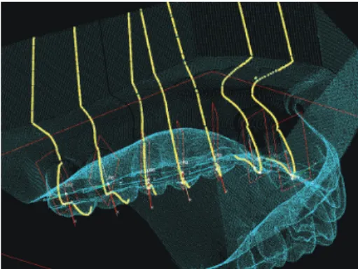
(4) Tooth Inclination in Elderly. 71. Table 1 Characteristics of the subject Number Men Women Total. 10 10 20. Age. Present teeth. Mean. SD. Mean. SD. 81.4 83.3 82.3. 1.9 2.9 2.6. 28.1 28.5 28.3. 1.7 1.9 1.8. Methods This study included 20 elderly persons (10 men, 10 women) aged 80 years or older with 20 or more present teeth who had been commended for achieving the 8020 goal in the 8020 contests sponsored by the Chiba Prefectural Dental Association in 2001, 2002 and 2005. The study inclusion criteria were 1) no prior orthodontic treatment, 2) no missing or restored tooth, 3) healthy tooth-supporting structures, 4) no TMJ symptoms and 5) pleasing facial profile relative to the E-plane. Their mean age was 82.3 years (81.4 years for men, 83.3 years for women) with a range of 80 to 89 years. Their mean number of present teeth was 28.3 (28.1 for men, 28.5 for women, Table 1). Standard orthodontic models with the base parallel to the occlusal plane were fabricated from the participants dental casts and digitized with a 3-D scanning system (VMS-100F, UNISN INC., Osaka, Japan). The occlusal plane was established by connecting the incisor edge of the central incisor and the distal buccal cusps of the right and left first molars. The labio or bucco-lingual crown inclination of each tooth was determined by the resulting angle between a line 90 degrees to the occlusal plane and a line tangential to the middle of the labial or buccal surface of the clinical crown (Fig. 1). The dental casts were digitized with a 3-D laser scanner (VMS-100F) for reconstruction into 3-D images. Labio or bucco-lingual crown inclination of each tooth was then measured with an analytical software (SURFLACER, UNISN INC. and IMAGEWARE 12, UGS PLM Solutions, MO, USA).. Fig. 1 Tooth inclination Tooth inclination was a complementary angle, where tangential line of clinical crown (long dots) was measured from line at 90 degrees to occlusal plane.. Measuring method of labio or bucco-lingual inclination of tooth in this study (Figs. 2–7): 1. The deepest point of the tooth cervical curve (White arrow) and the central point of the crown width (Red arrow) were marked. Next, the deepest point of the cervical curve and the central point of the crown width were connected. The tooth axis (Yellow line) was then determined (Fig. 2). 2. After the dental casts were digitized with a 3-D laser scanner to construct 3-D images the deepest point of the tooth cervical curve (Small yellow point) and the central point of the crown width were connected and the tooth axis (Light blue line) then determined. The tooth axis was the base line to determine the direction for each section (Fig. 3). 3. The Yellow line indicates the tooth axis and the curvature of the cutting section (Fig. 4). 4. The White line, as the curvature line, was projected onto the Red plane. The Yellow line, as the tooth axis, was projected onto the Red plane, which was vertical to the occlusal plane. The projected yellow line was changed to the White line (Fig. 5). 5. Tangential line (Blue line) on the central point of the White line. The Red asterisk (*) was the central point of the labial or buccal curvature of the clinical crown. The White line was projected onto the Red plane (Fig. 6). 6. The Pink line was drawn vertical to the occlusal plane on the Red plane. Labio, bucco-lingual tooth inclination was measured between the Blue line and the Pink line (Fig. 7)..
(5) 72. Fukagawa H et al.. Fig. 2 Yellow line represents tooth axis Deepest point of tooth cervical curve (White arrow) and central point of crown width (Red arrow) were marked. Next, the deepest point of cervical curve and central point of crown width were connected. Tooth axis (Yellow line) was then determined.. Fig. 3 Determination of tooth axis and direction for each section After dental casts were digitized with 3-D laser scanner to construct 3-D images deepest point of tooth cervical curve (Small yellow point) and central point of crown width were connected and tooth axis (Light blue line) determined. Tooth axis was base line to determine direction for section.. Fig. 4 Curvatures (Yellow line) of crown on tooth axis Yellow line is indicates tooth axis and curvature of cutting section.. Fig. 5 White line as curvature line was projected onto Red plane Yellow line, as tooth axis, was projected onto Red plane, which was vertical to occlusal plane. Projected Yellow line was changed to White line.. Fig. 6 Tangential line (Blue line) on central point of White line Red asterisk (*) is central point of labial or buccal curvature of clinical crown. White line was projected onto Red plane.. Fig. 7 Tooth inclination (arrow) Pink line was drawn vertical to occlusal plane on Red plane. Tooth inclination was measured between Blue line and Pink line..
(6) 73. Tooth Inclination in Elderly. Table 2 Results of labio or bucco-lingual inclination of the subjects Man (10). Maxillary Central incisor Lateral incisor Canine First premolar Second premolar First molar Second molar Mandibular Central incisor Lateral incisor Canine First premolar Second premolar First molar Second molar. Women (10). Total (20). t -test M⳯W. Mean. SD. Mean. SD. Mean. SD. 8.01 8.84 4.55 ⳮ6.49 ⳮ5.66 ⳮ4.73 ⳮ4.37. 2.57 2.23 1.99 2.06 2.12 1.98 1.84. 8.06 7.36 5.15 ⳮ6.86 ⳮ6.31 ⳮ5.56 ⳮ5.88. 2.30 2.28 2.13 2.50 3.07 2.87 3.13. 8.08 8.10 4.85 ⳮ6.68 ⳮ5.58 ⳮ5.15 ⳮ5.41. 2.41 2.35 2.06 2.28 2.63 2.47 2.74. n.s. * n.s. n.s. n.s. n.s. n.s.. 7.53 5.60 ⳮ5.27 ⳮ13.72 ⳮ20.47 ⳮ24.54 ⳮ27.54. 3.51 3.56 2.75 3.53 5.42 4.22 4.79. 6.04 4.14 ⳮ6.20 ⳮ13.76 ⳮ17.95 ⳮ22.97 ⳮ29.66. 2.58 2.46 2.11 3.22 4.56 5.53 4.77. 6.78 4.87 ⳮ5.73 ⳮ13.74 ⳮ19.21 ⳮ23.76 ⳮ28.63. 3.13 3.11 2.47 3.33 5.11 4.92 4.82. n.s. n.s. n.s. n.s. n.s. n.s. n.s.. *: p⬍5%, n.s.: non significant difference. Table 3 Comparison of labio, bucco-lingual tooth inclination in this study with those of Andrews1–3), Sebata21) and Mestriner et al.14). Maxillary Central incisor Lateral incisor Canine First premolar Second premolar First molar Second molar Mandibular Central incisor Lateral incisor Canine First premolar Second premolar First molar Second molar. Fukagawa. Sebata. Andrews. 8.08 8.10 4.85 ⳮ6.68 ⳮ5.58 ⳮ5.15 ⳮ5.41. 9.42 7.48 0.67 ⳮ6.46 ⳮ6.64 ⳮ1.73 ⳮ2.97. 6.11 4.42 ⳮ7.30 ⳮ8.50 ⳮ8.78 ⳮ11.50 ⳮ8.10. 6.78 4.87 ⳮ5.73 ⳮ13.74 ⳮ19.21 ⳮ23.76 ⳮ28.63. 3.55 1.66 ⳮ4.73 ⳮ14.80 ⳮ22.57 ⳮ26.17 ⳮ31.03. ⳮ1.71 ⳮ3.24 ⳮ12.70 ⳮ19.00 ⳮ23.60 ⳮ30.70 ⳮ36.00. A plus value indicated labio or buccal inclination and a minus value indicated lingual inclination.. Results Normal overbite (0–4 mm) was observed in 75% of the participants and deep bite. Mestriner. 2 ⳮ2 ⳮ9 ⳮ16 ⳮ22 ⳮ27 ⳮ32. (⬎4 mm) in 25%. No participant had open bite. Overjet was normal in 71.6% and maxillary protrusion was seen in 28.4%. No participant had anterior crossbite. Arch length discrepancy was less than 3 mm in 70% of the participants for the maxillary arch and 40% for the mandibular arch, and 3 mm or greater only in 5% for the maxillary arch and in 50% for the mandibular arch. Anterior spac-.
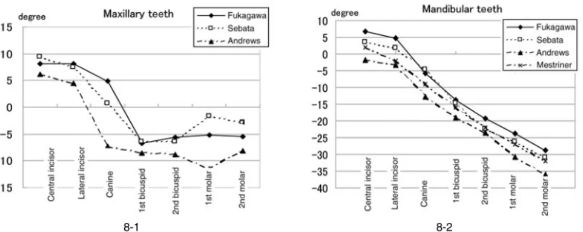
(7) 74. Fukagawa H et al.. 8-1. 8-2. Fig. 8 Comparison of tooth inclination of this study 8-1: With those of Andrews1–3) and Sebata21) (Maxillary teeth) 8-2: With those of Andrews1–3), Sebata21) and Mestriner et al.14) (Mandibular teeth). ing was observed in 25% of the participants for the maxillary arch and 10% for the mandibular arch. Average tooth inclinations (degrees) of the maxillary teeth of the 8020 male and female achievers were 8.08 for the central incisor, 8.10 for the lateral incisor, 4.85 for the canine, ⳮ6.68 for the first premolar, ⳮ5.58 for the second premolar, ⳮ5.15 for the first molar, and ⳮ5.41 for the second molar. The corresponding values for the mandibular teeth were 6.78, 4.87, ⳮ5.73, ⳮ13.74, ⳮ19.21, ⳮ23.76, and ⳮ28.63. There was a significant difference in the maxillary lateral incisor between men and women (p⬍0.05, Tables 2, 3).. Discussion With regard to Angle’s intended use of the line of occlusion, that is a line through the center of the crown, Ricketts20) pointed out that it would be necessary for an arch-wire attached to brackets. A bracket height or a labial or buccal surface has been proposed to obtain optimal final inclination. Andrews1–3) proposed that one of the keys to normal occlusion was crown inclination, which was expressed in plus or minus degrees, representing the angle formed by a line which. bears 90 degrees to the occlusal plane and a line that is tangential to the bracket site. This is in the middle of the labial or buccal long axis of the clinical crown. The clinical parameter for assessing the labio or bucco-lingual inclination of each tooth is the angle formed by its labial or buccal surface in relation to the occlusal plane1,2,6,14,21). A plus reading is given if the gingival portion of the tangential line (or of the crown) is lingual to the incisor portion in Fig 1; a minus reading is recorded when the gingival portion of the tangential line (or of the crown) is labial to the incisor portion. According to Sebata21), the plaster model was cut into 6 small blocks in order to face the labial or buccal surface to the screen of a profile projector. The curvature of the clinical crown and the occlusal plane were traced onto tracing paper, and ten points on the curvature were plotted with an x-y plotter. The tangent on each of the ten points was automatically calculated with a computer. Finally the tangent at the LACC, which was the center of the clinical crown, was determined as the inclination of each tooth. With this method, however, it is difficult to measure many anatomical positions such as angle or distance as the plaster model has be split up into small segments. This was the reason a 3-D procedure was used in this study. The 3-D.
(8) Tooth Inclination in Elderly. images used in our analysis offer the potential for further observations such as tooth rotation, tooth wear and arch form without cutting a plaster model. Van Loenen et al.24) noted that in order to obtain good treatment results with pre-adjusted brackets without any wire bending, two conditions have to be fulfilled. The brackets have to be placed 0.5 mm cervically from standard position on mandibular teeth according to Mestriner et al.14). They used 31 plaster models obtained from Caucasian Brazilian adults who had never received orthodontic treatment. He pointed that apart from mandibular incisors, which presented small differences in inclination from one another, the remaining average values were close to those found in the literature (Fig. 8-2 and Table 3). Watanabe and Hattori25) reported that occlusal wear of the posterior teeth led to changes in posterior occlusal support and increased occlusal load anteriorly. Masumoto et al.13) investigated the relationships between different facial types and both the buccolingual molar inclination and cortical bone thickness of the mandible by CT scanning of 31 dry skulls of Japanese adult men. It was found that the teeth of long-faced subjects were more lingually inclined than those of the short-faced subjects. Generally speaking, the short-faced type has stronger masticatory function then the long-faced type. Oda17) performed mathematical modeling of mandibular apical base forms and arrangement of the teeth based on CT radiographs and found neither sex-related nor right-left differences in the mean labio-lingual and bucco-lingual inclinations of the teeth. There was a significant difference (p⬍0.05) between men and women in the results for the maxillary lateral incisor. A tendency toward being small or cone-shaped is observed in the maxillary lateral incisor. Therefore, it is difficult to determine the center on a clinical crown in such teeth. Figures 8-1, 2 show a comparison of our results with those of Andrews1–3), Sebata21) and Mestriner et al.14) (Mandibular teeth only). Andrews1–3) reported six significant character-. 75. istics observed in a study of 120 casts of young non-orthodontic normal occlusion. He indicated the data for each inclination at the LA point, which was the center of the long axis on the clinical crown, relative to the occlusal plane by using a kind of protractor. Sebata21) observed tooth inclination in 41 young Japanese by computer calculation using the same concept as Andrews. Mestriner et al.14) measured the degree of inclination at the center of the clinical crown relative to the occlusal plane using mandibular plaster casts from 31 young Caucasian Brazilian adults with normal occlusion. Tooth inclination showed a progressive decrease from anterior to posterior. The decrease in the mandibular teeth was more regular than that in the maxillary teeth. It is impossible to compare them directly, because of the different ethnic groups involved, however, it is possible to infer from tendencies. Tooth inclination in these studies showed approximate values, especially mandibular values. This suggests that tooth inclination in the mandibular teeth may not be affected by aging. Harris9) reported that changes during adulthood occur most rapidly during the second and third decades of life, but do not stop thereafter by investigation of a longitudinal series of 60 adults aged from 20 to 55 years. Possible mechanisms driving these changes in tooth position are discussed. Dager et al.4) examined three stages in 40 patients: after the presumed cession of circumpugertal growth, at approximately 47 years of age and at least one decade later. They found changes reflected a decrease in arch width, depth and perimeter with a significant increase in the mandibular incisor irregularity index. Such longitudinal research is important in determining the effects of aging. Dental casts of 20 elderly participants with many remaining teeth were digitized with a 3-D laser scanner to construct 3-D images. Tooth inclination was measured with analytical software. Tooth inclination showed a progressive decrease from anterior to posterior. The decrease in the mandibular teeth was more regular than that in the maxillary teeth..
(9) 76. Fukagawa H et al.. References 1) Andrews LF (1972) The six keys to normal occlusion. Am J Orthod 62:296–309. 2) Andrews LF (1976) The straight-wire appliance, origin, controversy, commentary. J Clin Orthod 10:99–114. 3) Bennet JC, McLaughlin RP (1998) Orthodontic Management of the Dentition with the Preadjusted Appliance, 1st ed., pp. 47, 165, 193, 258, 283, 314, Isis Medical Media, Oxford. 4) Dager MM, McNamara JA, Baccetti T, Franch L (2008) Aging in the craniofacial complex. Angle Orthod 78:440–445. 5) Dempster WT, Adams WJ, Duddles RA, Mich AA (1963) Arrangement in the jaws of the roots of the teeth. J Am Dent Assoc 67:779– 797. 6) Ferrario VF, Sforza C, Colombo A, Ciusa V, Serrao G (2001) Three-dimensional inclination of the dental axes in healthy permanent dentition—A cross-sectional study in a normal population. Angle Orthod 71:257–264. 7) Fredericks CD (1974) A method for determining the maxillary incisor inclination. Angle Orthod 44:341–345. 8) Ghahferokhi AE, Elias L, Jonsson S, Rolfe B, Richmond S (2002) Critical assessment of a device to measure incisor crown inclination. Am J Orthod Dentofacial Orthop 121:185– 191. 9) Harris EF (1997) A longitudinal study of arch size and form in untreated adults. Am J Orthod Dentofacial Orthop 111:419–427. 10) Ishihara H (2000) Influence of occlusal contact on tooth displacement. J Stomatolog Disease 67:310–321. (in Japanese) 11) Knösel M, Engelke W, Attin R, KubeinMeesenburg D, Sadat-Khonsari R, GrippRudolph L (2008) A method for defining targets in contemporary incisor inclination correction. Eur J Orthod 30:374–380. 12) Knösel M, Kubein-Meesenburg D, SadatKhonsari R (2007) The third-order angle and the maxillary incisor’s inclination to the NA line. Angle Orthod 77:82–87. 13) Masumoto T, Hayashi I, Kawamura A, Tanaka K (2001) Relationships among facial type, buccolingual molar inclination, and cortical bone thickness of the mandible. Eur J Orthod 23:15–23. 14) Mestriner MA, Enoki C, Mucha JN (2006) Normal torque of the buccal surface of mandibular teeth and its relationship with bracket positioning: a study in normal occlusion. Braz Dent J 17:33–41.. 15) Miyazaki H, Motegi E, Yatabe K, Yamaguchi H, Maki Y (2005) A study of occlusion in elderly Japanese over 80 years with at least 20 teeth. Gerodontology 22:206–210. 16) Motegi E, Miyazaki H, Isshiki Y (1999) Dental arch and occlusion in 8020 achievers supported by Bunkyo Dental Association. J Jpn Dent Assoc 52:619–626. (in Japanese) 17) Oda Y (1998) Mandibular apical base form and arrangement of the teeth: Mathematical modeling and canonical correlations. J Osaka Univ Dent Soc 33:145–167. (in Japanese) 18) Richmond S (1987) Recording the dental cast in three dimension. Am J Orthod Dentofacial Orthop 92:199–206. 19) Richmond S, Klufas ML (1998) Assessing incisor inclination: a non-invasive technique. Eur J Orthod 20:721–726. 20) Ricketts RM (1978) A detailed consideration of the line of occlusion. Angle Orthod 48:274– 282. 21) Sebata E (1980) An orthodontic study of tooth and dental arch form on the Japanese normal occlusions. Shikwa Gakuho 80:945–969. (in Japanese) 22) Shimizu T, Motegi E, Nomura M, Kaneko Y, Takeuchi F, Yamaguchi T, Miyazaki H, Harazaki M, Hirai M, Kurihara S, Yamaguchi H (2006) Cephalometric study of elderly with nearly intact dental arches. Gerodontology 23:60–63. 23) Tokuda A (2004) Influence of occlusal contacts on tooth displacement for mesio-distal direction. J Oral Patho 71:18–26. (in Japanese) 24) Van Loenen M, Degriek J, De Pauw G, Demaut L (2005) Anterior tooth morphology and its effect on torque. Eur J Orthod 27:258–262. 25) Watanabe M, Hattori Y (1998) Aging of the teeth and occlusion. J Geriatric Dentistry 13: 3–7. (in Japanese) 26) Yasuda K, Masumoto T, Hirayama Y, Ono S, Enomoto Y, Imamura R, Kasai K (2004) Relationship between the lower incisor root position and the mandibular bone structure using CT scanning. J Tokyo Orthod Soc 14:28– 35. (in Japanese) Reprint requests to: Dr. Hiroko Fukagawa Division of Orthodontics, Department of Oral Health Clinical Science, Tokyo Dental College, 2-9-18 Misaki-cho, Chiyoda-ku, Tokyo 101-0061, Japan E-mail: [email protected].
(10)
図


関連したドキュメント
Then, an algorithm is established as the way of transformation of so called associated matrices, formed as a result of local inspection of patterns, into invariant ones which
Theorem 4.8 shows that the addition of the nonlocal term to local diffusion pro- duces similar early pattern results when compared to the pure local case considered in [33].. Lemma
Keywords: continuous time random walk, Brownian motion, collision time, skew Young tableaux, tandem queue.. AMS 2000 Subject Classification: Primary:
We present sufficient conditions for the existence of solutions to Neu- mann and periodic boundary-value problems for some class of quasilinear ordinary differential equations.. We
Then it follows immediately from a suitable version of “Hensel’s Lemma” [cf., e.g., the argument of [4], Lemma 2.1] that S may be obtained, as the notation suggests, as the m A
This paper gives a decomposition of the characteristic polynomial of the adjacency matrix of the tree T (d, k, r) , obtained by attaching copies of B(d, k) to the vertices of
This paper presents an investigation into the mechanics of this specific problem and develops an analytical approach that accounts for the effects of geometrical and material data on
While conducting an experiment regarding fetal move- ments as a result of Pulsed Wave Doppler (PWD) ultrasound, [8] we encountered the severe artifacts in the acquired image2.
