はじめに 海馬硬化と海馬外病変が同時にてんかん原性を有している 状態を二重病理(dual pathology)と称し,薬剤抵抗性てんか んの 5~20%を占める1).成因として,海馬外病変のために 繰返されたてんかん発作の結果として海馬硬化が生じるとす る説と2)3),海馬硬化も海馬外病変も同じ病因から生じるとす る説がある2).我々は,生後 9 ヶ月時に脳挫傷の既往があり, 7歳時から口部自動症を伴う複雑部分発作がみられ,薬剤抵 抗性に経過した 26 歳女性例を経験した.頭蓋内ビデオ脳波モ ニタリング(long-term video EEG monitoring; VEEG)にて, 左海馬起始の発作活動が外傷性瘢痕を含む左外側側頭葉皮質 へ早期に伝播することを捉え,外傷性皮質病変と海馬硬化を 伴う内側側頭葉てんかんと診断した.外傷性皮質病変に独立 したてんかん原性は認められず,本症例は二重病理の定義を 満たさない.しかし外傷性皮質病変がてんかん原性を有する 蓋然性が高い4)ことから,二重病理に準じて左扁桃体海馬切 除術および外傷性瘢痕を含む左側頭葉切除術を施行した.術 後は 3 年間の発作消失を維持している.てんかん診療におけ る包括的精査の重要性,海馬硬化及び二重病理の成因を考察 する上で貴重な症例と考えられたので報告する. 症 例 患者:26 歳,女性,矯正による右利き 主訴:口部自動症を伴う複雑部分発作 発育歴:周産期に異常なし.発育・発達に遅滞なし. 既往歴:熱性けいれんなし. 家族歴:特記事項なし. 生活歴:飲酒は機会飲酒のみ.喫煙なし. 現病歴:生後 9 ヶ月時,階段の 2 階部分から 2 m ほど転落 して,左後頭部を強打した.意識障害および嘔吐はなかった. 頭蓋骨骨折,脳挫傷の診断を受け,保存的加療で後遺症なく 軽快した.てんかん発作を疑わせるエピソードはなかったが, 予防的なバルプロ酸(VPA)内服を 6 歳頃まで継続された. 7歳時,自宅で兄弟と遊んでいる際に「口をモグモグ,クチャ クチャさせ,呼びかけても応答がない状態」が約 1 分間続い た.A クリニックを受診し,症候性てんかんの診断で薬剤調 整を経たが,同様の発作を月単位で繰返したため,精査加療 目的で当科に紹介された.当科初診時はカルバマゼピン (CBZ),ゾニサミド(ZNS),クロナゼパム(CZP)を内服中 であった. 入院時現症:身長 153 cm,体重 50 kg,血圧 98/65 mmHg, 脈拍 76/ 分・整,体温 36.3°C,SpO2 98%.一般身体所見およ び神経学的所見に特記すべき異常はなかった.神経心理学的
症例報告
外傷性皮質病変と海馬硬化を伴う内側側頭葉てんかんの 1 手術例
北澤 悠
1)2)*
神 一敬
1)岩崎 真樹
3)鈴木 博義
4)田中 章景
2)中里 信和
1) 要旨: 症例は 26 歳女性.生後 9 ヶ月時に脳挫傷,7 歳時から複雑部分発作を月単位で繰返し薬剤抵抗性に経過 した.病歴・画像所見から左側頭葉の外傷性皮質病変が発作焦点と疑われていたが,包括的精査の結果,海馬硬化 を伴う内側側頭葉てんかんと診断した.27 歳時に外科治療目的で再入院し,頭蓋内脳波で外傷性病変に独立した てんかん原性は証明されなかったが,海馬起始の発作活動が早期に伝播していたため,その蓋然性が高いと判断 し,二重病理に準じて左扁桃体海馬切除術および左側頭葉切除術を施行した.術後 3 年間の発作消失を維持して いる.薬剤抵抗性てんかんの治療方針決定には問診,脳波・画像・神経心理検査を含む包括的精査が重要である. (臨床神経 2017;57:698-704) Key words: 内側側頭葉てんかん,海馬硬化,外傷性皮質病変,二重病理,ビデオ脳波モニタリング *Corresponding author: 東北大学大学院医学系研究科てんかん学分野〔〒 980-8575 宮城県仙台市青葉区星陵町 2-1〕 1)東北大学大学院医学系研究科てんかん学分野 2)横浜市立大学大学院医学研究科神経内科学・脳卒中医学 3)国立精神・神経医療研究センター病院脳神経外科 4)国立病院機構仙台医療センター臨床検査科 / 病理診断科(Received March 21, 2017; Accepted August 1, 2017; Published online in J-STAGE on October 26, 2017) doi: 10.5692/clinicalneurol.cn-001029
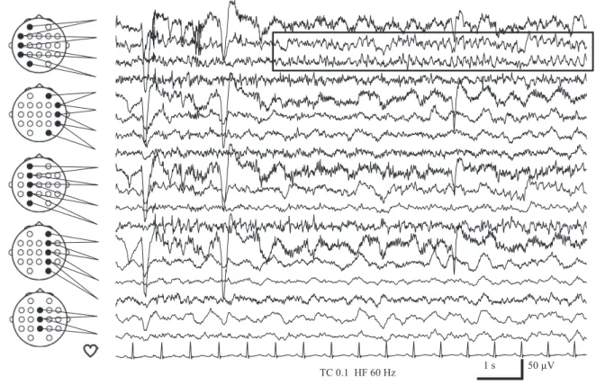
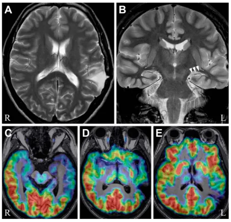
VEEG:発作間欠時には左側頭部に鋭波を認めた(Fig. 1). 明らかな前兆なく,「うー」という唸り声の後,口をモグモグ させてから反応が乏しい状態が約 80 秒間に渡り記録された. 上肢自動症およびジストニー肢位は明らかでなく,発作後に は数分間のもうろう状態を呈した.発作時脳波は,左優位の 不規則な全般性 δ 律動に始まり,約 20 秒後に左側頭部 θ 律動 が明瞭化し(Fig. 2),左半球性 θ 律動,続いて左半球性 δ 律 動に変化し,90 秒程度で脳波変化が終了した. 画像検査:脳 MRI で,左上側頭回後部~頭頂葉に周囲白質 の T2強調・FLAIR 画像で高信号を伴う楔状の欠損を認めた. 左海馬は T2強調・FLAIR 画像で高信号を呈し,萎縮してい た(Fig. 3A, B).左側頭葉は白質容積が全体的に右側より少 なく,皮髄境界が不明瞭化していた.脳 FDG-PET では,左 側頭頭頂葉の脳実質欠損に一致した集積欠失および左側頭葉 底部~外側に糖代謝低下を認めた(Fig. 3C~E). 脳磁図:左側頭部に鋭波を認め,等価電流双極子モデルに より左側頭葉前方に信号源が推定された(Fig. 4).
Fig. 1 Interictal EEG findings of long-term video EEG monitoring. A sharp wave was seen in the left anterior to middle temporal region (filled circle).
Fig. 2 Ictal EEG findings of long-term video EEG monitoring.
Rhythmic theta activities started in the left temporal region approximately 20 seconds after the ictal EEG onset in the left hemisphere with aural automatism.
入院後経過:発作症状・脳波所見から左内側側頭葉てんか んと診断した.MRI で左海馬萎縮・信号異常を認めることか ら,左海馬硬化がてんかん原性領域であると考えられた. FDG-PETや脳磁図も上記を示唆する結果であった.一方,左 外側側頭葉~頭頂葉皮質に外傷性瘢痕が認められたが,非侵 襲的術前評価の結果からは同病変がてんかん原性を有してい る所見が得られなかった.その後,クロバザムおよびレベチ ラセタム(LEV)が導入され最大用量まで増量されたが,発 作は抑制されず,27 歳時に外科治療目的で再入院した.脳磁 図と fMRI を用いた言語脳機能計測で,言語優位半球は右半 球に同定された.切除範囲決定目的に左外側側頭葉から頭頂 葉にかけて外傷性瘢痕を覆うように硬膜下電極を,左海馬に 深部電極を留置し,頭蓋内 VEEG を施行した.但し,外傷性 瘢痕内側部の脳波記録は行っていない.発作間欠時には左側 頭葉底部,左海馬,左側頭葉外側後方で棘波が記録された. 発作は合計 9 回記録され,発作起始はすべて左海馬であった. Fig. 3 MRI and FDG-PET images.
A: T2-weighted MR image showing a wedge-shaped deficit around the left temporo-parietal junction. B: T2-weighted MR image showing left hippocampal atrophy with increased T2 signal (arrowheads). C: FDG-PET image showing hypometabolism of the left mesial, frontal, and lateral temporal lobes. D and E: FDG-PET images showing absence of metabolism corresponding to the wedge-shaped brain deficit.
Fig. 4 Magnetoencephalography findings.
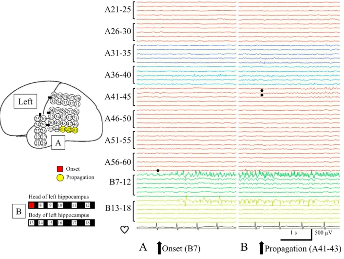
口部自動症を伴う複雑部分発作は 6 回記録され,脳波変化開 始の約 10 秒後に外傷性瘢痕の一部に発作活動が伝播してか ら意識減損・口部自動症が出現した(Fig. 5).上腹部不快感 を訴える単純部分発作が 3 回記録されたが,外傷性瘢痕への 伝播は認められなかった.以上より,外傷性皮質病変と海馬 硬化を伴う内側側頭葉てんかんと診断した.重症頭部外傷に よる皮質病変がてんかん原性を有する蓋然性が高いと判断 し,言語優位半球が右であったこと,知能検査で劣位半球側 頭葉全体の機能低下が疑われたことから,左扁桃体海馬切除 術に加えて左側頭葉切除術が施行された.側頭葉外側皮質の 切除線は Labbé 静脈より後方 3 cm 程度とし,外傷性瘢痕の うち,発作活動の伝播が認められた範囲のみが切除され,伝 播の認められなかった頭頂葉側の病変は残された. 病理所見:海馬は CA1~CA4 で高度な神経細胞の脱落と グリオーシスが認められ,特に CA4 で顕著であり(Fig. 6), Blümckeらの International League Against Epilepsy(ILAE)分 類5)により海馬硬化症 ILAE Type 1 と診断された.左外側側 頭葉皮質は全層,びまん性に中等度のアストロサイト増生と グリオーシスが認められたが,層構造の異常や異型ニューロ ンはみられず,この所見は外傷性変化に矛盾しなかった. 術後経過:LEV 単剤療法に移行したが,術後 2 年間の発作消 失を経て,その後は無投薬で 1 年間の発作消失を維持している. Fig. 5 Ictal intracranial EEG findings.
A: Ictal discharges starting at the depth electrodes targeted to the left hippocampus (B7). B: The discharges propagated to a posterior portion of the left superior temporal gyrus 10 seconds after onset (A41–43).
Fig. 6 Pathological findings of the left hippocampus. NeuN immunostaining showing marked neuronal cell loss in all sectors of cornu ammonis with CA4 predominance. Bar is 1,000 μm. CA, cornu ammonis; DG, dentate gyrus.
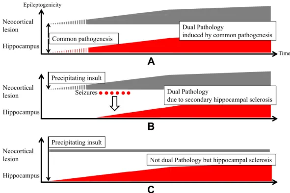
考 察 1.海馬硬化症と二重病理 海馬硬化と海馬外病変が同時にてんかん原性を有している 状態を二重病理と呼ぶ.海馬硬化症は,臨床的に先行損傷の 既往(熱性けいれん,熱性けいれん重積,外傷,低酸素脳症, 中枢神経感染症など)を有する症例が多い.しかし,海馬硬 化自体がてんかんの原因なのか結果なのか,未だに結論が得 られていない6).本症例をもとに,海馬硬化および二重病理 の成因について,以下の観点から考察した. (1)発症年齢 海馬硬化症による内側側頭葉てんかんの平均発症年齢は 9歳である7).本症例は 7 歳時に自動症を伴う複雑部分発作 で発症しているため,海馬硬化症として典型的な発症年齢で ある.しかし,実際には外傷性脳損傷を受傷してから 6 歳時 まで VPA の予防内服を継続していたため,より早期に発症し ていたてんかん発作が VPA で抑制されていた可能性がある. (2)頭蓋内脳波所見 頭蓋内脳波では,左海馬起始の発作活動が外傷性瘢痕を含 む左外側側頭葉皮質へ早期に伝播することを捉えた.しかし, 外傷性瘢痕またはその周囲に始まる発作活動は観察されず, 同部位に独立したてんかん原性は証明されなかった. (3)病理所見 本例の病理所見は海馬硬化症 ILAE Type 15)であった.熱性 けいれんなどの先行損傷を認めた海馬硬化症 ILAE Type 1 の 症例では,その先行損傷の時期が 5 歳以前と早期であった と報告されている5).本症例で脳挫傷を先行損傷と考えた際 に,生後 9 ヶ月という受傷時期はこの報告に矛盾しない.ま た,海馬硬化症 ILAE Type 1 では術後 1~2 年間の発作消失率 が 60~80%と良好であることも5)本症例の治療反応性と合致 する. (4)二重病理の術後発作予後 二重病理の 41 手術例を海馬外病変および内側側頭葉切除, 内側側頭葉切除のみ,海馬外病変切除のみの 3 群に分けて比 較した研究によると,90%以上の発作抑制が得られたのはそ れぞれ 86%,30%,31%と報告されている.海馬外病変およ び内側側頭葉の両方を切除しなければ,良好な予後は期待で きないことを示している8). (5)二重病理の成因 てんかん診断に際して,海馬硬化と海馬外病変の併存時に, 両者の関連を説明する仮説として次の 3 パターンを考えた (Fig. 7A~C).海馬硬化も海馬外病変も共通の病因から生じ, 同時期にてんかん原性を獲得するパターン A(二重病理),先 行損傷による海馬外病変のために繰り返されたてんかん発作
Fig. 7 Schematic diagram illustrating the relationship between hippocampal sclerosis and extrahippocampal lesion.
A: Dual pathology induced by common pathogenesis. Both neocortical lesion with epileptogenicity and hippocampal sclerosis are induced by common pathogenesis. B: Dual pathology due to secondary hippocampal sclerosis. Precipitating insult causes neocortical lesion with epileptogenicity, which further induces repeated seizures. Then, secondary epileptogenicity is induced in the hippocampus with a pathological diagnosis of hippocampal sclerosis. C: Not dual pathology but hippocampal sclerosis. Precipitating insult causes hippocampal sclerosis with epileptogenicity and neocortical lesion without epileptogenicity.
異常が皮質病変にも早期に伝播したことからパターン B の可 能性があり,厳密には二重病理ではないが,それに準ずる病 態と考えられた. 2.本症例の治療方針について 外科治療の検討対象としては,2~3 種類の適切な抗てんか ん薬を用いても無発作期間が 1 年以下で,発作のために患者 に不利益がある場合,とされており9),本症例はこれを満た していた.包括的精査の結果,外傷性皮質病変と海馬硬化を 伴う内側側頭葉てんかんと診断した.海馬外病変に独立した てんかん原性が証明されず,二重病理の定義は満たさないが, 重症頭部外傷に伴う皮質病変がてんかん原性を有する蓋然性 は高い4)とされる.神経心理学的には,IQ の軽度低下を認め たが,記憶は正常範囲内であった.これは劣位半球側頭葉全 体の機能低下で,言語優位半球の内側側頭葉機能は保たれて いると考えられた.本症例の言語優位半球は右であったこと から,発作活動が早期に伝播していた外側側頭葉皮質は切除 可能と判断された.最終的に左扁桃体海馬切除術および左側 頭葉切除術が施行され,発作消失が得られた.海馬外病変を 切除せずとも発作消失が得られた可能性は考慮されるが,病 変を安全な範囲で可及的に切除することが,術後発作消失率 を高めると考えられた. 結 語 脳挫傷の既往があり,病歴・画像所見から外傷性皮質病変 を発作起始とするてんかんが疑われていたが,包括的精査の 結果,海馬起始で外傷性皮質病変に伝播する発作を捉え,外 傷性皮質病変と海馬硬化を伴う内側側頭葉てんかんの診断に 至った.薬剤抵抗性てんかんの治療方針決定には,発作症状, 脳波・画像・神経心理学的所見を含めた包括的精査が重要で ある. します. ※本論文に関連し,開示すべき COI 状態にある企業,組織や団体 講演料 神一敬:ユーシービー・ジャパン株式会社,大塚製薬株式 会社,中里信和:第一三共株式会社,エーザイ株式会社 奨学寄付 中里信和:大塚製薬株式会社 寄付講座 中里信和:株式会社リコー 文 献 1) 中里信和.Dual Pathology(二重病変).日本てんかん学会編. てんかん学用語辞典.東京:日本てんかん学会;2006.p. 44. 2) Ho SS, Kuzniecky RI, Gilliam F, et al. Temporal lobe
developmental malformations and epilepsy: dual pathology and bilateral hippocampal abnormalities. Neurology 1998;50:748-754.
3) Cavazos JE, Sutula TP. Progressive neuronal loss induced by kindling : a possible mechanism for mossy fiber synaptic reorganization and hippocampal sclerosis. Brain Res 1990;527:1-6.
4) Annegers JF, Grabow JD, Groover RV, et al. Seizures after head trauma: a population study. Neurology 1980;30(7 Pt 1):683-689. 5) Blümcke I, Thom M, Aronica E, et al. International consensus
classification of hippocampal sclerosis in temporal lobe epilepsy: a Task Force report from the ILAE Commission on Diagnostic Methods. Epilepsia 2013;54:1315-1329.
6) Zarow C, Sitzer TE, Chui HC. Understanding hippocampal sclerosis in the elderly: epidemiology, characterization, and diagnostic issues. Curr Neurol Neurosci Rep 2008;8:363-370. 7) French JA, Williamson PD, Thadani VM, et al. Characteristics
of medial temporal lobe epilepsy: I. Results of history and physical examination. Ann Neurol 1993;34:774-780.
8) Li LM, Cendes F, Andermann F, et al. Surgical outcome in patients with epilepsy and dual pathology. Brain 1999;122(Pt 5):799-805.
9) 「てんかん治療ガイドライン」作成委員会編.てんかん治療 ガイドライン 2010.東京:医学書院;2010.p. 86-87.
Abstract
A surgical case of mesial temporal lobe epilepsy associated
with hippocampal sclerosis and traumatic neocortical lesion
Yu Kitazawa, M.D.
1)2), Kazutaka Jin, M.D., Ph.D.
1), Masaki Iwasaki, M.D., Ph.D.
3),
Hiroyoshi Suzuki, M.D., Ph.D.
4), Fumiaki Tanaka, M.D., Ph.D.
2)and Nobukazu Nakasato, M.D., Ph.D.
1)1)Department of Epileptology, Tohoku University Graduate School of Medicine
2)Department of Neurology and Stroke Medicine, Yokohama City University Graduate School of Medicine 3)Department of Neurosurgery, National Center Hospital, National Center of Neurology and Psychiatry
4)Department of Pathology and Laboratory Medicine, Sendai Medical Center
A 26-year-old right-handed woman, with a history of left temporal lobe contusion caused by a fall at the age of
9 months, started to have complex partial seizures with oral automatism at the age of 7 years. The seizures occurred
once or twice a month despite combination therapy with several antiepileptic agents. Her history and imaging studies
suggested the diagnosis of epilepsy arising from traumatic neocortical temporal lesion. Comprehensive assessment
including long-term video EEG monitoring, MRI, FDG-PET, MEG, and neuropsychological evaluation was performed at
the age of 26 years. The diagnosis was left mesial temporal lobe epilepsy associated with hippocampal atrophy and
traumatic temporal cortical lesion. The patient was readmitted for surgical treatment at the age of 27 years. Intracranial
EEG monitoring showed that ictal discharges started in the left hippocampus and spread to the traumatic lesion in the
left posterior superior temporal gyrus 10 seconds after the onset. This case could not be classified as dual pathology
exactly, because the traumatic left temporal cortical lesion did not show independent epileptogenicity. However, the
traumatic lesion was highly likely to be the source of the epileptogenicity, and she had right hemispheric dominance for
language and functional deterioration in the whole temporal cortex. Therefore, left amygdalo-hippocampectomy and left
temporal lobectomy including the traumatic lesion were performed according to the diagnosis of dual pathology.
Subsequently, she remained seizure-free for 3 years. Comprehensive assessment of seizure semiology, neurophysiology,
neuroradiology, and neuropsychology is important to determine the optimum therapeutic strategies for drug-resistant
epilepsy.
(Rinsho Shinkeigaku (Clin Neurol) 2017;57:698-704)
Key words: mesial temporal lobe epilepsy, hippocampal sclerosis, traumatic neocortical lesion, dual pathology, video EEG monitoring