Abstract. Since the prognosis of non-small cell lung cancer
(NSCLC) remains poor, the development of novel therapeutic approaches, including cancer vaccines, is highly desirable. In the current study, we conducted a phase II study of personalized peptide vaccination (PPV), in which a maximum of 4 peptides were selected based on pre-existing humoral immune responses and administered subcutaneously (weekly for 6 consecu- tive weeks and bi-weekly thereafter) in refractory NSCLC patients. Forty-one refractory NSCLC patients (4 stage IIIb, 22 stage IV and 15 recurrent), who had failed to respond to chemotherapy and/or targeted therapy (median number of regimens, 3; median duration, 10 months), were enrolled.
Median overall survival (OS) was 304 days with a one-year survival rate of 42% in the enrolled patients. The main toxicity of PPV was skin reactions at the injection sites, but no serious adverse events were observed. In order to identify potential biomarkers for predicting OS, pre-vaccination and post-vaccination clinical findings and laboratory data were retrospectively assessed and evaluated by multivariate Cox regression analysis. Among the pre-vaccination factors exam- ined, high C-reactive protein (CRP) level was a significant predictor of unfavorable OS [hazard ratio (HR)=10.115, 95%
confidence interval (CI)=2.447-41.806, P=0.001]. Among the post-vaccination factors, high CRP level and low frequency of CD3
+CD26
+cells were significant predictors of unfavorable OS (HR=23.127, 95% CI=2.919-183.233, P=0.003; HR=0.952, 95% CI=0.917-0.989, P=0.012). Taken together, our results suggest the feasibility of PPV for the treatment of refrac- tory NSCLC. Evaluation of the identified factors before or at an early stage of vaccination could be potentially useful for selecting NSCLC patients who would likely have better prognosis following PPV.
Introduction
Non-small cell lung cancer (NSCLC) is one of the most common causes of cancer death worldwide. Although recent advances in chemotherapy and/or targeted therapy have helped to improve the clinical outcomes of patients with refractory NSCLC (1-5), their prognosis still remains very poor with a median survival time of 6-8 months. Therefore, development of novel therapeutic approaches, including cancer vaccines, would be highly desirable.
We developed a new approach of peptide-based vaccination, named personalized peptide vaccination (PPV), in which vaccine antigens are selected and administered based on pre- existing host immunity before vaccination (6-14). We have shown promising results of PPV in various types of advanced cancers (6-9). For example, a recently conducted random- ized clinical trial of PPV for patients with advanced prostate cancer suggested a potentially favorable clinical outcome in the vaccinated group (9). However, to improve clinical efficacy further, prognostic biomarkers that would make it possible to select patients for whom cancer vaccines would be appropriate remain to be identified. In the present investi- gation, we conducted a small-scale phase II study to identify potential biomarkers that would be useful for prediction of overall survival (OS) before or at an early stage of vaccina- tion in refractory NSCLC patients. Our results suggested the feasibility of PPV for refractory NSCLC. The identified factors would be informative for predicting the subpopulation of NSCLC patients, who would likely have better prognosis following PPV.
Patients and methods
Patients. Patients with a histological diagnosis of NSCLC were eligible for inclusion in the present study, if they had failed to respond to previous chemotherapy and/or targeted therapy. They also had to show positive humoral responses to at least two of the 31 different candidate vaccine peptides (Table I), determined by both HLA class I type and the titer of IgG against each peptide. The other inclusion criteria, as well as the exclusion criteria, were not largely different from those of other previously reported clinical studies (6-9): patient age
Personalized peptide vaccination in patients with refractory non-small cell lung cancer
KOICHI YOSHIYAMA
1, YASUHIRO TERAZAKI
1, SATOKO MATSUEDA
2, SHIGEKI SHICHIJO
2, MASANORI NOGUCHI
3, AKIRA YAMADA
3, TAKASHI MINE
1, TETSUYA IOJI
2,
KYOGO ITOH
2, KAZUO SHIROUZU
1, TETSURO SASADA
2and SHINZO TAKAMORI
1Departments of
1Surgery and
2Immunology and Immunotherapy, Kurume University School of Medicine;
3
Research Center of Innovative Cancer Therapy, Kurume University, Kurume, Fukuoka, Japan Received November 8, 2011; Accepted December 30, 2011
DOI: 10.3892/ijo.2012.1351
Correspondence to:
Dr Tetsuro Sasada, Department of Immunology and Immunotherapy, Kurume University School of Medicine, 67 Asahi- machi, Kurume, Fukuoka 830-0011, JapanE-mail: [email protected]
Key words:
non-small cell lung cancer, peptide vaccine, biomarkerbetween 20 and 80 years; an Eastern Cooperative Oncology Group (ECOG) performance status of 1 or 2; positive status for HLA-A2, -A3, -A11, -A24, -A26, -A31, or -A33; life expectancy of at least 12 weeks; negative status for hepatitis virus B and C; adequate hematologic, renal, and hepatic func- tion. Patients with lymphocyte counts of <1000 cells/µl were excluded from the study, since we had previously reported that pre-vaccination lymphopenia is a predictor of unfavorable OS in cancer patients receiving PPV (12). Other exclusion criteria included pulmonary, cardiac, or other systemic diseases; acute infection; a history of severe allergic reactions; pregnancy or nursing; or other inappropriate conditions for enrollment as judged by clinicians. The protocol was approved by the Kurume University Ethics Committee, and was registered in the UMIN Clinical Trials Registry (UMIN no. 1839). After a full explanation of the protocol, written informed consent was obtained from all patients before enrollment.
Clinical protocol. This was an open-label phase II study in which the primary and secondary endpoints were to identify potential biomarkers for OS and to evaluate the safety of PPV in NSCLC patients, respectively. Thirty-one peptides, whose safety and immunological effects had been confirmed in previously conducted clinical studies (6-9, 13), were employed for vaccination [12 peptides for HLA-A2, 14 peptides for HLA-A24, 9 peptides for HLA-A3 supertype (-A3, -A11, -A31, and -A33), 4 peptides for HLA-A26] (Table I). The peptides were prepared under the conditions of good manufacturing practice (GMP) by the PolyPeptide Laboratories (San Diego, CA) and American Peptide Company (Vista, CA). Appropriate peptides for vaccination in individual patients were selected in consideration of pre-existing host immunity before vacci- nation, assessed from the titers of IgG specific to each of the 31 different vaccine candidates, as described previously (14).
Combined chemotherapy and/or targeted therapy were allowed during the vaccination period, unless patients were unable to tolerate combined chemotherapies or declined them (Table II).
A maximum of 4 peptides (3 mg/each peptide), which were selected on the basis of HLA typing and peptide-specific IgG titers, were administrated subcutaneously with incomplete Freund’s adjuvant (Montanide ISA51; Seppic, Paris, France) once a week for 6 consecutive weeks. After the first cycle of 6 vaccinations, up to 4 antigen peptides, which were re-selected according to the titers of peptide-specific IgG in every cycle of 6 vaccinations, were administered every 2 weeks. Adverse events were monitored according to the National Cancer Institute Common Terminology Criteria for Adverse Events version 3.0 (NCI-CTC Ver3). Complete blood counts and serum biochemistry tests were performed after every 6 vaccinations.
The clinical responses were evaluated using the Response Evaluation Criteria in Solid Tumors (RECIST) in the vacci- nated patients, for whom computed tomography (CT) scan or magnetic resonance imaging (MRI) data were available before and after the first cycle of vaccinations.
Measurement of humoral and T cell responses. The humoral responses specific to each of the 31 candidate peptides (Table I), including those employed and not employed for vaccination, were determined by the peptide-specific IgG levels using the Luminex system (Luminex, Austin, TX), as reported previously
(14). If the plasma titers of peptide-specific IgG in response to at least one of the vaccinated peptides after vaccination were
>2-fold higher than those before vaccination, the changes were considered to be significant.
T cell responses specific to the vaccine peptides were evaluated by interferon (IFN)-
γElispot using peripheral blood mononuclear cells (PBMCs), which were separated by density gradient centrifugation from peripheral blood (30 ml) with Ficoll-Paque Plus (GE Healthcare; Uppsala, Sweden) and stored frozen until analysis. After thawing, PBMCs (2.5x10
4cells/well) were incubated in 384-well microculture plates (Iwaki, Tokyo, Japan) with 25 µl of medium (OpTmizer™ T Cell Expansion SFM; Invitrogen, Carlsbad, CA) containing 10% FBS (MP Biologicals, Solon, OH), IL-2 (20 IU/ml; AbD Serotec, Kidlington, UK), and each peptide (10 µM). Half of the medium was removed and replaced with new medium containing a corresponding peptide (20 µM) after culture for 3 days. After incubation for a further 6 days, the cells were harvested and tested for their ability to produce IFN-
γin response to either the corresponding peptides or a negative control peptide from human immunodeficiency virus (HIV) sequence (SLYNTYATL for HLA-A2; RYLRQQLLGI for HLA-A24; RLRDLLLIVTR for HLA-A3 supertype; EVIPMFSAL for HLA-A26).
Antigen-specific IFN-
γsecretion after 18 h of incubation was determined by Elispot, in accordance with the manufacturer's instructions (MBL, Nagoya, Japan). All assays were carried out in triplicate, and analyzed with the Zeiss Elispot reader (Carl Zeiss MicroImaging Japan, Tokyo, Japan). Antigen-specific T cell responses were evaluated by the difference between the numbers of spots produced in response to each corresponding peptide and that produced in response to the control peptide; a difference of at least 30 spots per 10
5PBMCs was considered positive.
Measurement of C-reactive protein (CRP), serum amyloid A (SAA), and cytokines. CRP, SAA, and IL-6 in plasma were exam- ined by ELISA using kits from R&D Systems (Minneapolis, MN), Invitrogen, and eBioscience (San Diego, CA), respectively.
Multiplexed bead-based Luminex assays were used to measure Th1/Th2 cytokines, including IL-2, IL-4, IL-5, and IFN-
γ(Invitrogen). Frozen plasma samples were thawed, diluted, and assayed in duplicate in accordance with the manufacturer's instructions.
Flow cytometric analysis of immune subsets among PBMCs.
A suppressive immune subset, myeloid-derived suppressor cells (MDSCs), among PBMCs were examined by flow cyto- metry. For analysis of MDSCs, PBMCs (0.5x10
6) suspended in PBS containing 2% FBS were incubated with the following monoclonal antibodies (Abs) for 30 min at 4˚C: anti-CD3- FITC, anti-CD56-FITC, anti-CD19-FITC, anti-CD33-APC, anti-HLA-DR-PE/Cy7, and anti-CD14-APC/Cy7. In the cell subset negative for lineage markers (CD3, CD19, CD56, CD14) and HLA-DR, MDSCs were identified as positive for CD33.
The frequency of MDSCs in the lymphocyte gate defined by
forward scatter and side scatter was calculated. In addition,
the expression of CD26 in PBMCs was also analyzed, since
the expression level of this gene assessed by cDNA microarray
analysis has been shown to be predictive of OS in patients with
prostate cancer receiving PPV (Sasada et al, unpublished data).
PBMCs were stained with anti-CD26-PE and anti-CD3-FITC Abs. The frequency of the CD26
+subset among CD3
+cells was calculated. The samples were run on a FACSCanto II (BD Biosciences, San Diego, CA), and data were analyzed using the Diva software package (BD Biosciences). All Abs were purchased from Biolegend (San Diego, CA).
Statistical analysis. The two-sided Wilcoxon test was used to compare differences between pre- and post-vaccination measurements at a significance level of P<0.05. OS time was calculated from the first day of peptide vaccination until the date of death or the last date when the patient was known to be alive. The survival curve was estimated by the Kaplan-Meier method. Predictive factors for OS were evaluated by univariate and multivariate analyses with the Cox proportional hazards regression model. Statistically significant (P<0.05) variables
in the univariate analysis were included in the multivariate analysis. Spearman rank correlation index was also utilized to choose the variables for multivariate analysis. All statistical analyses were conducted using the JMP version 8 or SAS version 9.1 software package (SAS Institute Inc., Cary, NC).
Results
Patient characteristics. Between December 2008 and October 2010, 41 patients with refractory NSCLC were enrolled in this study. Table II shows the clinicopathological characteristics of the enrolled patients. There were 19 male and 22 female subjects with a median age of 63 years, ranging from 37 to 76 years.
Histologically, the tumors comprised 32 adenocarcinomas, 5 squamous cell carcinomas, 2 adenosquamous cell carcinomas, 1 large cell carcinoma, and 1 pleomorphic carcinoma. The Table I. Peptide candidates for cancer vaccination.
Symbol for peptide Protein Position of peptide Amino acid sequence HLA type
CypB-129 Cyclophilin B 129-138 KLKHYGPGWV A2, A3sup
aLck-246 p56Lck 246-254 KLVERLGAA A2
Lck-422 p56Lck 422-430 DVWSFGILL A2, A3sup
MAP-432 ppMAPkkk 432-440 DLLSHAFFA A2, A26
WHSC2-103 WHSC2 103-111 ASLDSDPWV A2, A3sup, A26
HNRPL-501 HNRPL 501-510 NVLHFFNaPL A2, A26
UBE-43 UBE2V 43-51 RLQEWCSVI A2
UBE-85 UBE2V 85-93 LIADFLSGL A2
WHSC2-141 WHSC2 141-149 ILGELREKV A2
HNRPL-140 HNRPL 140-148 ALVEFEDVL A2
SART3-302 SART3 302-310 LLQAEAPRL A2
SART3-309 SART3 309-317 RLAEYQAYI A2
SART2-93 SART2 93-101 DYSARWNEI A24
SART3-109 SART3 109-118 VYDYNCHVDL A24, A3sup, A26
Lck-208 p56Lck 208-216 HYTNaSDGL A24
PAP-213 PAP 213-221 LYCESVHNF A24
PSA-248 PSA 248-257 HYRKWIKDTI A24
EGFR-800 EGF-R 800-809 DYVREHKDNI A24
MRP3-503 MRP3 503-511 LYAWEPSFL A24
MRP3-1293 MRP3 1293-1302 NYSVRYRPGL A24
SART2-161 SART2 161-169 AYDFLYNYL A24
Lck-486 p56Lck 486-494 TFDYLRSVL A24
Lck-488 p56Lck 488-497 DYLRSVLEDF A24
PSMA-624 PSMA 624-632 TYSVSFDSL A24
EZH2-735 EZH2 735-743 KYVGIEREM A24
PTHrP-102 PTHrP 102-111 RYLTQETNKV A24 SART3-511 SART3 511-519 WLEYYNLER A3sup
SART3-734 SART3 734-742 QIRPIFSNR A3sup
Lck-90 p56Lck 90-99 ILEQSGEWWK A3sup
Lck-449 p56Lck 449-458 VIQNLERGYR A3sup
PAP-248 PAP 248-257 GIHKQKEKSR A3sup
aA3sup, HLA-A3 supertype (A3, A11, A31, and A33).
Table II. Characteristics of the enrolled patients with refractory NSCLC (n=41). Previous treatment --- Patient Histology HLA Gender Age Stage PS No. of Period Combined No. of Treatment OS no. type regimens (months) therapy vaccination response (days) 1 Ad A24 F 67 IV 0 1 2 CBDCA + PTX 24 SD 683 2 Ad A26 F 56 R 0 5 16 S-1 24 SD 691 3 Ad A1 1/A31 M 70 IV 0 1 5 - 6 PD 58 4 Ad A24 F 69 IV 1 4 4 - 15 PD 225 5 Adsq A2/A24 M 68 IIIb 0 3 5 Erlotinib 7 PD 95 6 Adsq A24/A33 F 52 R 0 2 18 Erlotinib 6 NA 467 7 Ad A2/A33 M 63 IV 0 1 1 - 4 NA 41 8 Ad A2/A24 F 53 R 1 5 24 GEM 9 PD 159 9 Pleo A24 M 55 R 0 2 6 DOC 3 NA 41 10 Ad A2/A26 M 50 R 0 1 11 CBDCA + PTX 6 NA 422 11 Ad A2/A24 M 57 IIIb 0 1 6 - 18 SD 354 12 Ad A24 M 72 IV 0 1 4 - 22 SD 596
b13 Sq A1 1/A33 F 53 IV 0 2 8 Gefitinib 6 SD 573
b14 Ad A26 M 75 R 0 2 10 - 17 SD 366 15 Ad A2 F 59 IV 0 3 10 Gefitinib 8 PD 291 16 Ad A2 F 54 IV 1 4 24 CDDP + PEM 2 NA 304 17 Ad A24 F 72 IV 0 1 25 - 11 SD 266 18 Ad A2/A33 F 69 R 0 6 23 - 5 NA 51 19 Ad A2/A31 F 76 R 0 3 4 - 6 NA 503
b20 Ad A2/A1 1 M 61 IV 0 1 4 DOC 6 NA 431 21 Ad A2/A1 1 F 65 R 0 1 3 Gefitinib 20
aSD 412
b22 Ad A2/A1 1 M 50 IV 0 1 2 - 14 NA 356 23 Ad A24/A33 M 67 R 0 3 9 - 17 SD 398
b24 Ad A2/A3 M 70 IV 0 2 12 - 6 NA 230 25 Ad A24/A33 F 68 IV 1 4 9 - 7 PD 81 26 Ad A26/A33 F 65 IV 0 6 30 - 5 NA 208 27 Ad A2/A26 F 70 IV 0 3 21 Erlotinib 11 SD 258 28 Ad A24/A26 M 53 R 0 4 13 - 11 NA 189 29 Ad A24 M 54 IV 0 5 13 - 8 PD 77 30 Ad A24 M 37 R 0 2 10 PEM 14 PD 239
b31 Sq A2/A24 M 64 IIIb 0 3 6 VNR 14
aNA 232
b32 Ad A2/A24 F 59 R 0 3 43 Gefitinib 16
aSD 251
b33 Ad A24 F 73 IIIb 0 10 72 - 11 PD 246
bpatients' cancers were at the refractory stage (stage IIIb, n=4;
stage IV, n=22; recurrent, n=15) when they had failed to respond to one (n=11), two (n=7), three (n=11), or >4 (n=12) regimen(s) of chemotherapy, targeted therapy, and/or a combination of them. The median duration of these preceding regimens prior to PPV was 10 months, ranging from 1 to 72 months.
Performance status at the time of enrollment was grade 0 (n=36) or grade 1 (n=5). The numbers of peptides used for vaccination of the patients during the first cycle were 4 peptides in 31 patients, 3 in 5 patients, and 2 in 5 patients. Among the 41 patients, 35 completed the first cycle of 6 vaccinations, whereas the remaining 6 patients failed to do so due to rapid disease progression. The median number of vaccinations was 11, with a range of 2 to 24. Among the 25 vaccinated patients for whom both pre- and post-vaccination radiological findings were available, none had a complete response (CR) or partial response (PR). The best response, seen in 14 patients, was stable disease (SD); the remaining 11 patients had progressive disease (PD).
Toxicities. Toxicities are shown in Table III. The most frequent adverse events were skin reactions at the injection sites (n=28) and hypoalbuminemia (n=21). One grade 4 serious adverse event (SAE), anemia, was noted. Grade 3 SAEs comprised injection site reaction (n=2), fever (n=1), hemoptysis (n=1), anemia (n=1), lymphopenia (n=1), and thrombocytopenia (n=1). According to evaluation by the independent safety evaluation committee for this trial, all of these SAEs, except for two cases of grade 3 injection site reaction, were concluded to be not directly associated with the vaccinations, but with cancer progression or other causes.
Immune responses to the vaccine peptides. Both humoral and T cell responses specific to the vaccine peptides were analyzed using blood samples obtained before and after the PPV. Plasma samples were obtained from 41, 35 and 18 patients before vaccination and at the end of the fi rst (6 vacci- nation and at the end of the fi rst (6 vacci- tion and at the end of the first (6 vacci- nations) and second (12 vaccinations) cycles, respectively. Due to disease progression, 6 patients failed to complete the first cycle of 6 vaccinations. For monitoring of humoral immune responses, peptide-specific IgG reactive with each of the 31 different peptides, including those employed and not employed for vaccination, were measured by bead-based multiplex assay. The IgG responses specific to at least one of the vaccine peptides were augmented in 17 of 35 patients (49%) and in all of the 18 patients (100%) examined at the end of the first and second cycles of vaccination, respectively (data not shown).
T cell responses to the vaccine peptides were measured by IFN-
γElispot assay. PBMCs from 36, 32 and 9 patients were available for this assay before and at the end of the first (6 vacci- nations) and second (12 vaccinations) cycles, respectively. In the pre-vaccination samples, antigen-specific T cell responses were detectable in only 8 patients (22%). Among the 32 patients at the end of the first cycle of vaccinations, 11 (34%) showed T cell responses to the vaccine peptides. Among the 9 samples at the end of the second cycle of vaccinations, T cell responses were observed in 5 patients (56%) (data not shown).
Collectively, an increase of peptide-specific IgG titers was
observed in about half and in all of the vaccinated patients at
the end of the first and second cycles, respectively. In contrast,
Table II. Continued. Previous treatment --- Patient Histology HLA Gender Age Stage PS No. of Period Combined No. of Treatment OS no. type regimens (months) therapy vaccination response (days) 34 Sq A2/A24 F 62 IV 0 1 2 - 4 NA 50 35 Ad A26/A33 F 54 IV 0 3 17 Gefitinib 14
aNA 239
b36 Sq A24/A1 1 M 60 IV 0 3 12 - 15
aNA 237
b37 LCC A24/A26 M 70 IV 0 6 19 - 14
aSD 190
b38 Sq A2 M 66 R 1 3 6 - 10 PD 127 39 Ad A2/A30 F 57 IV 0 4 32 PEM + Gefitinib 13
aPD 181
b40 Ad A24/A26 F 44 R 0 3 23 Erlotinib 12 SD 176
b41 Ad A2/A26 F 57 IV 0 2 11 - 12
aSD 176
b aUnder treatment, bpatients alive. NSCLC, non-small cell lung cancer; Ad, adenocarcinoma; Adsq, adenosquamous carcinoma; LCC, large cell carcinoma; Ple, Pleomorphic carcinoma; Sq, squamous cell carcinoma: M, male; F, female; R. recurrent; PS, performance status: CBDCA, carboplatin; PTX, paclitaxel; GEM, gemcitabine; DOC, docetaxel; CDDP, cisplatin; PEM, pemetrexed; VNR, vinorelbine; SD, stable disease; PD, progressive disease; NA, not assessed; OS, overall survival.antigen-specific T cell responses were induced in only limited patients even after vaccination.
Cytokines and inflammation markers. We then measured cytokines (IL-2, IL-4, IL-5, IL-6, IFN-
γ) and inflammation markers (CRP and SSA) in plasma before and at the end of the first cycle of vaccinations. IL-6 was detectable in 23 of 41 patients before vaccination, with a median level of 1 pg/ml, ranging from 0 to 103 pg/ml. Among the 35 plasma samples available at the end of the first cycle of vaccination, IL-6 levels were increased, decreased, and unchanged in 13, 7, and 15 patients, respectively. There was no significant difference in the
level of IL-6 before and after vaccination (P=0.614, Wilcoxon test). However, the 22 patients who showed a decrease or no change in IL-6 levels after vaccination had a tendency to have a better prognosis than the remaining 13 patients who showed an increase in IL-6 (P=0.068, log-rank test). Other cytokines, including IL-2, IL-4, IL-5, and IFN-
γ, were rarely detectable in either pre- or post-vaccination plasma (data not shown).
The inflammation marker, CRP, was detectable in pre- vaccination plasma from the majority of patients (40 of the 41 patients), with a median level of 0.39 mg/dl (ranging from 0 to 1.11 mg/dl). Among the 35 plasma samples tested at the end of the first cycle of vaccination, plasma CRP levels were increased and decreased in 30 and 5 patients, respectively.
Another inflammation marker, SAA, was also detected in pre- vaccination plasma from the majority of patients (40 of the 41 patients), with a median level of 6.21 mg/dl (ranging from 0 to 14.12 mg/dl). Among the 35 plasma samples available at the end of the first cycle of vaccination, plasma SAA levels were increased and decreased in 25 and 10 patients, respectively.
There were significant increases in the levels of CRP (P<0.001, Wilcoxon test) as well as SAA (P=0.005, Wilcoxon test) after vaccination, compared with those before vaccination. However, there were no significant associations between changes in CRP or SAA levels and clinical outcomes in the vaccinated patients (data not shown).
Flow cytometric analysis of immune subsets among PBMCs.
Immune cell subsets among both pre-vaccination and post- vaccination PBMCs were examined by flow cytometry. The median frequency of MDSCs among pre- and post-vaccination PBMCs was 0.4% (range, 0.1-3.4%, n=33) and 0.3% (range, 0.1-2.0%, n=33), respectively. There was a significant decrease in the frequencies of MDSCs after vaccination (P=0.002, Wilcoxon Table III. Toxicities.
Toxicity type Grade 1 Grade 2 Grade 3 Grade 4
Skin reactions at injection sites (n=28) 10 16 2 0
Constitutional symptom
Fever (n=3) 1 1 1 0
Pulmonary/upper respiratory
Dyspnea (n=3) 1 2 0 0
Hemoptysis (n=1) 0 0 1 0
Blood/bone marrow
Anemia (n=11) 9 0 1 1
Leukocytopenia (n=7) 5 2 0 0
Neutropenia (n=4) 3 1 0 0
Lymphopenia (n=12) 10 1 1 0
Thrombocytopenia (n=2) 1 0 1 0
Laboratory
Hyperbilirubinemia (n=3) 1 2 0 0
AST elevation (n=3) 2 1 0 0
ALT elevation (n=4) 3 1 0 0
Hypoalbuminemia (n=21) 17 4 0 0
Creatinine elevation (n=1) 1 0 0 0
Figure 1. Kaplan-Meier survival analysis in the NSCLC patients receiving PPV. The median overall survival of patients who received PPV (n=41, solid line) was 304 days and the one-year survival rate was 42%. Dotted lines show 95% confidence intervals.
test). The median frequency of CD3
+CD26
+cells among pre- and post-vaccination PBMCs was 18.8% (range, 7.4-47.0%, n=35) and 18.3% (range, 3.3-61.8%, n=35), respectively. There was no significant difference in the frequencies of CD3
+CD26
+cells (P=0.965, Wilcoxon test) before and after vaccination. There were no significant associations between changes in the frequen- cies of MDSCs or CD3
+CD26
+cells and clinical outcomes in the vaccinated patients (data not shown).
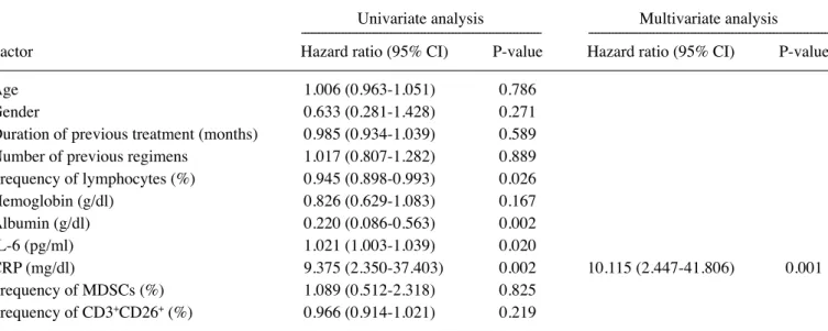
Relationship between clinical findings or laboratory data and OS. The median OS for the 41 patients was 304 days, with a one- year survival rate of 42% (Fig. 1). The Cox proportional hazards model was used to identify factors that were significantly associated with OS from clinical findings or laboratory data before vaccination. Univariate analysis using pre-vaccination data showed that albumin, CRP, SAA, IL-6, and the frequency
of lymphocytes in whole blood (P=0.002, P=0.002, P=0.004, P=0.020, and P=0.026, respectively) were significantly predic- tive of OS (Table IV). However, none of other factors examined, including age, gender, performance status, duration of chemo- therapy or target therapy before vaccination, number of previous regimens, or other laboratory data (hemoglobin, creatinine, frequencies of regulatory T cells, MDSCs, or CD3
+CD26
+cells), were significantly correlated with OS (data not shown). In addition, multivariate Cox regression analysis was performed to evaluate the influence of each of the factors that had been shown to be significantly associated with OS in the univariate analysis (P<0.05), after adjusting for possible confounding factors. Albumin, CRP, IL-6, and the frequency of lymphocytes in whole blood were included in the multivariate Cox regression analysis. SAA was excluded in this analysis, since the level of SAA was highly correlated with that of CRP (Spearman rank Table IV. Univariate and multivariate analysis with pre-vaccination clinical findings or laboratory data.
Univariate analysis Multivariate analysis --- ---
Factor Hazard ratio (95% CI) P-value Hazard ratio (95% CI) P-value
Age 1.006 (0.963-1.051) 0.786
Gender 0.633 (0.281-1.428) 0.271
Duration of previous treatment (months) 0.985 (0.934-1.039) 0.589 Number of previous regimens 1.017 (0.807-1.282) 0.889 Frequency of lymphocytes (%) 0.945 (0.898-0.993) 0.026
Hemoglobin (g/dl) 0.826 (0.629-1.083) 0.167
Albumin (g/dl) 0.220 (0.086-0.563) 0.002
IL-6 (pg/ml) 1.021 (1.003-1.039) 0.020
CRP (mg/dl) 9.375 (2.350-37.403) 0.002 10.115 (2.447-41.806) 0.001
Frequency of MDSCs (%) 1.089 (0.512-2.318) 0.825
Frequency of CD3
+CD26
+(%) 0.966 (0.914-1.021) 0.219
CI, confidence interval; CRP, C-reactive protein; MDSCs, myeloid-derived suppressor cells.Table V. Univariate and multivariate analysis with post-vaccination clinical findings or laboratory data.
Univariate analysis Multivariate analysis
--- ---
Factor Hazard ratio (95% CI) P-value Hazard ratio (95% CI) P-value
Skin reactions at injection sites 0.861 (0.287-2.585) 0.789 Increase in T cell responses 0.708 (0.227-2.203) 0.551 Increase in humoral responses 1.042 (0.407-2.669) 0.932 Frequency of lymphocytes (%) 0.953 (0.909-0.999) 0.048
Hemoglobin (g/dl) 0.745 (0.546-1.017) 0.064
Albumin (g/dl) 0.169 (0.064-0.445) <0.001
IL-6 (pg/ml) 1.055 (1.023-1.087) <0.001
CRP (mg/dl) 13.250 (2.095-83.794) 0.006 23.127 (2.919-183.233) 0.003
Frequency of MDSCs (%) 0.833 (0.183-3.785) 0.813
Frequency of CD3
+CD26
+(%) 0.956 (0.916-0.998) 0.042 0.952 (0.917-0.989) 0.012
CI, confidence interval; CRP, C-reactive protein; MDSCs, myeloid-derived suppressor cells.correlation coefficient, 0.819; P<0.001). As shown in Table IV, higher CRP level in pre-vaccination plasma was significantly predictive of unfavorable OS [hazard ratio (HR)=10.115, 95%
confidence interval (CI)=2.447-41.806, P=0.001]. However, the other factors showed no significant association.
Similarly, the Cox proportional hazards model was used to identify factors associated with OS from clinical findings or laboratory data at the end of the first cycle of vaccination.
Univariate analysis showed that albumin, IL-6, SAA, CRP, frequency of CD3
+CD26
+cells, and frequency of lymphocytes in whole blood were predictive of OS at the end of the first cycle of vaccination (P<0.001, P<0.001, P=0.004, P=0.006, P=0.042, and P=0.048, respectively) (Table V). None of the other factors, including other laboratory data, increase in IgG or T cell responses to the vaccine peptides, and skin reactions at the injection sites, were significantly correlated with OS. Albumin, IL-6, CRP, frequency of CD3
+CD26
+cells, and frequency of lymphocytes were included in the multivariate Cox regression analysis. SAA was excluded in this analysis, since the level of SAA was highly correlated with that of CRP (Spearman rank correlation coefficient, 0.698; P<0.001). Multivariate Cox regression analysis demonstrated that higher CRP level and lower frequency of CD3
+CD26
+cells in post-vaccination samples were predictive of unfavorable OS (HR=23.127, 95%
CI=2.919-183.233, P=0.003; HR=0.952, 95% CI=0.917-0.989, P=0.012) (Table V).
Discussion
Since only a subset of patients obtain clinical benefits from peptide-based cancer vaccines, it would be critical to identify biomarkers for selection of suitable patients (15-17). With regard to post-vaccination biomarkers, we have shown that an increase in peptide-specific IgG responses after PPV is well associated with improved OS in patients with certain types of cancers (12,18). In addition, several factors, including cytotoxic T lymphocytes (CTL) responses, Th1 responses, delayed type hypersensitivity (DTH), and autoimmunity, have also been reported to be associated with clinical responses in some clinical trials (16,17,19,20), although these results have not always been reproducible. Notably, there are currently no validated pre-vaccination biomarkers, predictive of clinical responses, in widespread use. Therefore, in the present study, we searched for clinically useful predictive markers for PPV in patients with NSCLC. Multivariate analysis of pre- vaccination factors showed that higher level of plasma CRP was predictive of unfavorable OS. Among post-vaccination factors, higher level of plasma CRP and lower frequency of CD3
+CD26
+cells were predictive of unfavorable OS.
Although more data are still needed to validate our findings, evaluation of the factors identified here could be useful for selecting patients with NSCLC who would potentially benefit from cancer vaccines.
Elevated CRP level was shown to be also a predictor of unfavorable OS in NSCLC patients receiving chemotherapy or targeted therapy (21,22), suggesting that it might not necessarily be unique to vaccinated patients. In contrast, the frequency of CD3
+CD26
+cells among PBMCs has not been reported previously as a biomarker in NSCLC patients. CD26 is a cell surface glycoprotein that functions as a proteolytic
enzyme, dipeptidyl peptidase IV, and plays a critical role in signal transduction (23). Since it is highly expressed on acti- nal transduction (23). Since it is highly expressed on acti- l transduction (23). Since it is highly expressed on acti- vated T cells (23), increased frequency of CD3
+CD26
+might reflect the immune activation induced by vaccination. The role of CD26
+activated T cells induced by PPV in NSCLC thus remains to be determined.
MDSCs are a heterogeneous population of immature myeloid cells that inhibit the functions of other immune cells and promote tumor progression (24,25). MDSCs can facilitate tumor growth by inducing angiogenesis at tumor sites or by suppressing anti-tumor immune cells, such as antigen-specific T cells (24,25). Notably, the frequencies of MDSCs were significantly decreased after PPV. In addition, the patients who showed a decrease or no change in IL-6 after vaccination had a tendency to have better outcome. IL-6 is a multifunctional cytokine that regulates various aspects of cancer development, such as tumor cell growth and suppression of anti-tumor immune cells, including CTL and NK cells (26). The roles of these immune suppressive cells and/or cytokine, MDSCs and IL-6, in immune responses to cancer vaccines remain to be examined.
The prognosis of refractory NSCLC patients remains very poor, with a median survival time of 6-8 months (1-5). In contrast, the median OS of the 41 NSCLC patients who received PPV was 304 days (>10 months), with a one-year survival rate of 42%, in the current study. The main toxicity of PPV was skin reactions at the injection sites, but no SAEs were observed. Our previous trials of PPV for various types of cancers have also confirmed its safety (13). Considering the disease conditions of the patients enrolled in the current study, all of whom had already been resistant to or ineligible for conventional chemo- nal chemo- l chemo- therapeutic and targeted agents before enrollment, our findings suggest the feasibility of PPV for refractory NSCLC, even though OS was not the main objective of the current study.
Nevertheless, since this is a retrospective study with a limited number of patients, clinical utility of PPV should be further verified in larger-scale, prospective trials conducted in defined patient populations with or without receiving PPV.
Acknowledgements
This study was supported by the grants from the Regional Innovation Cluster Program of the Ministry of Education, Culture, Sports, Science and Technology of Japan (K. Itoh);
Kurozumi Medical Foundation (T. Sasada), and Osaka Cancer Research Foundation (T. Sasada).
References
1. de Marinis F and Grossi F: Clinical evidence for second- and third-line treatment options in advanced non-small cell lung cancer. Oncologist 13 (Suppl 1): 14-20, 2008.
2. Janku F, Stewart DJ and Kurzrock R: Targeted therapy in non- small-cell lung cancer - is it becoming a reality? Nat Rev Clin Oncol 7: 401-414, 2010.
3. Adjei AA, Mandrekar SJ, Dy GK, et al: Phase II trial of peme- trexed plus bevacizumab for second-line therapy of patients with advanced non-small-cell lung cancer: NCCTG and SWOG study N0426. J Clin Oncol 28: 614-619, 2010.
4. Krzakowski M, Ramlau R, Jassem J, et al: Phase III trial comparing vinflunine with docetaxel in second-line advanced non-small-cell lung cancer previously treated with platinum- containing chemotherapy. J Clin Oncol 28: 2167-2173, 2010.
5. Okamoto I, Yoshioka H, Morita S, et al: Phase III trial comparing oral S-1 plus carboplatin with paclitaxel plus carboplatin in chemotherapy-naïve patients with advanced non-small-cell lung cancer: results of a west Japan oncology group study. J Clin Oncol 28: 5240-5246, 2010.
6. Terasaki M, Shibui S, Narita Y, et al: Phase I trial of a personalized peptide vaccine for patients positive for human leukocyte antigen- A24 with recurrent or progressive glioblastoma multiforme. J Clin Oncol 29: 337-344, 2011.
7. Yanagimoto H, Shiomi H, Satoi S, et al: A phase II study of personalized peptide vaccination combined with gemcitabine for non-resectable pancreatic cancer patients. Oncol Rep 24:
795-801, 2010.
8. Hattori T, Mine T, Komatsu N, et al: Immunological evaluation of personalized peptide vaccination in combination with UFT and UZEL for metastatic colorectal carcinoma patients. Cancer Immunol Immunother 58: 1843-1852, 2009.
9. Noguchi M, Kakuma T, Uemura H, et al: A randomized phase II trial of personalized peptide vaccine plus low dose estramustine phosphate (EMP) versus standard dose EMP in patients with castration resistant prostate cancer. Cancer Immunol Immunother 59: 1001-1009, 2010.
10. Itoh K, Yamada A, Mine T and Noguchi M: Recent advances in cancer vaccines: an overview. Jpn J Clin Oncol 39: 73-80, 2009.
11. Itoh K and Yamada A: Personalized peptide vaccines: a new therapeutic modality for cancer. Cancer Sci 97: 970-976, 2006.
12. Noguchi M, Mine T, Komatsu N, et al: Assessment of immuno- logical biomarkers in patients with advanced cancer treated by personalized peptide vaccination. Cancer Biol Ther 10: 1266-1279, 2011.
13. Yoshida K, Noguchi M, Mine T, et al: Characteristics of severe adverse events after peptide vaccination for advanced cancer patients: Analysis of 500 cases. Oncol Rep 25: 57-62, 2011.
14. Komatsu N, Shichijo S, Nakagawa M and Itoh K: New multi- plexed flow cytometric assay to measure anti-peptide antibody: a novel tool for monitoring immune responses to peptides used for immunization. Scand J Clin Lab Invest 64: 535-545, 2004.
15. Sasada T, Komatsu N, Suekane S, Yamada A, Noguchi M and Itoh K: Overcoming the hurdles of randomised clinical trials of therapeutic cancer vaccines. Eur J Cancer 46: 1514-1519, 2010.
16. Disis ML: Immunologic biomarkers as correlates of clinical response to cancer immunotherapy. Cancer Immunol Immunother 60: 433-442, 2011.
17. Hoos A, Eggermont AM, Janetzki S, et al: Improved endpoints for cancer immunotherapy trials. J Natl Cancer Inst 102: 1388-1397, 2010.
18. Mine T, Sato Y, Noguchi M, et al: Humoral responses to peptides correlate with overall survival in advanced cancer patients vaccinated with peptides based on pre-existing, peptide-specific cellular responses. Clin Cancer Res 10: 929-937, 2004.
19. Amos SM, Duong CP, Westwood JA, et al: Autoimmunity associated with immunotherapy of cancer. Blood 118: 499-509, 2011.
20. López MN, Pereda C, Segal G, et al: Prolonged survival of dendritic cell-vaccinated melanoma patients correlates with tumor-specific delayed type IV hypersensitivity response and reduction of tumor growth factor beta-expressing T cells. J Clin Oncol 27: 945-952, 2009.
21. Koch A, Fohlin H and Sörenson S: Prognostic significance of C-reactive protein and smoking in patients with advanced non-small cell lung cancer treated with first-line palliative chemotherapy. J Thorac Oncol 4: 326-332, 2009.
22. Masago K, Fujita S, Togashi Y, et al: Clinical significance of pretreatment C-reactive protein in patients with advanced nonsquamous, non-small cell lung cancer who received gefitinib.
Oncology 79: 355-362, 2010.
23. Ohnuma K, Dang NH and Morimoto C: Revisiting an old acquaintance: CD26 and its molecular mechanisms in T cell function. Trends Immunol 29: 295-301, 2008.
24. Gabrilovich DI and Nagaraj S: Myeloid-derived suppressor cells as regulators of the immune system. Nat Rev Immunol 9:
162-174, 2009.
25. Peranzoni E, Zilio S, Marigo I, et al: Myeloid-derived suppressor cell heterogeneity and subset definition. Curr Opin Immunol 22:
238-244, 2010.
26. Naugler WE and Karin M: The wolf in sheep’s clothing: the role of interleukin-6 in immunity, inflammation and cancer. Trends Mol Med 14: 109-119, 2008.