Acta Med. Nagasaki 38:285 - 287
Esophagectomy in Combination with a Resection of Involved Lung for Esophageal Cancer
Masao Tomita, Hiroyoshi Ayabe, Katsunobu Kawahara, Yutaka Tagawa, Masayuki Obatake, Akihiro Nakamura, Nobufumi Sasaki, Hiroshi Shingu, Takeshi Nagayasu, Kazuhiko Hatano, Masashi Muraoka, Satoshi Yamamoto and Seiichirou Ide
The First Department of Surgery, Nagasaki University School of Medicine
The combined resection with involved lung for esophageal carcinoma was evaluated in terms of surgical indication and outcome in the 6 patients who underwent subtotal esophagectomy with pulmonary resection. It was con- firmed that the operation was technically feasible but the surgical results were unsatisfactory. It was reasoned that grave surgical insult and adjuvant therapy to prevent recur- rence result in immunodepressive status of the host and tends to accompany postoperative complications related to operative death.
In conclusion, prevention of immunosuppression for the host is required by meticulous cares of nutrition and elimi- nation of surgical stress by staged operation in order to obtain satisfactory result after surgery.
Introduction
The surgical outcomes of esophageal carcinomas were unsatisfactory even though their pathogenesis and etiology had been elucidated. The reason is attributable to far ad- vanced extension of carcinoma on the basis of anatomical specificity of the esophagus which is adjacent to the organs surrounding the loose tissue and devoid of the serosal layer, probably playing a role in the barrier of extension of cancer infiltration.
In advanced esophageal carinomas, it is not infrequently encountered that cancer infiltration extends outside the wall of the esophagus and involves the neighbouring organs. At
that time, surgeons attempt to perform a combined resec- tion with involved organs in an effort to accomplish opera- tive radicality despite grave insult surgery.
The combined resection with involved organ should be striven for establishment of surgical safety in terms of immunodepression of the patient by surgical stress, deter- mination of surgical indication for the poor risk patients and prevention of surgical complications related to opera- tive death.
The aim of this study is to clarify the validity of the combined resection of the esophageal lesion with the in- volved lung on the basis of the result of our clinical experience.
Patients
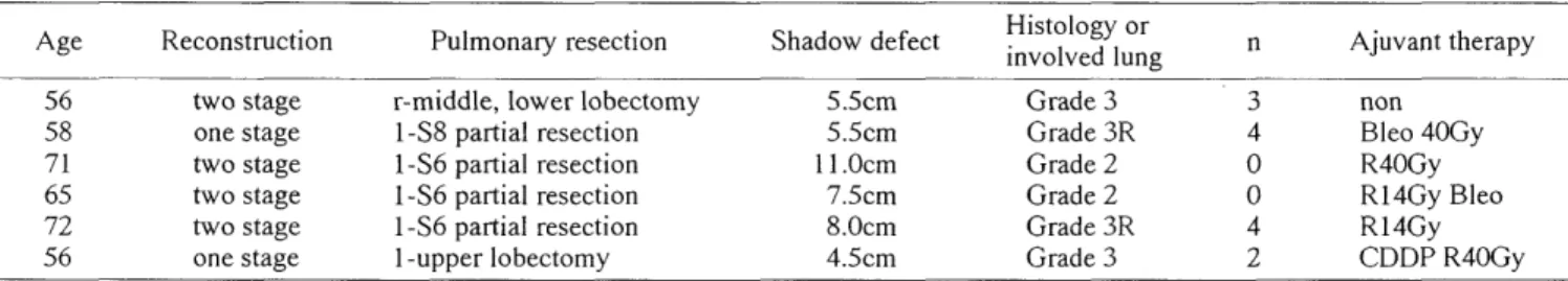
Six patients were eligible to this study (Table 1). They underwent the combined resection of the esophagus with the lung for thoracic esophageal carcinomas.
The ages ranged from 56 to 72 with an average of 63.2 years The ratio of men to women was equivalent. The histologic type was squamous cell carcinoma in all. The tumor locations were Im in 3, Im + lu in 2 and Im + Ei in one, respectively.
The length of shadow defect varied from 4.5cm to 11.0cm. The histologic findings of resected lungs were graded as the following categories (Fig. 1, 2, 3), Grade 0:
no evidence of histologic cancer invasion to the lung with
Table 1. Patient Profile
Age Reconstruction Pulmonary resection Shadow defect Histology or i n Ajuvant therapy nvolved lung
56 two stage r-middle, lower lobectomy 5.5cm Grade 3 3 non
58 one stage 1-S8 partial resection 5.5cm Grade 3R 4 Bleo 40Gy
71 two stage 1-S6 partial resection 11.0cm Grade 2 0 R40Gy
65 two stage 1-S6 partial resection 7.5cm Grade 2 0 R14Gy Bleo
72 two stage 1-S6 partial resection 8.0cm Grade 3R 4 R14Gy
56 one stage 1-upper lobectomy 4.5cm Grade 3 2 CDDP R40Gy
r:right, l:left, R:preoperative irradiation
Fig. 1. Histology of Grade 1 showing involvement of the visceral pleura
Fig. 2. Histology of Grade 2 revealing preferable involvement of small bronchi and vessels
cancer infiltration preferable to the bronchial and vessel walls in the pulmonary parenchym. Grade 3: dense cancer infiltration to the lung regardless of the alveolar and the interstitial spaces. Although preoperative shadow defect ranged from 4.5cm to 11.0cm in length, a longer shadow defect was not proportional to the degree and the range of the involved lung.
Two out of 6 had no evidence of histologic cancer invasion to the lung. These received preoperative radiation of 14 Gy, and bleomycin was prescribed in one. It is not acertained as to whether involved lungs were benefited from irradiation therapy. Histologic finding revealed that Grade 3 was seen in 4, in which two received preoperative irradiation therapy. Grade 2 was indicated in 2. A n- category was also histologically assessed. Two patients were categorized in n4, one in n3, one in n2 and the other two in no, the respectively. Preoperative irradiation therapy was done in 5, radiation dosis ranging from 40 Gy to 14 Gy, and preoperative chemotherapy was bleomycin in two and CDDP in one, respectively. The operative procedure was subtotal esophagectomy in all in whom reconstruction was performed by the two staged operation in three. The operative procedures concurrently used were bilobectomy in one, lobectomy in one and partial resection in 4. As postoperative complications we have encountered respi- ratory failure in one, heart failure in one and minor anasto- motic insufficiency in 3. The two operative deaths, of heart failure and acute respiratory failure, were encountered on the first day and on the 12th day of surgery, respectively.
The other 4 patients have expired of pneumonia in one and cancer recurrence in 3, at 4 to 7 months after surgery.
The surgical outcome was unsatisfactory. The reason for the unsatisfactory result remains obscure but main reasons are grave operative insult and far advanced carcinoma of the disease stages.
Fig. 3. Histology of Grade 3 illustrating a dense cancer infil- tration regardless less of alveolar and interstitial spaces