Systemic impact on secondary brain aggravation due to ischemia/reperfusion injury in post-cardiac arrest syndrome: a prospective observational study using high-mobility group box 1 protein
日本大学医学部救急医学系救急集中治療医学分野 杉田 篤紀
2017 年
指導教員 木下 浩作
R E S E A R C H Open Access
Systemic impact on secondary brain
aggravation due to ischemia/reperfusion injury in post-cardiac arrest syndrome:
a prospective observational study using high-mobility group box 1 protein
Atsunori Sugita, Kosaku Kinoshita, Atsushi Sakurai*, Nobutaka Chiba, Junko Yamaguchi, Tsukasa Kuwana, Nami Sawada and Satoshi Hori
Abstract
Background:Ischemia/reperfusion injury (I/R) is an important pathophysiology of post-cardiac arrest syndrome (PCAS) against multiple organ dysfunction and mortality. The inflammatory response in PCAS causes systemic I/R.
The purpose of this study was to demonstrate the pathophysiology of systemic I/R for secondary brain damage using the biomarkers high-mobility group box 1 (HMGB1), neuron-specific enolase (NSE), and interleukin-6 (IL-6).
Methods:This study was designed as a single-institution prospective observational study. Subjects were observed for 90 days, and neurological outcome was classified according to the Glasgow-Pittsburgh Cerebral Performance Categories Scale (CPC). Serum HMGB1, NSE, and IL-6 were evaluated for variability, correlation with each biomarker, or the Sequential Organ Function Assessment (SOFA) score and CPC at return of spontaneous circulation at 0, 24, 48, and 168 h.
Results:A total of 128 patients were enrolled in this study. Initial HMGB1 correlated with CPC (ρ= 0.27,p= 0.036) and SOFA score (ρ= 0.33,p< 0.001). The early phase of HMGB1 (0–24 h), all phases of IL-6, and the delayed phase of NSE (24–168 h) manifested poor neurological outcome. HMGB1 showed a significant correlation with NSE (ρ= 0.29, p= 0.002 at 0 h; ρ= 0.42, p< 0.001 at 24 h) and IL-6 (ρ= 0.36, p< 0.001 at 24 h).
Conclusions: Serum HMGB1 for first 24 h after cardiac arrest was significantly correlated with SOFA score, NSE, and IL-6. This result suggests that systemic I/R may contribute to secondary brain aggravation. It is expected that research on HMGB1 focused on systemic I/R will help prevent aggravating neurological outcomes.
Keywords:HMGB1, IL-6, NSE, SOFA score, Post-cardiac arrest syndrome, Systemic ischemia/reperfusion injury, Secondary brain injury
* Correspondence:[email protected]
Division of Emergency and Critical Care Medicine, Department of Acute Medicine, Nihon University School of Medicine, 30-1 Oyaguchi Kamimachi, Itabashi-ku, Tokyo 173-8610, Japan
© The Author(s). 2017Open AccessThis article is distributed under the terms of the Creative Commons Attribution 4.0 International License (http://creativecommons.org/licenses/by/4.0/), which permits unrestricted use, distribution, and reproduction in any medium, provided you give appropriate credit to the original author(s) and the source, provide a link to the Creative Commons license, and indicate if changes were made. The Creative Commons Public Domain Dedication waiver (http://creativecommons.org/publicdomain/zero/1.0/) applies to the data made available in this article, unless otherwise stated.
Background
The inflammatory response in patients with post-cardiac arrest syndrome (PCAS) causes systemic ischemia/reper- fusion injury (I/R), which may lead to multiple organ dysfunction and mortality, and is described as “sepsis- like syndrome” [1]. Inflammatory cytokines play an important role in systemic I/R and are promoted by high-mobility group box 1 protein (HMGB1) [2]. Previ- ous pilot studies showed that elevated serum HMGB1 is related to poor neurological outcome in PCAS [3, 4].
However, this does not sufficiently explain how systemic I/R affects neurological outcome on the basis of the limited data available. HMGB1 protein is passively re- leased by necrotic or damaged cells and actively secreted by innate immune cells. Extracellular HMGB1 promotes production of systemic inflammatory cells such as mac- rophages, monocytes, and dendritic cells [5]. HMGB1 is also regarded as a proinflammatory cytokine which act on systemic organ I/R [2]. I/R also causes oxidative stress on vital organs, such as the heart, liver, kidneys, and brain, accompanied by accumulation of HMGB1 [6].
Interleukin (IL)-6 is also activated by the inflammatory response to systemic I/R [7]. Several studies have shown that IL-6 is an inflammatory factor in PCAS [1, 8]. IL-6, which has an established measurement method, is known to be promoted by HMGB1. Consequently, IL-6 is considered to be useful in comparison with HMGB1.
Early release of HMGB1 in brain tissue after brain ische- mia has been reported [9] and is released into the extra- cellular space of brain tissue [10]. It is also related to increased permeability of the blood-brain barrier (BBB) [11]. In this context, serum concentration of brain pro- teins such as the neuron-derived enzyme neuron- specific enolase (NSE), which is released after stroke [12] and cardiac arrest [13], has been assessed as a method of predicting secondary brain injury. NSE is an established and well-known measurement method used to predict poor neurological outcome in PCAS. We speculate that these markers can identify possible car- diac arrest survivors and prognosis. We focused on sys- temic inflammation after cardiac arrest that induces whole-body ischemia, including the brain. The purpose of this study was to demonstrate the pathophysiology of systemic I/R due to secondary brain damage with analysis of the variability of three biomarkers (HMGB1, NSE, and IL-6) in an early phase of PCAS.
Methods Study design
This study was designed as a single-institution prospect- ive observational study and was conducted from January 2011 to July 2013. Inclusion criteria were (1) patients who achieved return of spontaneous circulation (ROSC) from out-of-hospital cardiac arrest (OHCA) and who
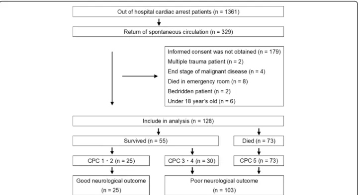
were brought to the emergency and critical care depart- ment, regardless of cardiac or noncardiac etiology; and (2) patients who did not meet the exclusion criteria. The exclusion criteria were (1) informed consent not obtained, (2) cases of multiple trauma, (3) end-stage malignancy, (4) death in the emergency room, (5) bedridden prior to hospitalization, and (6) less than 18 years old (Fig. 1).
Patients brought to the emergency and critical care de- partment were registered and tabulated using the medical record and emergency medical service (EMS) report based on the Utstein style: (1) age, (2) sex, (3) witness present, (4) cardiopulmonary resuscitation (CPR) performed by a bystander, (5) shockable waveform observed on the electro- cardiogram (ECG) at the time of EMS arrival, (6) time from receipt of the emergency call to ROSC, (7) adrenaline administration, and (8) cardiac etiology [14]. A shockable waveform is a wave pattern on the ECG that indicates arrhythmia necessitating defibrillation, such as ventricular fibrillation or pulseless ventricular tachycardia.
The following factors related to treatment and evalu- ation after hospitalization were also considered: (1) whether coronary angiography and percutaneous coron- ary intervention (PCI) were performed and (2) whether therapeutic hypothermia (TH) was performed. Three biomarkers (HMGB1, IL-6, and NSE) were compared.
The primary endpoint was set as neurological outcome at 90 days after ROSC.
Clinical protocol
Basic life support was performed by public and co-medics, including use of an automated external defibrillator. Defib- rillation, airway protection including intubation, and adren- aline infusion were performed by EMS personnel [15].
Advanced cardiovascular life support [16, 17] and emer- gency cardiovascular care [18, 19] were performed prior to admission to the intensive care unit (ICU). TH was performed according to the following exclusion criteria:
(1) unstable hemodynamics even with the use of vasopres- sors (mean blood pressure < 60 mm Hg or systolic blood pressure < 90 mm Hg), (2) inadequate oxygenation (ratio of partial pressure of arterial oxygen to fraction of inspired oxygen < 200), (3) end stage of a chronic disease, and (4) informed consent not obtained from the patient’s family [20]. Patients were subjected to a modulated temperature of 34 °C for 24 h during TH and gradually rewarmed from 34 °C to 36 °C for 24 h using an external cooling device.
During TH, patients were placed under anesthesia using midazolam hydrochloride as sedation, fentanyl citrate as analgesia, and rocuronium bromide as a muscle relaxant.
Sample collection
Physical findings and clinical measurements were re- corded in the medical chart. Blood samples collected from
the peripheral artery were evaluated using clinical vari- ables within 6 h of ROSC and 24 h, 48 h, and 7 days after ROSC (abbreviated as 0 h, 24 h, 48 h, and 168 h, respectively). Serum samples for biomarkers (HMGB1, IL-6, and NSE) were centrifuged at 3000 cycles for 15 mi- nutes at 4 °C. Separated samples were preserved frozen in a−80 °C refrigerator until analysis. Blood parameters were measured in the clinical laboratory department by coagula- tion test, chemical examination, and complete cell count.
Measurements
Serum concentration of HMGB1 was measured using an enzyme-linked immunosorbent assay (ELISA; Shino- Test Corporation, Kanagawa, Japan). The lower sensitivity of HMGB1 was 0.2 ng/ml, and the cross-reactivity of HMGB2 was < 2%. IL-6 and NSE were measured using the Quantikine® ELISA kit (R&D Systems, Minneapolis, MN, USA). The minimum detectable doses of IL-6 and NSE were typically < 0.70 pg/ml and < 0.038 ng/ml, respectively.
Mortality, multiple organ dysfunction, and outcome Survival time and mortality in the early phase of PCAS was evaluated. Multiple organ dysfunction was evaluated according to individual organ dysfunction subscales of the Sequential Organ Failure Assessment (SOFA) [21].
SOFA scores were measured at ROSC in the early phase of PCAS according to (1) Glasgow Coma Scale (GCS) score, (2) blood pressure with vasopressor, (3) platelet count, (4) total bilirubin, and (5) creatinine. To rule out
the influence of first brain insult by cardiac arrest, SOFA scores excluding GCS scores between two neurological outcome groups were compared.
Neurological outcome was evaluated according to the Glasgow-Pittsburgh Cerebral Performance Categories Scale (CPC) as follows: CPC 1 (good recovery), CPC 2 (moderate disability), CPC 3 (severe disability), CPC 4 (vegetative state), and CPC 5 (death). Subjects were divided into two groups by CPC category: a good neuro- logical outcome group (CPC 1 or 2) and a poor neuro- logical outcome group (CPC 3–5).
Biomarkers, multiple organ dysfunction, and neurological outcome
SOFA score was evaluated according to CPC and HMGB1 at 0 h. The variability of three biomarkers (HMGB1, IL-6, and NSE) was assessed, and each peak level was compared. Serum levels of the three bio- markers were compared between the two neurological outcome groups, and correlations were evaluated.
Subanalysis
In this study, patients were divided into groups for sub- group analysis, namely good or poor neurological out- come, cardiac etiology or noncardiac etiology, and with PCI or without PCI, in order to assess biomarker vari- ability at various points in time.
Fig. 1Flowchart of the study design and results of the number of patients. Outcome is shown at 90 days after return of spontaneous circulation.
CPCGlasgow-Pittsburgh Cerebral Performance Categories Scale
Statistical analysis
Statistical analysis was performed using the IBM SPSS Statistics version 22 statistical software program (IBM, Armonk, NY, USA). The Kaplan-Meier method was per- formed to evaluate mortality. Collected measurements were analyzed for normal distribution using the Shapiro- Wilk test. Median and IQR (first quartile to third quar- tile) statistics were used for nonparametric measure- ments, and average and SD were used for parametric measurements.
The Mann-Whitney U test was performed for non- parametric data, and Student’s ttest was performed for parametric data. The chi-square test and Fisher’s exact test were performed for categorical data. The Kruskal- Wallis test was performed for multiple data compari- sons. Spearman’s rank correlation test was performed to evaluate correlations. Simple logistic regression analysis was carried out. An explanatory variable was set as HMGB1 at 0 h, and response variables included two neurological outcome groups.
Results
Patient characteristics
A total of 128 patients were enrolled in this study after the exclusion criteria were applied. Of 1361 patients with OHCA who were brought to the emergency room, 329 pa- tients achieved ROSC. Among these of 329 patients, 201 satisfied the exclusion criteria, including informed consent not being obtained (n= 179), cases of multiple trauma (n= 2), end-stage malignancy (n= 4), death in the emer- gency room (n= 8), bedridden prior to hospitalization (n= 2), and being less than 18 years old (n= 6).
Median survival time was 25 days, and the worst mor- tality was 11% within the first 24 h. Survival numbers and times are as follows: 114 after 24 h, 101 after 48 h, 82 after 7 days, and 55 after 90 days. In total, 73 patients died within the follow-up interval of 90 days. Twenty- five patients had a good neurological outcome (CPC 1, n= 24; CPC 2, n= 1), and 103 patients had a poor neurological outcome (CPC 3, n= 13; CPC 4, n= 17;
CPC 5,n= 73). Seventy-two patients classified as CPC 5 (n= 73) died without recovery from the comatose state even once within the follow-up period. One patient clas- sified as CPC 5 recovered from the comatose state but died within 90 days as a result of cardiac complications.
Characteristics of all patients are shown in Table 1.
Seventy-three (57%) patients had a cardiac etiology, and 55 (43%) had a noncardiac etiology. The ratio of patients with a cardiac etiology was higher in the good neuro- logical outcome group. Noncardiac etiology included airway obstruction due to a foreign body (n= 19), acute stroke (n= 7), pneumonia (n= 7), chronic obstructive pulmonary disease (n= 4), hyperkalemia (n= 4), gastro- intestinal hemorrhage (n= 3), neck hanging (n= 3), sepsis
(n= 3), drug intoxication (n= 1), diabetic ketoacidosis (n= 1), heatstroke (n= 1), and pulmonary embolus (n= 1).
A significant difference in sex and age was observed, but witness and bystander CPR were similar in the two groups. Patients in the poor neurological outcome group had a significantly longer interval from receipt of the emergency call to ROSC, as well as to nonshockable wave, and a higher dose of required adrenaline adminis- tration than patients in the good neurological outcome group.
Emergency cardiac angiography was performed on 63 patients. Patients in the good neurological outcome group achieved a higher PCI than those in the poor neurological outcome group. TH was performed on 70 (55%) patients and was similar between the two groups.
Correlation with SOFA score was significant in the poor neurological outcome group, especially for circulation instability, liver dysfunction, and coagulation dysfunction (Table 1).
Biomarker variability
A significant sequential difference in the levels of initial serum HMGB1 or NSE after ROSC was observed. When comparing values at 0 h with those at 24 or 48 h, we observed a gradual decrease (HMGB1; Fig. 2a) or increase (NSE; Fig. 2b). However, there was no significant change in serum HMGB1 or NSE levels between 48 and 168 h (Fig. 2a, b). Regarding IL-6 levels, no significant difference was observed for ROSC at 24, 48, or 168 h (Fig. 2c).
SOFA score was positively and significantly correlated with CPC (ρ= 0.44, p< 0.001) and HMGB1 (ρ= 0.33, p< 0.001) at 0 h. A significant positive correlation be- tween HMGB1 and neurological outcomes (CPC 1, 2, 3, 4, or 5) was observed, and a high HMGB1 indicated a weak but significant correlation with a poor neuro- logical outcome (ρ= 0.27,p= 0.036). The relationships between HMBG1 and IL-6 or NSE at the same point in time indicated that HMGB1 had a partial but significant positive correlation with NSE (0 h,ρ= 0.29, p= 0.002; 24 h, ρ= 0.42, p< 0.001; 48 h, ρ= 0.17, p= 0.135) and IL-6 (0 h,ρ= 0.14, p= 0.126; 24 h, ρ= 0.36, p< 0.001; 48 h,ρ= 0.11,p= 0.337).
Biomarkers and outcome
The correlation between biomarkers and patient out- come is shown in Fig. 3. Serum HMGB1 (Fig. 3a) was significantly higher in the poor neurological outcome group than in the good neurological outcome group at 0 and 24 h. In the poor neurological outcome group, serum NSE (Fig. 3b) and IL-6 (Fig. 3c) values were significantly higher, with a wide dispersion, than those in the good neurological outcome group from 0 to 168 h, but there was no difference between these outcome groups for NSE at 0 h. Serum NSE was similar in the
two groups at 0 h and began to increase after 24 h. In the poor neurological outcome group, NSE peaked with a median value at 48 h, later than HMGB1 and IL-6 peaked.
On the basis of single-variable logistic regression ana- lysis, a significant poor outcome for PCAS was identified by observing good neurological outcomes in patients with low HMGB1 (adjusted OR 0.963, 95% CI 0.933–
0.994, p= 0.021) according to changes in each single value. Of these patients, a significant correlation was observed between HMGB1 and NSE or IL-6 at 0 h (NSE, ρ= 0.29, p= 0.002; IL-6, ρ= 0.14, p= 0.126) or 24 h (NSE, ρ= 0.42, p< 0.001; IL-6, ρ= 0.36, p< 0.001).
In the subanalysis of this study, no significant correlation between HMGB1 and NSE at 0 h (ρ= 0.089, p= 0.680), 24 h (ρ=−0.13, p= 0.586), or 48 h (ρ= 0.30, p= 0.195) in the good neurological outcome group was observed.
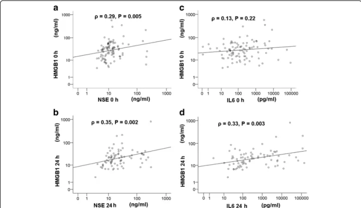
However, a significant positive correlation between HMGB1 and NSE at 0 h (ρ= 0.29, p= 0.005) and 24 h (ρ= 0.35, p= 0.002) was observed, but not at 48 h, in the poor neurological outcome group (Fig. 4).
However, the correlation between neurological out- come and biomarkers (HMGB1, NSE, and IL-6) according to cardiac etiology (Fig. 5) was similar to that of cases that included cardiac and noncardiac etiology.
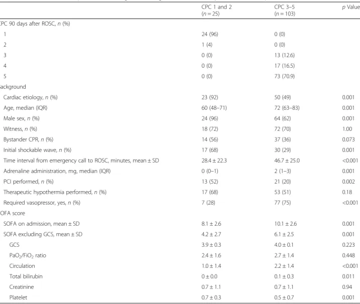
Table 1Characteristics of all patients according to neurological outcome 90 days after return of spontaneous circulation CPC 1 and 2
(n= 25)
CPC 3–5
(n= 103) pValue
CPC 90 days after ROSC,n(%)
1 24 (96) 0 (0)
2 1 (4) 0 (0)
3 0 (0) 13 (12.6)
4 0 (0) 17 (16.5)
5 0 (0) 73 (70.9)
Background
Cardiac etiology,n(%) 23 (92) 50 (49) 0.001
Age, median (IQR) 60 (48–71) 72 (63–83) 0.001
Male sex,n(%) 24 (96) 64 (62) 0.001
Witness,n(%) 18 (72) 72 (70) 1.00
Bystander CPR,n(%) 14 (56) 37 (36) 0.073
Initial shockable wave,n(%) 17 (68) 30 (29) 0.001
Time interval from emergency call to ROSC, minutes, mean ± SD 28.4 ± 22.3 46.7 ± 25.0 <0.001
Adrenaline administration, mg, median (IQR) 0 (0–1) 2 (1–3) 0.001
PCI performed,n(%) 13 (52) 21 (20) 0.002
Therapeutic hypothermia performed,n(%) 17 (68) 53 (51) 0.18
Required vasopressor, yes,n(%) 7 (28) 77 (75) <0.001
SOFA score
SOFA on admission, mean ± SD 8.1 ± 2.6 10.1 ± 2.6 0.001
SOFA excluding GCS, mean ± SD 4.2 ± 2.7 6.1 ± 2.5 0.001
GCS 3.9 ± 0.3 4.0 ± 0.1 0.223
PaO2/FiO2ratio 2.4 ± 1.6 2.7 ± 1.4 0.448
Circulation 1.0 ± 1.4 2.2 ± 1.4 <0.001
Total bilirubin 0 ± 0.0 0.1 ± 0.3 0.011
Creatinine 0.7 ± 1.1 0.7 ± 1.1 0.94
Platelet 0.7 ± 0.3 0.5 ± 0.7 0.001
Abbreviations: ROSCReturn of spontaneous circulation,CPCGlasgow-Pittsburgh Cerebral Performance Categories Scale,CPRCardiopulmonary resuscitation, PCIPercutaneous coronary Intervention,THTherapeutic hypothermia, SOFA Sequential Organ Failure Assessment,PaO2/FiO2Ratio of partial pressure of arterial oxygen to fraction of inspired oxygen,GCSGlasgow Coma Scale
Required vasopressorrefers to patients who needed an intravenous vasopressor after initial resuscitation on the first hospital day. Parametric data are presented as mean ± SD. Nonparametric data are presented as median with IQR. Categorical data are presented asn(%). The percentages in parentheses are the ratios for poor outcome or good outcome. Statistical significance was set atp< 0.05
Serum biomarkers according to cardiac or noncardiac etiology demonstrated that serum HMGB1 was similar between the two patterns of etiology, but NSE and IL-6 increases were higher in noncardiac etiology cases than in cardiac etiology cases (Fig. 6).
With PCI or without PCI, serum HMGB1 levels (median [IQR]) did not show a significant difference for with PCI vs without PCI (0 h, 24.0 [12.5 – 34.7]
vs 28.9 [12.6–54.6],p= 0.56; 24 h, 16.1 [9.86–32.2] vs 12.9 [8.5–27.7],p= 0.75).
Discussion
Some pilot studies focused on HMGB1 after cardiac arrest have been done regarding the influence on neuro- logical outcome [3, 4]. However, the pathophysiology of elevated serum HMGB1 affecting poor neurological
outcome is not sufficiently clear. The correlation be- tween systemic I/R and secondary brain aggravation has not been discussed. In the present study, serum HMGB1 correlated with serum NSE in patients with PCAS. This phenomenon may indicate a possible mechanism of a secondary brain injury process with systemic I/R after cardiac arrest.
Increasing serum HMGB1 at ROSC means that initial HMGB1 was passively released by necrotic or damaged cells due to cardiac arrest. The increase of HMGB1 affecting the neurological outcome lasts 24 h and shows a correlation with IL-6, indicating an inflammatory re- sponse. Serum elevation of HMGB1 for the first 24 h may include active excretion by inflammatory cells.
These two patterns of pathways to release of HMGB1 may promote exacerbation to inflammation as a
Fig. 2Variability of biomarkers (high-mobility group box 1 protein [HMGB1], neuron-specific enolase [NSE], and interleukin [IL]-6) from return of spontaneous circulation (ROSC). Biomarker variability was analyzed from ROSC to 7 days using box plots for HMGB1 (a), NSE (b), and IL-6 (c).
Measurement points were at 0, 24, 48, and 168 h (7 days) after ROSC. This plot is presented on a logarithmic scale. Statistical significance was set atp< 0.05 (*) andp< 0.001 (**) in the box plot. This box plot consists ofboxes,whiskers,open circles, andasterisksusing a logarithmic scale. The horizontal bold linein the middle of the box is the median value. Theboxis the IQR from the first quartile to the third quartile.Whiskersare the range of maximum and minimum values between 1.5 times IQR above the third quartile and 1.5 times IQR below the first quartile.Open circles are the outliers between 1.5 and 3 times IQR either above the third quartile or below the first quartile.Asterisksare the outliers three times the IQR either above the third quartile or below the first quartile. Number of patients in the graphs are 0 h (n= 128), 24 h (n= 114), 48 h (n= 101), and 168 h (n= 82)
systemic I/R in PCAS. The correlation with HMGB1 and SOFA score indicates that excessive inflammation in the early phase of PCAS contributes to the organ damage.
Interestingly, our study indicates a positive correlation between serum HMGB1 and NSE in the early phase, which is correlated with neurological outcome. This phenomenon was significantly observed in patients with a poor neurological outcome, but not in patients with a good neurological outcome.
The molecular weights of NSE and HMGB1 are ap- proximately 80,000 Daltons [22] and 30,000 Daltons [3], respectively. Systemic I/R, including in the brain, may lead to NSE or HMGB1 leaking into the cerebrospinal fluid and systemic blood [11], increasing the permeabil- ity of the BBB [23]. Recent studies have shown that
HMGB1, not only in cerebrospinal fluid but also in blood, can induce a brain inflammatory response and contribute to brain injury [24, 25]. Although these bio- markers have limited ability to cross the BBB, the in- flammatory response in the brain is thought to relate to neurological outcome after post-cardiac arrest hypoxia, and brain inflammation as a secondary aggravation process may result from systemic I/R due to the change in the ability to cross the BBB after cerebral ischemia [26, 27]. IL-6 may also affect infiltration of inflammatory cells and induce organ damage. These systemic inflam- mations may play an important role in the postinflam- matory effect on systemic organ damage, including brain tissue, as estimated by SOFA score [28]. These condi- tions might also be related to serum HMGB1 elevation and neurological outcome in PCAS.
Fig. 3Box plot comparing biomarkers by outcome. Mann-WhitneyUtest was performed. Analysis results are shown by a box plot in a logarithm scale. Serum level of high-mobility group box 1 protein (HMGB1) (a), neuron-specific enolase (NSE) (b), and interleukin (IL)-6 (c) were compared with group Glasgow-Pittsburgh Cerebral Performance Categories Scale (CPC) 3–5 and CPC 1 or 2 at 0, 24, 48, and 168 h (7 days). Statistical significance was set atp< 0.05 (*) andp< 0.001 (**) above the box plot. Thehorizontal bold linein the middle of the box is the median value. Theboxis the IQR from the first quartile to the third quartile.Whiskersare the range of maximum and minimum values between 1.5 times IQR above the third quartile and 1.5 times IQR below the first quartile.Open circlesare the outliers between 1.5 and 3 times IQR either above the third quartile or below the first quartile.Asterisksare the outliers three times IQR either above the third quartile or below the first quartile. Number of patients in the graphs (a–c) are 0 h (n= 25), 24 h (n= 25), 48 (n= 25), and 168 (n= 25) for CPC 1 or 2; and 0 h (n= 103), 24 h (n= 89), 48 h (n= 76), and 168 h (n= 57) for CPC 3 or 4
It is undeniable that an inflammatory response after resuscitation comes from an etiology before cardiac ar- rest. In subanalysis, serum elevation of HMGB1, IL-6, and NSE contributed to neurological outcome in the case of cardiac etiology, which has less influence on in- flammation and brain injury than a noncardiac etiology.
A sequential response of post-cardiac arrest including HMGB1 can be observed in common, regardless of car- diac or noncardiac etiology.
Serum HMGB1 has been reported to be independ- ently associated with increased mortality in patients with ST elevation myocardial infarction treated with PCI [29]. Regarding serum level of HMGB1, a signifi- cant difference between patients receiving PCI and patients not receiving PCI was not observed in our study. This might indicate that the cause of increased HMGB1 includes other factors (whole-body ischemia including the brain) in PCAS.
Most physicians consider whole brain anoxia/hyp- oxia as a major pathogenesis of poor neurological outcome in PCAS. Although the main cause of poor neurological outcome is the primary anoxic brain in- jury, the length of time until ROSC after cardiac
arrest may be the most important factor for predic- tion of final outcome [30, 31]. However, the question remains whether the secondary brain injury process after ROSC influences neurological outcome in pa- tients with PCAS. Systemic I/R, cardiac dysfunction, and persistent pathophysiology, in addition to pri- mary anoxia/hypoxia brain injury after cardiac arrest, should be considered in the pathogenesis of PCAS to poor neurological outcome [32]. Treatment focus- ing on I/R after cardiac arrest is not considered, although target temperature management [33], in- cluding with brain hypothermia, may be effective for systemic inflammation after cardiac arrest [34]. Be- cause the main treatment goal for patients with PCAS is focused on a secondary brain injury process, our results suggest that the next step in treatment strategy should be consideration of sys- temic I/R. On the basis of these results, a correlation between neurological outcome of PCAS and early systemic inflammatory response leading to exacerba- tion of inflammatory balance [35] is suggested and may possibly be associated with secondary brain in- jury processes after systemic I/R.
Fig. 4Scatterplot showing the correlation between high-mobility group box 1 protein (HMGB1) and neuron-specific enolase (NSE) or interleukin (IL)-6 in the poor outcome group. Spearman’s rank correlation test was performed to analyze the correlation between HMGB1 and NSE or IL-6 in the poor outcome group. Correlation between HMGB1 and NSE at return of spontaneous circulation (ROSC) (a) and 24 h after ROSC (b) is represented on a logarithmic scale. Correlation between HMGB1 and IL-6 at ROSC (c) and 24 h after ROSC (d) is represented on a logarithmic scale. The measurement points at ROSC and 24 h are represented as 0 h and 24 h, respectively, in this graph. The coefficient of correlation is shown byρ, andp< 0.05 is defined as statistically significant above the scatterplot. The reference line indicates positive correlation. The numbers of patients represented in the graphs aren= 103 (a),n= 89 (b),n= 103 (c), andn= 89 (d)
Although it is unclear whether the origin of serum HMGB1 is from a peripheral organ or brain tissue, NSE is mainly considered to be of brain origin [36].
If HMGB1 increases in the brain extracellular space after ROSC with an injured and disturbed BBB and brain autoregulation caused by a more severe I/R [37], brain damage can be further aggravated during the injury processes. Taking this information together, we speculate that an aggravation of brain injury processes in patients with PCAS could be estimated by measuring serum HMGB1 in the early phase of PCAS, preceding the upregulation of proinflammatory cytokines.
This study has some limitations. Healthy volunteers did not participate in this study. Normal levels of the
biomarkers were not measured, and the contribution of primary brain hypoxia on neurological outcome was not evaluated in the small number of patients.
The origin of HMGB1 remains unclear because the mediators could have passed through the BBB after brain vascular permeability was altered.
In addition to NSE, S100β and glial fibrillary acidic protein (GFAP) are known as biomarkers reflecting brain damage in PCAS. S100β predicts poor neuro- logical outcome with NSE. S100β has a short half-life of about 30 minutes: The serum level rapidly decreases within 1 h [38], and thus it was not suitable for long-term measurement in our study (this study followed 7 days). However, the predictive outcome value of GFAP is not established [39, 40]. Because
Fig. 5Box plot comparing biomarkers by outcome in cardiac etiology. The Mann-WhitneyUtest was performed. Analysis results are shown by box plot on a logarithmic scale. Serum level of high-mobility group box 1 protein (HMGB1) (a), neuron-specific enolase (NSE) (b) and interleukin (IL)-6 (c) were compared with group Glasgow-Pittsburgh Cerebral Performance Categories Scale (CPC) 3–5 and CPC 1 or 2 at 0, 24, 48, and 168 h (7 days) in cardiac etiology for subanalysis. Statistical significance was set atp< 0.05 (*) andp< 0.001 (**) above the box plot. Thehorizontal bold linein the middle of the box is the median value. Theboxis the IQR from the first quartile to the third quartile.Whiskersare the range of maximum and minimum values between 1.5 times IQR above the third quartile and 1.5 times IQR below the first quartile.Open circlesare the outliers between 1.5 and 3 times IQR either above the third quartile or below the first quartile.Asterisksare the outliers three times the IQR either above the third quartile or below the first quartile. Numbers of patients in the graphs are 0 h (n= 23), 24 h (n= 23), 48 h (n= 23), 168 h (n= 23) for CPC 1 or 2; and 0 h (n= 50), 24 h (n= 45), 48 h (n= 39), 168 h (n= 27) for CPC 3 or 4
S100β and GFAP were not selected for analysis, glial disintegration by cardiac arrest was not assessed in this study.
Increased serum HMGB1 does not show high speci- ficity in PCAS. The influence of serum HMGB1 be- fore and after cardiac arrest could not be completely excluded. However, a significant difference between cardiac arrest etiologies (cardiac or noncardiac) was not observed.
In this study, increasing HMGB1 correlated with SOFA score and poor neurological outcome in the early phase.
HMGB1 is, however, reported to play a role in repairing damaged tissue as well as promoting inflammation [41]. Because the beneficial aspect of regeneration by HMGB1 was not analyzed in this study, whether inhi- biting HMGB1 would mitigate tissue damage remains unknown.
Conclusions
Our study indicates that serum HMGB1 for first 24 h after cardiac arrest significantly correlates with SOFA score, NSE, and IL-6. This result was ob- served in the poor neurological outcome group, which shows that systemic I/R after cardiac arrest may contribute to secondary brain aggravation, de- pending on the severity with increasing BBB perme- ability. It is expected that research on HMGB1 focusing on systemic I/R will help to prevent aggra- vating neurological outcome.
Abbreviations
BBB:Blood-brain barrier; CPC: Glasgow-Pittsburgh Cerebral Performance Categories Scale; CPR: Cardiopulmonary resuscitation;
ECG: Electrocardiogram; ELISA: Enzyme-linked immunosorbent assay;
EMS: Emergency medical service; GCS: Glasgow Coma Scale; GFAP: Glial fibrillary acidic protein; HMGB1: High-mobility group box 1 protein; I/
R: Ischemia/reperfusion injury; ICU: Intensive care unit; IL-6: Interleukin-6;
Fig. 6Box plot comparing biomarkers by cardiac etiology or noncardiac etiology. The Mann-WhitneyUtest was performed. Analysis results are shown by a box plot on a logarithmic scale. Serum levels of high-mobility group box 1 protein (HMGB1) (a), neuron-specific enolase (NSE) (b), and interleukin (IL)-6 (c) were compared with cardiac etiology and noncardiac etiology at 0, 24, 48, and 168 h (7 days). Statistical significance was set atp< 0.05 (*) andp< 0.001 (**) above the box plot. Thehorizontal bold linein the middle of the box is the median value. Theboxis the IQR from the first quartile to the third quartile.Whiskersare the range of maximum and minimum values between 1.5 times IQR above the third quartile and 1.5 times the IQR below the first quartile.Open circlesare the outliers between 1.5 and 3 times the IQR either above the third quartile or below the first quartile.Asterisksare the outliers three times the IQR either above the third quartile or below the first quartile. Numbers of patients in the graphs are 0 h (n= 73), 24 h (n= 68), 48 h (n= 62), 168 h, (n= 50) in the cardiac group; and 0 h (n= 55), 24 h (n=46), 48 h (n= 39), 168 h (n= 32) in the noncardiac group
IQR: Interquartile range; NSE: Neuron-specific enolase; OHCA: Out-of-hospital cardiac arrest; PaO2/FiO2: Ratio of partial pressure of arterial oxygen to fraction of inspired oxygen; PCAS: Post-cardiac arrest syndrome;
PCI: Percutaneous coronary intervention; ROSC: Return of spontaneous circulation; SOFA: Sequential Organ Failure Assessment; TH: Therapeutic hypothermia
Acknowledgements
We thank Kumiko Oobayashi, research assistant.
Funding Not applicable.
Availability of data and materials
The datasets used and/or analyzed during the present study are available from the corresponding author on reasonable request.
Authors’contributions
ASu designed the study; carried out sample collection as well as data analysis, including clinical aspects; and wrote the manuscript. TK, NS, and SH participated in sample collection and data analysis. NC and JY participated in analyzing samples and data, including clinical aspects. KK and ASa analyzed samples and revised the manuscript. All authors read and approved the final manuscript.
Ethics approval and consent to participate
Approval was obtained from the Clinical Research Institutional Review Board of Nihon University School of Medicine Itabashi Hospital (RK-120511-5).
Informed consent was obtained from the appropriate person, usually the patient’s immediate family or relative.
Consent for publication Not applicable.
Competing interests
The authors declare that they have no competing interests.
Publisher’s Note
Springer Nature remains neutral with regard to jurisdictional claims in published maps and institutional affiliations.
Received: 5 May 2017 Accepted: 29 August 2017
References
1. Adrie C, Adib-Conquy M, Laurent I, Monchi M, Vinsonneau C, Fitting C, et al.
Successful cardiopulmonary resuscitation after cardiac arrest as a“sepsis-like” syndrome. Circulation. 2002;106:562–8.
2. Andersson U, Wang H, Palmblad K, Aveberger AC, Bloom O, Erlandsson-Harris H, et al. High mobility group 1 protein (HMG-1) stimulates proinflammatory cytokine synthesis in human monocytes. J Exp Med. 2000;192:565–70.
3. Oda Y, Tsuruta R, Fujita M, Kaneda K, Kawamura Y, Izumi T, et al. Prediction of the neurological outcome with intrathecal high mobility group box 1 and S100B in cardiac arrest victims: a pilot study. Resuscitation. 2012;83:
1006–12.
4. Omura T, Kushimoto S, Yamanouchi S, Kudo D, Miyagawa N. High-mobility group box 1 is associated with neurological outcome in patients with post- cardiac arrest syndrome after out-of-hospital cardiac arrest. J Intensive Care.
2016;4:37.
5. Andrassy M, Volz HC, Igwe JC, Funke B, Eichberger SN, Kaya Z, et al. High- mobility group box-1 in ischemia-reperfusion injury of the heart. Circulation.
2008;117:3216–26.
6. Tang D, Kang R, Zeh 3rd HJ, Lotze MT. High-mobility group box 1, oxidative stress, and disease. Antioxid Redox Signal. 2011;14:1315–35.
7. Meng ZH, Dyer K, Billiar TR, Tweardy DJ. Essential role for IL-6 in postresuscitation inflammation in hemorrhagic shock. Am J Physiol Cell Physiol. 2001;280:C343–51.
8. Sablik AS, Sablik Z, Gaszynski W. The role of the immuno-inflammatory response in patients after cardiac arrest. Arch Med Sci. 2011;4:619–26.
9. Qiu J, Nishimura M, Wang Y, Sims JR, Qiu S, Savitz SI, et al. Early release of HMGB-1 from neurons after the onset of brain ischemia. J Cereb Blood Flow Metab. 2008;28:927–38.
10. Kim JB, Sig Choi J, Yu YM, Nam K, Piao CS, Kim SW, et al. HMGB1 a novel cytokine-like mediator linking acute neuronal death and delayed neuroinflammation in the postischemic brain. J Neurosci. 2006;26:6413–21.
11. Pedrazzi M, Raiteri L, Bonanno G, Patrone M, Ledda S, Passalacqua M, et al.
Stimulation of excitatory amino acid release from adult mouse brain glia subcellular particles by high mobility group box 1 protein. J Neurochem.
2006;99:827–38.
12. Zandbergen EG, Hijdra A, Koelman JH, Hart AA, Vos PE, Verbeek MM, et al.
Prediction of poor outcome within the first 3 days of postanoxic coma.
Neurology. 2006;66:62–8.
13. Auer J, Berent R, Weber T, Porodko M, Lamm G, Lassnig E, et al. Ability of neuron-specific enolase to predict survival to hospital discharge after successful cardiopulmonary resuscitation. CJEM. 2006;8:13–8.
14. Cummins RO, Chamberlain DA, Abramson NS, Allen M, Baskett PJ, Becker L, et al. Recommended guidelines for uniform reporting of data from out-of- hospital cardiac arrest: the Utstein style. A statement for health professionals from a Task Force of the American Heart Association, the European Resuscitation Council, the Heart and Stroke Foundation of Canada, and the Australian Resuscitation Council. Circulation. 1991;84:960–75.
15. Travers AH, Perkins GD, Berg RA, Castren M, Considine J, Escalante R, et al.
Part 3: Adult basic life support and automated external defibrillation. 2015 International Consensus on Cardiopulmonary Resuscitation and Emergency Cardiovascular Care Science with Treatment Recommendations. Circulation.
2015;132(16 Suppl 1):S51–83.
16. Morrison LJ, Deakin CD, Morley PT, Callaway CW, Kerber RE, Kronick SL, et al.
Part 8: Advanced life support. 2010 International Consensus on Cardiopulmonary Resuscitation and Emergency Cardiovascular Care Science with Treatment Recommendations. Circulation. 2010;122(16 Suppl 2):S345–421.
17. Callaway CW, Soar J, Aibiki M, Böttiger BW, Brooks SC, Deakin CD, et al. Part 4:
Advanced life support. 2015 International Consensus on Cardiopulmonary Resuscitation and Emergency Cardiovascular Care Science with Treatment Recommendations. Circulation. 2015;132(16 Suppl 1):S84–S145.
18. Peberdy MA, Callaway CW, Neumar RW, Geocadin RG, Zimmerman JL, Donnino M, et al. Part 9: Post–cardiac arrest care. 2010 American Heart Association Guidelines for Cardiopulmonary Resuscitation and Emergency Cardiovascular Care. Circulation [published errata appear in Circulation. 2011;
123:e237; and Circulation. 2011;124:e403]. 2010;122(18 Suppl 3):S768–86.
19. Callaway CW, Donnino MW, Fink EL, Geocadin RG, Golan E, Kern KB, et al.
Part 8: Post–cardiac arrest care. 2015 American Heart Association Guidelines Update for Cardiopulmonary Resuscitation and Emergency Cardiovascular Care. Circulation. 2015;132(8 Suppl 2):S465–82.
20. Sakurai A, Kinoshita K, Moriya T, Utagawa A, Ebihara T, Furukawa M, et al.
Reduced effectiveness of hypothermia in patients lacking the wave V in auditory brainstem responses immediately following resuscitation from cardiac arrest. Resuscitation. 2006;70:52–8.
21. Rittenberger JC, Tisherman SA, Holm MB, Guyette FX, Callaway CW. An early, novel illness severity score to predict outcome after cardiac arrest.
Resuscitation. 2011;82:1399–404.
22. Marangos PJ, Schmechel D, Parma AM, Clark RL, Goodwin FK. Measurement of neuron-specific (NSE) and non-neuronal (NNE) isoenzymes of enolase in rat, monkey and human nervous tissue. J Neurochem. 1979;33:319–29.
23. Selakovic V, Raicevic R, Radenovic L. The increase of neuron-specific enolase in cerebrospinal fluid and plasma as a marker of neuronal damage in patients with acute brain infarction. J Clin Neurosci. 2005;12:542–7.
24. Haruma J, Teshigawara K, Hishikawa T, Wang D, Liu K, Wake H, et al. Anti-high mobility group box-1 (HMGB1) antibody attenuates delayed cerebral vasospasm and brain injury after subarachnoid hemorrhage in rats. Sci Rep. 2016;6:37755.
25. Zhu XD, Chen JS, Zhou F, Liu QC, Chen G, Zhang JM. Relationship between plasma high mobility group box-1 protein levels and clinical outcomes of aneurysmal subarachnoid hemorrhage. J Neuroinflammation. 2012;9:194.
26. Banks WA, Kastin AJ, Gutierrez EG. Penetration of interleukin-6 across the murine blood-brain barrier. Neurosci Lett. 1994;179:53–6.
27. Remmers M, Schmidt-Kastner R, Belayev L, Lin B, Busto R, Ginsberg MD. Protein extravasation and cellular uptake after high-dose human-albumin treatment of transient focal cerebral ischemia in rats. Brain Res. 1999;827:237–42.
28. Utagawa A, Truettner JS, Dietrich WD, Bramlett HM. Systemic inflammation exacerbates behavioral and histopathological consequences of isolated traumatic brain injury in rats. Exp Neurol. 2008;211:283–91.
29. Sørensen MV, Pedersen S, Møgelvang R, Skov-Jensen J, Flyvbjerg A. Plasma high-mobility group box 1 levels predict mortality after ST-segment elevation myocardial infarction. JACC Cardiovasc Interv. 2011;4:281–6.
30. Komatsu T, Kinoshita K, Sakurai A, Moriya T, Yamaguchi J, Sugita A, et al.
Shorter time until return of spontaneous circulation is the only independent factor for a good neurological outcome in patients with postcardiac arrest syndrome. Emerg Med J. 2014;31:549–55.
31. Soga T, Nagao K, Sawano H, Yokoyama H, Tahara Y, Hase M, et al.
Neurological benefit of therapeutic hypothermia following return of spontaneous circulation for out-of-hospital non-shockable cardiac arrest.
Circ J. 2012;76:2579–85.
32. Neumar RW, Nolan JP, Adrie C, Aibiki M, Berg RA, Böttiger BW, et al. Post– cardiac arrest syndrome: epidemiology, pathophysiology, treatment, and prognostication. A consensus statement from the International Liaison Committee on Resuscitation (American Heart Association, Australian and New Zealand Council on Resuscitation, European Resuscitation Council, Heart and Stroke Foundation of Canada, InterAmerican Heart Foundation, Resuscitation Council of Asia, and the Resuscitation Council of Southern Africa); the American Heart Association Emergency Cardiovascular Care Committee; the Council on Cardiovascular Surgery and Anesthesia; the Council on Cardiopulmonary, Perioperative, and Critical Care; the Council on Clinical Cardiology; and the Stroke Council. Circulation. 2008;118:2452–83.
33. Nielsen N, Wetterslev J, Cronberg T, Erlinge D, Gasche Y, Hassager C, et al.
Targeted temperature management at 33 °C versus 36 °C after cardiac arrest. N Engl J Med. 2013;369:2197–206.
34. Bro-Jeppesen J, Kiaergaard J, Wanscher M, Nielsen N, Friberg H, Bjerre M, et al. Systemic inflammatory response and potential prognostic implications after out-of-hospital cardiac arrest: a substudy of the Target Temperature Management trial. Crit Care Med. 2015;43:1223–32.
35. Geppert A, Zorn G, Karth GD, Haumer M, Gwechenberger M, Koller-Strametz J, et al. Soluble selectins and the systemic inflammatory response syndrome after successful cardiopulmonary resuscitation. Crit Care Med. 2000;28:2360–5.
36. Lima JE, Takayanagui OM, Garcia LV, Leite JP. Use of neuron-specific enolase for assessing the severity and outcome in patients with neurological disorders. Braz J Med Biol Res. 2004;37:19–26.
37. Park JS, Svetkauskaite D, He Q, Kim JY, Strassheim D, Ishizaka A, et al.
Involvement of Toll-like receptors 2 and 4 in cellular activation by high mobility group box 1 protein. J Biol Chem. 2004;279:7370–7.
38. Ghanem G, Loir B, Morandini R, Sales F, Lienad D, Eggermont A, et al. On the release and half-life of S100βprotein in the peripheral blood of melanoma patients. Int J Cancer. 2001;94:586–90.
39. Mörtberg E, Zetterberg H, Nordmark J, Blennow K, Rosengren L, Rubertsson S. S-100B is superior to NSE, BDNF and GFAP in predicting outcome of resuscitation from cardiac arrest with hypothermia treatment. Resuscitation.
2011;82:26–31.
40. Kaneko T, Kasaoka S, Miyauchi T, Fujita M, Oda Y, Tsuruta R, et al. Serum glial fibrillary acidic protein as a predictive biomarker of neurological outcome after cardiac arrest. Resuscitation. 2009;80:790–4.
41. Nakamura Y, Suzuki S, Shimizu T, Miyata M, Shishido T, Ikeda K, et al. High mobility group box 1 promotes angiogenesis from bone marrow-derived endothelial progenitor cells after myocardial infarction. J Atheroscler Thromb. 2015;22:570–81.
• We accept pre-submission inquiries
• Our selector tool helps you to find the most relevant journal
• We provide round the clock customer support
• Convenient online submission
• Thorough peer review
• Inclusion in PubMed and all major indexing services
• Maximum visibility for your research Submit your manuscript at
www.biomedcentral.com/submit
Submit your next manuscript to BioMed Central and we will help you at every step:
1
Ⅰ. 緒言
脳傷害には直接傷害を受ける一次性脳損傷とその後に傷害が加わる二次性脳 損傷が知られている。心停止後脳傷害は心停止蘇生後の転帰を決定する重篤な 病態であり心停止中の全脳虚血による一次性脳損傷と、蘇生後の再灌流傷害に よる二次性脳損傷からなると考えられている。Damage-associated molecular
pattern molecules(DAMPs)の一種であるHigh mobility group box 1 (HMGB1)
は炎症性サイトカインを活性化させ、様々な疾患で心臓、肝、腎、脳などの重要 臓器に傷害を来すと報告されているが、心停止後脳傷害に対する報告は少ない。本研究では心停止蘇生後のHMGB1とInterleukin-6(IL-6)、Neuron specific
enolase (NSE)との関連を経時的に検討することにより、HMGB1に関連する心停
止後脳傷害の病態について考察することを目的とした。Ⅱ. 対象と方法
本研究は
2011
年1
月から2013
年7
月の期間中に日本大学医学部附属板橋病 院で行った前向き観察研究であり、日本大学医学部附属板橋病院の臨床研究審 査委員会の承認と患者本人、家族からの同意を得て行った。HMGB1、IL-6、NSE
を蘇生後6
時間以内(0時間)と24
時間、48時間、168時 間で採血した。血清の測定はEnzyme-Linked Immuno Sorbent Assay(ELISA)で行
なった(HMGB1; シノテスト社、神奈川、日本 / IL-6・NSE;R&B社、ミネアポ リス、USA)。多臓器傷害の評価には Sequential Organ Failure Assessment (SOFA) スコ ア、神経学的転帰は
Pittsburgh cerebral-performance categories (CPC)
を用 いた。神経学的転帰により良好群 (CPC 1、2)と不良群 (CPC 3-5)の2
群に分け て検討を行った。サブ解析として対象を心原性群と非心原性群の
2
群に分け、心原性心停止で のバイオマーカーの検討を行った。また、0
時間群と定義された6
時間以内での 症例で検討できる症例に関し0、3、6
時間の3
群に分けてHMGB1
の動態を検討 した。統計手法として
Mann-Whitney
のU
検定、Kruskal-Wallis 検定、Spearmanの 順位相関分析を使用した。Ⅲ. 結果
128
例が対象となり25
例が良好群、103
例が不良群であった。73
例(53%)が心 原性心停止、55例(43%)が非心原性心停止であった(補足表1)。自己心拍再開時
2
間で転帰不良群が有意に高値であった。バイオマーカーの変動では
HMGB1
は6
時 間以内が最も高値であり以後漸減した。IL-6は24
時間、NSE は48
時間でピー ク値となった。24時間後のHMGB1
は48
時間後のNSE
と正の相関を示した(補足 図1)。転帰不良群でのみ 0、24
時間でHMGB1
とIL-6、NSE
と有意に正の相関が みられた。心原性群で
HMGB1、NSE、IL-6
の値を神経学的転帰別に比較したところ、心原 性、非心原性の区別をせずに行った解析結果と同様であった。検討可能な
47
例で0、3、6
時間群のHMGB1
を検討したところ0
と6
時間で有 意に低下が見られた(補足図2)。
Ⅴ. 考察
脳梗塞では、
DAMPs
であるHMGB1
は最初に発現して炎症を惹起し、以後の脳傷 害や脳損傷修復機転に関連する事が報告されている。今回の検討では自己心拍 再開直後のHMGB1
がSOFA
スコアやCPC
に正の相関を認めたことから、心拍再開直後の
HMGB1
は全身の臓器傷害や脳傷害に関連している事が示唆された。バイオマーカーのピークは
HMGB1
が6
時間、IL-6 が24
時間、NSE が48
時間である ことより、心停止と自己心拍再開による脳を含めた全身の虚血再灌流によりDAMPs
であるHMGB1
が出現して炎症を惹起したためIL-6
が発現し、最終的に48
時間後に脳傷害が出現していると考えられた。転帰不良群で初期のHMGB1
やIL-
6、後期の NSE
が有意に高値である事もこれを裏付けていると考えられた。転帰不良群でのみ
0
時間、24時間でHMGB1
とIL-6、NSE
が有意に正の相関が あることより、心停止による強い脳傷害を受けると、HMGB1は蘇生後から24
時 間で炎症を惹起して脳傷害を進行させている可能性がある。24 時間後のHMGB1
と
48
時間後のHMGB1
が正の相関を示すこともこの可能性を裏付けている。これは、
HMGB1
により炎症を介して心停止後脳傷害の二次性脳損傷が進行していることを示唆している。
心停止の原因疾患によっては心停止前より炎症反応が存在している可能性が 考えられるため、心原性心停止のみで検討を行ったところ同様の結果が得られ た。DAMPs としての
HMGB1
の6
時間以内における急性期の動きは非常に興味深 い。今回は限られたデータではあるが、0
時間より6
時間後においてHMGB1
の低 下がみられ、心停止直後から血清中のHMGB1
は急激に低下することが示唆され た。実験的検討では心停止蘇生後の脳室に
HMGB1
を注入すると脳傷害が進行する ことが報告されている。脳血液関門が心停止蘇生後の虚血再還流により破堤し3
な流れの概念図(補足図
3)を示す。
本研究では炎症性マーカーとして測定しているサイトカインは
IL-6
のみであ り、心停止蘇生後における他の炎症性サイトカインとの比較ができていない。他 のサイトカイン等の炎症マーカーを検討することにより、HMGB1
放出後の炎症の 病態生理を検討できる可能性が示唆されるため今後の検討課題とする。Ⅵ. 結論
本研究の結果より心停止蘇生後の虚血再灌流傷害による
HMGB1
の発現が炎症 を通して脳傷害を進行させる可能性が示唆された。心停止蘇生後早期の血清中の
HMGB1
の検討により心停止後脳傷害の神経学的転帰悪化を防ぐ治療開発の手掛かりとなり得ると考えられた。
4
P = < 0.05 で統計学的有意差あり。
CPC; Pittsburgh cerebral-performance categories SOFA; Sequential Organ Failure Assessment
GCS; Glasgow coma scale IQR;
Interquartile rangeSD; S
tandard deviation5
HMGB; High mobility group box 1 NSE; Neuron specific enolase
心停止後
24
時間でのHMGB1
は48
時間後のNSE
と正の相関を示した(n=103)。グラフは
high-mobility group box 1 (HMGB1)と neuron-specific enolase
(NSE)の散布図を示し、HMGB1
とNSE
の相関関係を表す。統計処理はSpearman’s rank correlation test を用いた。ρは相関係数を示し、統計学
的有意差
(P < 0.05)
があることで相関関係にあることを示す。対照線は正の相関関係にあることを表す。24時間後の
HMGB1
が高い程、時相のことなる48
時間後のNSE
値も高くなることを示し、先に血清中に出ているHMGB1
が脳傷害 に影響を与えている可能性を示唆している。6
HMGB1; High mobility group box 1
グラフの箱髭図は
HMGB1
の0、3、6
時間における値を示す(n = 47)。箱髭図の中央の横線は中央値、箱の上端と下端の横線は四分位範囲を示す。
髭の範囲は四分位範囲から
1.5
倍以内の範囲にあることを示し、〇は四分位範 囲より1.5
から3
倍の範囲にあることを示す。箱髭図上の*は外れ値であるこ とを示す。P < 0.05で統計学的有意差があることを示し、グラフ上の●はP
≧ 0.05、*は
P < 0.05、**は P < 0.001
を示す。HMGB1
は0
時間と6
時間の間で有意に低下している。HMGB1の血清値は蘇生直後がピークで急速に低下する可能性を示している。
● ●
● P ≧0.05
* P < 0.05
** P < 0.001
7
心停止の全身の虚血と自己心拍再開での再灌流が炎症をきたし多臓器傷害をき たすことが報告されてきた(概念図上部)。今回の検討では、心停止蘇生後の 病態として、心停止で
![Fig. 2 Variability of biomarkers (high-mobility group box 1 protein [HMGB1], neuron-specific enolase [NSE], and interleukin [IL]-6) from return of spontaneous circulation (ROSC)](https://thumb-ap.123doks.com/thumbv2/123deta/6026705.2073864/7.892.87.804.131.619/variability-biomarkers-mobility-protein-specific-interleukin-spontaneous-circulation.webp)