Tumor-suppressive effects of natural-type interferon- b through CXCL10 in melanoma
Hikaru Kobayashi, Yoshimasa Nobeyama
*, Hidemi Nakagawa
Department of Dermatology, The Jikei University School of Medicine, 25-8 Nishi-shimbashi 3-chome, Minato-ku, Tokyo, Japan
a r t i c l e i n f o
Article history:
Received 16 June 2015 Accepted 18 June 2015 Available online 20 June 2015
Keywords:
CXCL10
Chemokine (C-X-C motif) ligand 10 Melanoma
Interferon-b
Tumor microenvironment
a b s t r a c t
Introduction: Type 1 interferon is in widespread use as adjuvant therapy to inhibit melanoma progres- sion. Considering the tumor-suppressive effects of local administration of interferon-
b
(IFN-b
) on lymphatic metastasis, the present study was conducted to identify melanoma-suppressive molecules that are up-regulated by IFN-b
treatment of lymphatic endothelial cells.Materials and methods: Lymphatic endothelial cells,fibroblasts, and melanoma cells were treated with natural-type IFN-
b
, and melanoma cells were treated with CXCL10. Genome-wide oligonucleotide microarray analysis was performed using lymphatic endothelial cells with or without IFN-b
treatment.Quantitative real-time reverse transcription-PCR and an enzyme-linked immunosorbent assay were performed to examine CXCL10 expression. A proliferation assay was performed to examine the effects of IFN-
b
and CXCL10 in melanoma cells.Results:Genome-wide microarray analyses detectedCXCL10as a gene encoding a secretory protein that was up-regulated by IFN-
b
in lymphatic endothelial cells. IFN-b
treatment significantly induced CXCL10 in dermal lymphatic endothelial cells and melanoma cells that are highly sensitive to IFN-b
. CXCL10 reduced melanoma cell proliferation in IFN-b
-sensitive cells as well as resistant cells. Melanoma cells in which CXCL10 was knocked down were sensitive to IFN-b
.CXCR3-B, which encodes the CXCL10 receptor, was up-regulated in melanoma cells with high sensitivity to IFN-b
and down-regulated in melanoma cells with medium to low sensitivity.Conclusions: Our data suggest that IFN-
b
suppresses proliferation and metastasis from the local lymphatic system and melanoma cells via CXCL10. Down-regulation of CXCR3-B by IFN-b
may be associated with resistance to IFN-b
.©2015 Elsevier Inc. All rights reserved.
1. Introduction
Melanoma is a malignant tumor that develops by trans- formation of melanocytes, which are present in various organs or tissues including the skin, retina, cranial pia mater, and intestine
[1]. Melanoma is curable in the early stages; however, its prognosisis sometimes poor when the tumor progresses to advanced stages
[2]. Melanoma often metastasizes through the lymphatic system,resulting in progression from early stages to advanced stages.
Recently, therapy for melanoma has greatly improved with the use of immune checkpoint modulators such as anti-programmed death receptor-1 antibodies and anti-cytotoxic T-lymphocyte- associated protein 4 antibodies. Although BRAF inhibitors and MEK inhibitors are now available for unresectable and distant metastatic
melanomas, the advanced melanomas often show dismal outcomes (the median overall survival is 15.9 months)
[3]; therefore, adjuvanttherapies to inhibit recurrence and metastases are still needed.
Type 1 interferon (IFN) is in widespread use as an adjuvant therapy, mainly for stage II and resectable stage III melanoma (ac- cording to the staging system of American Joint Committee on Cancer)
[4]. Prospective, randomized, multicenter treatment trialshave demonstrated that high-dose IFN- a -2b and pegylated IFN can improve relapse-free survival
[5]. Some reports have indicated thatlocal administration of natural-type IFN- b may improve the prog- nosis including the 5-year survival rate
[6]and the recurrence rate
[7,8]. On the other hand, Yamamoto et al. reported that high-doseIFN- b (310
e4012 IU/g) accumulates in the regional lymph nodes 2 h after local administration of 3 10
6IU IFN- b but only a low dose (0
e17.5 IU/ml) is found in peripheral blood
[9].Considering the tumor-suppressive effects of local administra- tion of natural-type IFN- b on lymphatic metastasis of melanoma
*Corresponding author.
E-mail address:[email protected](Y. Nobeyama).
Contents lists available atScienceDirect
Biochemical and Biophysical Research Communications
j o u r n a l h o m e p a g e :w w w . e l s e v ie r . c o m / l o c a t e / y b b rc
http://dx.doi.org/10.1016/j.bbrc.2015.06.122 0006-291X/©2015 Elsevier Inc. All rights reserved.
Biochemical and Biophysical Research Communications 464 (2015) 416e421
and high-dose accumulation in regional lymph nodes after local administration of IFN- b , we hypothesized that natural-type IFN- b induces expression of molecules in lymphatic endothelial cells (LECs) in the tumor microenvironment and contributes to inhibi- tion of lymphatic metastasis including regional lymph node metastasis and in-transit metastasis.
The present study was conducted to identify melanoma- suppressive molecules that are up-regulated in lymphatic vessels following treatment with natural-type IFN- b . As a result, we iden- ti
fied chemokine (C-X-C motif) ligand 10 (CXCL10), which was signi
ficantly induced by treatment of LECs with IFN- b .
2. Materials and methods
2.1. Cells and cell culture
Human melanoma cell lines SK-MEL-1, SK-MEL-5, A375, and RPMI-7951 were purchased from the American Type Culture Collection (Manassas, VA). The human melanoma cell line G-361 was purchased from the Riken BioResources Center (Tsukuba, Japan). Human dermal LECs were purchased from PromoCell (Heidelberg, Germany), and human skin
fibroblasts (FBs), NB1-RGB, were purchased from Riken Gene Bank (Tsukuba, Japan). All cells were cultured in a humidi
fied atmosphere (5% CO
2) at 37
C ac- cording to the manufacturer's instructions.
2.2. Treatment with IFN- b and CXCL10
Natural-type IFN- b protein with sugar chains produced by hu- man
fibroblasts (Feron) was kindly provided by Toray Medical (Tokyo, Japan). LECs, FBs, and melanoma cells were treated with IFN- b at a
final concentration of 10
3international units/ml (IU/ml) in complete culture medium. Melanoma cells (RPMI-7951 cells and G-361 cells) were treated with CXCL10 recombinant protein (R
&D Systems, Minneapolis, MN) at a
final concentration of 0, 50, or 250 m g/ml in complete culture medium.
2.3. Oligonucleotide microarray analysis
Using the RNeasy Plus Mini Kit (QIAGEN, Hilden, Germany), total RNA was extracted from LECs that were treated or not treated with 10
3IU/ml IFN- b for 48 h. For expression pro
filing, oligonucleotide microarray analysis was performed at Hokkaido System Science (Sapporo, Japan) using Agilent Human Genomic microarray 8 60 K Ver.2.0 (Agilent Technologies, Santa Clara, CA). The raw data were normalized with a locally weighted scatterplot smoothing linear algorithm using the software Feature Extraction 10.7.3.1 (Agilent Technologies).
2.4. Quantitative real-time reverse transcription-PCR (RT-PCR)
Total RNA was extracted from cultured cells using the RNeasy Plus Mini Kit (QIAGEN). cDNA was synthesized using the Super- Script III First-Strand Synthesis System (Life Technologies, Rock- ville, MD). Quantitative RT-PCR was performed with the SYBR Green PCR Master Mix (Toyobo, Osaka, Japan) and a 7300 Real Time PCR System (Applied Biosystems, Foster City, CA). The mRNA quantity of each gene was normalized to that of glyceraldehyde-3- phosphate dehydrogenase (GAPDH). The primers and PCR condi- tions are shown in
Supplementary Table 1.2.5. Enzyme-linked immunosorbent assay (ELISA)
LECs, FBs, and melanoma cells were treated with IFN- b , which was added at a
final concentration of 10
3IU/ml in the complete
culture medium on day 1 after the medium was changed. The concentration of CXCL10 in the culture supernatant at 24 h after addition of IFN- b was determined with an ELISA using the Human CXCL10/IP-10 Quantikine ELISA kit (R
&D Systems) according to the manufacturer's instructions. The luminescence intensity was quanti
fied with the DTX 800 Multimode Detector plate reader (Beckman Coulter, Fullerton, CA) at an optical density of 450 nm.
2.6. Plasmid transfection and generation of stable clones
To generate stable clones of A375 cells in which CXCL10 was knocked down, HuSH shRNA plasmids against CXCL10 (5
0- AACTGCGATTCTGATTTGCTGCCTTATCT-3
0) along with the gene encoding puromycin N-acetyl-transferase (OriGene, Rockville, MD) were used. A non-effective 29-mer scrambled shRNA cassette was used as a control. Transfections were performed with TurboFection 8.0 reagent (OriGene) according to the manufacturer's instructions.
Stable control and CXCL10-knocked down clones were selected and maintained with 1.0 m g/ml puromycin.
2.7. Invasion assay
The invasion assay was performed using the CytoSelect 96-Well Cell Invasion Assay Kit (Cell Biolabs, San Diego, CA) according to the manufacturer's instructions. Invasive melanoma cells were stained, extracted, and quanti
fied with a plate reader (Wallac ARVO MX, Perkin Elmer, Waltham, MA) at an optical density of 560 nm.
2.8. Statistical analysis
Statistical analysis was performed with commercially available software, SPSS version 18 (SPSS Japan, Tokyo, Japan). Signi
ficant differences were analyzed with the Kruskal
eWallis test, one-way analysis of variance, and multiple comparisons test (Bonferroni method). P
<0.05 was considered statistically signi
ficant.
3. Results
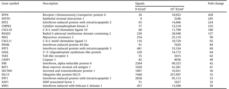
3.1. Genome-wide microarray indicates up-regulation of CXCL10
To detect genes up-regulated by IFN- b in LECs, genome-wide oligonucleotide microarray analyses that can examine expression of 56,792 genes were performed using LECs that were treated or not treated with IFN- b . The microarray analyses identi
fied 893 up- regulated genes and 665 down-regulated genes in treated LECs compared to non-treated LECs. Of the 893 up-regulated genes, 20 genes were up-regulated
>30-fold (Table 1). Among these 20 genes, three genes encode secretory molecules that may affect melanoma cells adjacent to lymphatic systems. The product of one of these three genes, CXCL10, is closely involved in the inhibition of mela- noma proliferation and invasiveness
[10]. Consequently, we furtherexamined the gene product.
3.2. CXCL10 is signi
ficantly induced by IFN- b in LECs compared to FBs
To con
firm the induction of CXCL10 in LECs following treatment with IFN- b , mRNA expression at 0, 3, 6, 12, 24, and 48 h after treatment with IFN- b in LECs and FBs was analyzed with RT-PCR.
CXCL10 mRNA was signi
ficantly induced in LECs compared to FBs
(Fig. 1a). We next performed ELISA tests using the culture super-
natant 24 h after treatment with IFN- b to con
firm the induction of
the secretory protein following IFN- b treatment. We con
firmed that
the secretory protein was signi
ficantly induced by IFN- b treatment
in LECs compared to FBs (Fig. 1b).
3.3. The melanoma cell lines exhibit a wide spectrum of sensitivity to IFN- b
To evaluate the sensitivity of melanoma cells to IFN- b , the sensitivity of each melanoma cell line to IFN- b was examined using the proliferation assay following treatment with IFN- b . The mela- noma cell lines exhibited a wide spectrum of sensitivity in the order of RPMI-7951, A375, G-361, SK-MEL-5, and SK-MEL-1 (high to low sensitivity;
Supple. Fig. 1a); the cells could be classified into two groups: cell lines with high sensitivity to IFN- b including RMPI-
7951 and A375 cells, and cell lines with medium to low sensi- tivity to IFN- b including G-361, SK-MEL-5, and SK-MEL-1 cells.
3.4. CXCL10 is signi
ficantly induced by IFN- b in cells with high sensitivity compared to medium to low sensitivity
To elucidate the association between CXCL10 expression levels and sensitivities of melanoma cells to IFN- b , the mRNA expression at 0, 3, 6, 12, 24, and 48 h after treatment with IFN- b was analyzed with RT-PCR. The peak CXCL10 expression levels in each melanoma cell line (RPMI-7951
>A375
>G-361
>SK-MEL-5
>SK-MEL-1 cells) were completely consistent with the order of sensitivity to IFN- b (Fig. 2a). We next performed ELISA tests using the culture super- natant 24 h after treatment with IFN- b to con
firm induction of the secretory protein by IFN- b treatment. We con
firmed that the con- centration levels of the secretory protein in each melanoma cell line (RPMI-7951
>A375
>SK-MEL-5
>G-361
>SK-MEL-1 cells) were nearly consistent with the order of sensitivity to IFN- b (Fig. 2b).
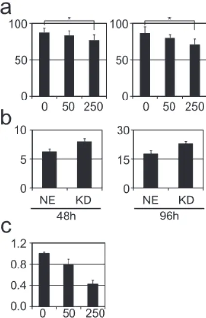
3.5. CXCL10 reduces proliferation of IFN- b -sensitive cells as well as resistant cells
To elucidate the role of CXCL10 in melanoma proliferation, a cell proliferation assay was performed after treatment of RPMI-7951 and G-361 cells (as representatives of relatively high and medium to low sensitivity to IFN- b , respectively) with 0, 50, or 250 m g/ml CXCL10 recombinant protein. Cell proliferation of RPMI-7951 and G-361 cells at day 5 was similarly suppressed by treatment with CXCL10 recombinant protein in a dose-dependent manner. We noted a signi
ficant difference in each graph (Kruskal
eWallis test, P
¼0.01 and
<0.001 in the graph of RPMI-7951 and G-361 cells, respectively) and a signi
ficant difference between the cell numbers following treatment with 0 and 250 m g/ml CXCL10 recombinant protein in both cell lines (non-parametric multiple comparison test, P
¼0.007 in RPMI-7951 cells and P
<0.001 in G-361 cells) (Fig. 3a).
3.6. CXCL10 knockdown renders melanoma cells less sensitive to IFN- b
To con
firm the tumor-suppressive effects of CXCL10 in mela- noma, we performed the cell proliferation assay using A375 cells in
Table 1List of genes up-regulated by>30-fold following IFN-btreatment of LECs.
Gene symbol Description Signals Fold change
0 IU/mla 103IU/mla
RTP4 Receptor (chemosensory) transporter protein 4 26 10,922 428
EPSTI1 Epithelial stromal interaction 1 9 2246 245
IFIT2 Interferon-induced protein with tetratricopeptide 2 65 14,466 224
CMPK2 Cytidine monophosphate kinase 2 57 12,218 216
CXCL10 C-X-C motif chemokine ligand 10 66 11,790 180
RSAD2 Radial S-adenosyl methionine domain containing 2 228 28,948 127
MX2 Myxovirus resistance 2 254 25,119 99
CXCL11 C-X-C motif chemokine ligand 11 116 10,729 92
IFI44L Interferon-induced protein 44-like 91 7629 84
IFIT3 Interferon-induced protein with tetratricopeptide 3 481 33,334 69
OASL 20-50-oligoadenylate synthetase-like protein 220 14,172 64
TLR3 Toll-like receptor 3 55 3415 62
CASP1 Caspase 1 82 4036 49
IFI6 Interferon, alpha-inducible protein 6 2364 99,323 42
BST1 Bone marrow stromal cell antigen 1 1020 41,281 41
SECTM1 Secreted and transmembrane protein 1 1007 35,921 36
ISG15 Ubiquitin-like protein ISG15 7440 257,997 35
IFIT1 Interferon-induced protein with tetratricopeptide 1 2858 95,113 33
XAF1 XIAP associated factor 1 51 1627 32
IFIH1 Interferon induced with helicase C domain 1 457 13,500 30
aFinal concentration of IFN-b.
Fig. 1.Induction of CXCL10 by IFN-btreatment in LECs and FBs. (a) mRNA expression of CXCL10at 0, 3, 6, 12, and 24 h after IFN-btreatment was measured with RT-PCR.
Horizontal, depth, and vertical axes indicate treatment times (hours), type of cells, and theCXCL10expression level divided byGAPDHexpression, respectively. The ver- tical axis of the right bar graph is indicated in one-hundredth scale of the vertical axis of the left bar graph to illustrate the relatively low expression values.*Scale out data.
(b) Protein expression of CXCL10 at 24 h after IFN-btreatment was measured with ELISA. Horizontal, depth, and vertical axes indicate the absence () and presence (þ) of IFN-btreatment, type of cells, and the CXCL10 protein concentration level (pg/ml/cell) in the culture supernatant, respectively. The vertical axis of the right bar graph is indicated in one-hundredth scale of the vertical axis of the left bar graph to illustrate the relatively low expression values.*Scale out data.
which CXCL10 were knocked down as representative of relatively high sensitivity to IFN- b . A375 cells in which CXCL10 was knocked down were less sensitive to IFN- b than A375 cells transduced with non-effective shRNA at both 48 and 96 h after treatment (Fig. 3b).
3.7. CXCL10 reduces the invasive capacity of melanoma cells
To evaluate the effects of CXCL10 on the invasive capacity of melanoma cells, a Matrigel invasion assay was performed using A375 cells treated with 0, 50, or 250 m g/ml CXCL10 recombinant protein. The invasive capacity was reduced in treated A375 cells in a dose-dependent manner compared to non-treated A375 cells (Fig. 3c).
3.8. CXCR3-B is up-regulated in cells with high sensitivity to IFN- b and down-regulated in cells with medium to low sensitivity
To elucidate the association between the expression of CXCR3-B, which encodes the CXCL10 receptor, in melanoma cells and the sensitivity to IFN- b , we used RT-PCR to examine CXCR3-B mRNA expression in melanoma cells after treatment with IFN- b . The expression levels in RPMI-7951 and A375 cells, which are highly sensitive to IFN- b , were up-regulated at both 6 and 24 h after treatment compared to non-treated cells. Conversely, the levels in G-361 and SK-MEL-1 cells, which have medium to low sensitivity, were down-regulated at both 6 and 24 h (Fig. 4).
4. Discussion
The present study demonstrated the tumor-suppressive effects of natural-type IFN- b on melanoma cells via CXCL10, which is derived from melanoma cells and the local lymphatic system.
Previous reports described that IFN- b treatment induces CXCL10 in metastatic melanoma cells and FBs
[11,12]and that CXCL10 re- duces melanoma proliferation and invasiveness
[10]. The presentstudy demonstrated that (i) IFN- b induces a signi
ficantly higher
amount of CXCL10 in LECs; (ii) The sensitivity to IFN- b was signif- icantly correlated with the level of CXCL10 induction following IFN- b treatment in melanoma cells; (iii) The sensitivity to CXCL10 was independent of the sensitivity to IFN- b in melanoma cells; (iv) The sensitivity to IFN- b was correlated with the expression of CXCR3-B in melanoma cells. These previously known and novel
findings potentially suggested that (i) the sensitivity of melanoma cells to IFN- b is closely correlated with the level of CXCL10 induction by
Fig. 2.Induction of CXCL10 by IFN-btreatment of melanoma cells. (a) mRNA expres-sion ofCXCL10at 0, 3, 6, 12, 24, and 48 h after IFN-btreatment was measured with RT- PCR. Horizontal, depth, and vertical axes indicate treatment times (hours), type of cells, and theCXCL10expression level divided byGAPDHexpression, respectively. The ver- tical axis of the right bar graph is indicated in one-hundredth scale of the vertical axis of the left bar graph to illustrate the relatively low expression values. (b) Protein expression of CXCL10 24 h after IFN-btreatment was measured with ELISA. Horizontal, depth, and vertical axes indicate the absence () and presence (þ) of IFN-btreatment, type of cells, and the CXCL10 protein concentration level (pg/ml/cell) in the culture supernatant, respectively. The vertical axis of the right bar graph is indicated in one- hundredth scale of that of the left bar graph to illustrate the relatively low expres- sion values.
Fig. 3.CXCL10 effects on melanoma cells. (a) Cell proliferation assay for RPMI-7951 and G-361 cells as representative cell lines with relatively high and medium to low sensitivity to IFN-b, respectively. The cell lines were treated with 0, 50, or 250mg/ml CXCL10 recombinant protein, and the cells were counted at day 5. CXCL10 recombinant protein was added in medium on days 1 and 3 after the medium was changed. The assay was performed 10 times, and the standard deviation is indicated in the bar graph. The vertical and horizontal axes indicate the number of cells relative to the number at day 1 and the CXCL10 concentration (mg/ml), respectively. Significant dif- ferences are indicated with asterisks (*). (b) Cell proliferation assay using A375 cells in whichCXCL10was knocked down (KD) and A375 cells transduced with non-effective shRNA (NE). Left and right bar graphs indicate the averages of triplicate cell counts at 48 and 96 h after the treatment, respectively. The vertical and horizontal axes indicate the number of cells relative to the number at day 1 and the type of cells, respectively. (c) Matrigel invasion assay using A375 cells treated with 0, 50, or 250mg/
ml CXCL10 recombinant protein. Vertical and horizontal axes indicate the cell number invading through Matrigel consisting of a gelatinous protein mixture and the con- centrations of CXCL10 recombinant protein (mg/ml), respectively (Fig. 3c).
Fig. 4.CXCR3-Bexpression in melanoma cells treated with IFN-b. mRNA expression of CXCR3-Bat 6 and 24 h after IFN-btreatment was measured with RT-PCR. The vertical axis indicates the expression level relative to the expression level in non-treated cells.
The horizontal axis indicates melanoma cells treated (þ) and not treated () with IFN- bfor various times (hours).
IFN- b and the CXCR3-B response to IFN- b in melanoma cells, and (ii) IFN- b suppresses lymphatic metastasis by inducing a high amount of CXCL10 from the local lymphatic system.
CXCL10 is a member of the CXC chemokine family and binds to the chemokine (C-X-C motif) receptor (CXCR3) to exert its biolog- ical effects
[13]. CXCR3 is classified into three splice variants:
CXCR3-A, CXCR3-B, and CXCR3-alt. Activation of CXCR3-A induces chemotaxis and proliferation in various cell types, whereas acti- vation of CXCR3-B inhibits cell proliferation and migration and induces apoptosis
[13]. Based on these data, CXCR3-B may playcritical roles in the tumor-suppressive effects of CXCL10 on mela- noma cells. This encouraged us to focus on CXCR3-B expression in melanoma cells treated with IFN- b .
Interestingly, CXCR3-B expression was down-regulated in mel- anoma cells with medium to low sensitivity to IFN- b . This indicates that a certain type of melanoma is not suppressed by IFN- b treat- ment, even though IFN- b induces a high amount of CXCL10 from the local lymphatic system and melanoma cells. IFN treatment may have a poor effect on melanoma in some cases, suggesting that CXCR3-B on melanoma cells in such cases may be down-regulated by IFN treatment as shown in the present study. On the other hand, the present study demonstrated that CXCL10 suppressed the pro- liferation of G-361 cells with medium to low sensitivity to IFN- b as well as RPMI-7951 cells with high sensitivity, suggesting that CXCL10 exerts tumor-suppressive effects on melanoma regardless of the sensitivity to IFN- b . This fact suggests the possibility of choosing local administration of CXCL10 recombinant protein for IFN- b treatment-resistant melanoma cases.
The present study demonstrated that CXCL10 is signi
ficantly induced by IFN- b treatment in LECs compared to FBs, although a previous report showed
“strong and sustained
”CXCL10 induction from FBs following IFN- b treatment
[12]. These facts suggest thatCXCL10 is induced from LECs by IFN- b treatment in remarkably high amounts. This high amount of CXCL10 from the local lymphatic system following IFN- b treatment may suppress mela- noma progression in combination with CXCL10 from melanoma cells when CXCR3-B is up-regulated on melanoma cells. Conversely, CXCL10 secreted from the local lymphatic system and melanoma cells following IFN- b treatment only minimally suppresses mela- noma proliferation when CXCR3-B is down-regulated by IFN- b treatment of melanoma cells.
Our data showed two melanoma cell lines with low sensitivity to IFN- b treatment that were derived from lymphatic system- metastasized lesions (Supple. Fig. 2), possibly because IFN- b - resistant melanoma cells after IFN treatment may be selected as melanoma cells derived from lymphatic system-metastasized le- sions. Marder et al. reported that p53 mutation attenuates the tumor-suppressive effects of IFN- b
[14]. Conversely, our datashowed that all the melanoma cell lines with high sensitivity to IFN- b treatment have p53 mutation (Supple. Fig. 2). Although the reason for this discrepancy is not clear, IFN- b may exhibit p53- independent tumor-suppressive effects on melanoma cells, simi- larly as IFN- a and IFN- g stimulate a p53-independent apoptotic pathway
[15,16].In the present study, IFN- b treatment was performed with a
final concentration of 10
3IU/ml for LECs, FBs, and melanoma cells. This IFN- b dose was determined based on the fact that 310-4,012 IU/g IFN- b accumulates in regional lymph nodes after local adminis- tration of a clinically used dose of IFN- b
[9]. Based on these ob-servations, the in vitro conditions of this study are comparable to the in vivo tumor microenvironment of the local lesion treated with IFN- b .
A limitation of the present study is the in vitro conditions.
CXCL10 is involved in various biological functions including chemotaxis, apoptosis, and angiostatic effects
[17], in addition toregulation of tumor proliferation and invasion. This indicates the need for in vivo research to con
firm the roles of CXCL10 in melanoma.
In conclusion, our data suggest that IFN- b may suppress mela- noma progression by inducing CXCL10 from the local lymphatic system and melanoma cells. Down-regulation of CXCR3-B by IFN- b may be associated with resistance to IFN- b treatment.
Conflicts of interests
We obtained a grant for clinical research from Toray Medical Co., Ltd.
Transparency document
Transparency document related to this article can be found online at
http://dx.doi.org/10.1016/j.bbrc.2015.06.122.Appendix A. Supplementary data
Supplementary data related to this article can be found at
http://dx.doi.org/10.1016/j.bbrc.2015.06.122.
References
[1] A.J. Miller, M.C. Mihm Jr., Melanoma, N. Engl. J. Med. 355 (2006) 51e65.
[2] H. Tsao, M.B. Atkins, A.J. Sober, Management of cutaneous melanoma, N. Engl.
J. Med. 351 (2004) 998e1012.
[3] J.A. Sosman, K.B. Kim, L. Schuchter, R. Gonzalez, A.C. Pavlick, J.S. Weber, G.A. McArthur, T.E. Hutson, S.J. Moschos, K.T. Flaherty, P. Hersey, R. Kefford, D. Lawrence, I. Puzanov, K.D. Lewis, R.K. Amaravadi, B. Chmielowski, H.J. Lawrence, Y. Shyr, F. Ye, J. Li, K.B. Nolop, R.J. Lee, A.K. Joe, A. Ribas, Survival in BRAF V600-mutant advanced melanoma treated with vemurafenib, N. Engl.
J. Med. 366 (2012) 707e714.
[4] A.M. Eggermont, A. Testori, J. Marsden, P. Hersey, I. Quirt, T. Petrella, H. Gogas, R.M. MacKie, A. Hauschild, Utility of adjuvant systemic therapy in melanoma, Ann. Oncol. 20 (Suppl. 6) (2009) vi30e4.
[5] A.M. Eggermont, S. Suciu, M. Santinami, A. Testori, W.H. Kruit, J. Marsden, C.J. Punt, F. Sales, M. Gore, R. Mackie, Z. Kusic, R. Dummer, A. Hauschild, E. Musat, A. Spatz, U. Keilholz, Adjuvant therapy with pegylated interferon alfa-2b versus observation alone in resected stage III melanoma:final results of EORTC 18991, a randomised phase III trial, Lancet 372 (2008) 117e126.
[6] A. Yamamoto, K. Ishihara, Clinical study of DAVþIFN-btherapy (combination adjuvant therapy with intravenous DTIC, ACNU and VCR, and local injection of IFN-b) for malignant melanoma, Int. J. Immunother. 12 (1996) 73e78.
[7] Y. Fujisawa, F. Otsuka, Jyutugo hojyoryouhou (DAVFeron, Feron ryouhou, Feron iji ryouhou) ha akuseikokusyokusyu suteeji II, III kannjyano yogowo kaizennsuruka: 831reinokaiseki (Dose adjuvant therapy improve the prog- nosis of stage II and III melanoma patients? An analysis of 831 cases) (in Japanese), Jpn. J. Dermatol. 122 (2012) 2305e2311.
[8] A. Yamamoto, Meranooma feron ijiryouhou tashisetsu-kyoudoukennkyuu no kekkahoukoku (Multicenter study of Feron maintenance therapy for mela- noma) (in Japanese), Skin. Cancer 29 (2014) 251e257.
[9] A. Yamamoto, A. Sumi, Y. Kato, Y. Urata, S. Endo, S. Mori, N. Minowa, Aku- seikokusyokusyu niokeru kyokutyuu IFN-b no rinnpa ikousei nituite (Lymphatic transferability of local injection of IFN-bin malignant melanoma patients) (in Japanese), Skin. Cancer 1 (1986) 47e53.
[10] F. Antonicelli, J. Lorin, S. Kurdykowski, S.C. Gangloff, R. Le Naour, J.M. Sallenave, W. Hornebeck, F. Grange, P. Bernard, CXCL10 reduces mela- noma proliferation and invasiveness in vitro and in vivo, Br. J. Dermatol. 164 (2011) 720e728.
[11] L.T. Dengel, A.G. Norrod, B.L. Gregory, E. Clancy-Thompson, M.D. Burdick, R.M. Strieter, C.L. Slingluff Jr., D.W. Mullins, Interferons induce CXCR3-cognate chemokine production by human metastatic melanoma, J. Immunother. 33 (2010) 965e974.
[12] A. Antao-Menezes, E.A. Turpin, P.C. Bost, J.P. Ryman-Rasmussen, J.C. Bonner, STAT-1 signaling in human lungfibroblasts is induced by vanadium pentoxide through an IFN-beta autocrine loop, J. Immunol. 180 (2008) 4200e4207.
[13] C. Billottet, C. Quemener, A. Bikfalvi, CXCR3, a double-edged sword in tumor progression and angiogenesis, Biochim. Biophys. Acta 1836 (2013) 287e295.
[14] S. Madar, E. Harel, I. Goldstein, Y. Stein, I. Kogan-Sakin, I. Kamer, H. Solomon, E. Dekel, P. Tal, N. Goldfinger, G. Friedlander, V. Rotter, Mutant p53 attenuates the anti-tumorigenic activity offibroblasts-secreted interferon beta, PLoS One 8 (2013) e61353.
[15] C. Porta, R. Hadj-Slimane, M. Nejmeddine, M. Pampin, M.G. Tovey, L. Espert, S. Alvarez, M.K. Chelbi-Alix, Interferons alpha and gamma induce p53-
dependent and p53-independent apoptosis, respectively, Oncogene 24 (2005) 605e615.
[16] N.K. Ossina, A. Cannas, V.C. Powers, P.A. Fitzpatrick, J.D. Knight, J.R. Gilbert, E.M. Shekhtman, L.D. Tomei, S.R. Umansky, M.C. Kiefer, Interferon-gamma
modulates a p53-independent apoptotic pathway and apoptosis-related gene expression, J. Biol. Chem. 272 (1997) 16351e16357.
[17] M. Liu, S. Guo, J.K. Stiles, The emerging role of CXCL10 in cancer (Review), Oncol. Lett. 2 (2011) 583e589.