Identification of a Novel Deactivating Small-Molecule Compound for Fibrogenic
Hepatic Stellate Cells
Takuki Sakaguchi,*† Yohei Kono,* Noriko Itaba,* Minoru Morimoto,‡ Hajime Isomoto† and Goshi Shiota* *Department of Genetic Medicine and Regenerative Therapeutics, Graduate School of Medical Sciences, Tottori University, Yonago 683-8503, Japan, †Division of Medicine and Clinical Science, Department of Multidisciplinary Internal Medicine, School of Medicine, Tottori University Faculty of Medicine, Yonago 683-8503, Japan, and ‡Research Initiative Center, Tottori University, Tottori 680-8550, Japan
ABSTRACT
Background Liver fibrosis progresses to decompen-sated liver cirrhosis, for which medical needs remain unmet. We recently developed IC-2, a small-molecule compound that suppresses Wnt/β-catenin signaling, and found that IC-2 also suppresses liver fibrosis. In this study, we performed three-step screening of newly synthesized IC-2 derivatives to identify other small-molecule compounds that suppress liver fibrosis.
Methods The screening system consisted of three steps: a cell viability assay, a transcription factor 4 (TCF4) reporter assay, and induction of α-smooth mus-cle actin (α-SMA) and collagen 1α1 (Col1A1) expression in response to each compound. Screening using human LX-2 hepatic stellate cells (HSCs) was performed to target HSCs, which are the driver cells of liver fibrosis.
Results In the first step, since 9b and 9b-CONH2 at 100 μM did not have any effects on cell viability, they were omitted in the next screening. Additionally, the conditions that led to > 40% inhibition of the controls were also excluded in subsequent screening. The second step was performed under 31 conditions for 19 small-molecule compounds. Sixteen small-small-molecule com-pounds caused significant reduction of TCF4 activity relative to that of 0.1% DMSO. Of the 16 compounds, the 10 showing the greatest suppression of TCF4 activity were selected for the third step. Expressions of mRNA for α-SMA and Col1A1 were significantly reduced by seven and three small-molecule compounds, respectively. The greatest reductions in the α-SMA and Col1A1 mRNA expressions were observed in the cells treated with IC-2-F. Protein expressions of α-SMA and Col1A1 caused by IC-2-F were also comparable to those caused by IC-2.
Conclusion IC-2-F was identified as a novel deac-tivating small-molecule compound for HSCs in vitro. These data suggest that IC-2-F is a promising medicine for liver fibrosis.
Key words deactivation; hepatic stellate cells; liver fibrosis; small-molecule compound
Liver fibrosis is caused by chronic infection with the hepatitis B virus or hepatitis C virus, alcohol intoxica-tion, or non-alcoholic steatohepatitis.1 Progressive liver
fibrosis leads to decompensated liver cirrhosis (LC), for which medical needs remain unmet, and results in > 1 million deaths annually worldwide.2, 3 Therefore, there
is a great need for development of a novel therapy for liver fibrosis.
Myofibroblasts are the driver of tissue fibrogenesis in multiple organs, including the liver.1 A myofibroblast
is a fibroblast-like cell with contractile properties and is typically derived from hepatic stellate cells (HSCs) via activation.4 Activated HSCs produce a large amount of
collagen types I and III, which constitute fibrous tissues of pathological liver fibrosis.5 Several paracrine factors,
such as platelet-derived growth factor (PDGF), trans-forming growth factor-β (TGF-β), and connective tissue growth factor (CTGF) activate HSCs and maintain its activated state. Thus, since activation of HSCs is the major mechanism of liver fibrosis, the target of therapy for liver fibrosis proves to be HSCs.
Wnt/β-catenin signaling is upregulated in activated HSCs.6 The crosstalk between Wnt/β-catenin and
TGF-β signaling highlights the role of Wnt/β-catenin signaling in liver fibrosis. Importantly, Wnt antagonism inhibits HSCs activation and liver fibrosis.7, 8 These data
suggest that development of small-molecule compounds to target the Wnt/β-catenin pathway is an intriguing strategy for therapy of liver fibrosis.
We previously reported that genes downstream of Wnt/β-catenin signaling are downregulated during he-patic differentiation of human mesenchymal stem cells9 Corresponding author: Goshi Shiota, MD, PhD
[email protected] Received 2019 December 16 Accepted 2020 February 6 Online published 2020 February 20
Abbreviations: α-SMA, α-smooth muscle actin; Col1A1, col-lagen 1α1; CTGF, connective tissue growth factor; GAPDH, glyceraldehyde 3-phosphate dehydrogenase; HCC, hepatocel-lular carcinoma; HSCs, hepatic stellate cells; LC, liver cirrhosis; PDGF, platelet-derived growth factor; TCF4, transcription factor 4; TGF-β, transforming growth factor-β
T. Sakaguchi et al.
and that suppression of Wnt/β-catenin signaling causes hepatic differentiation of human mesenchymal stem cells.10 Indeed, it has been demonstrated that
mesen-chymal stem cell sheets engineered by a Wnt/β-catenin signal inhibitor accelerated liver regeneration in mice.11
Furthermore, we identified IC-2, a derivative of ICG-001, as a potent inhibitor of Wnt/β-catenin signaling in human mesenchymal stem cells.12 Transplantation
of IC-2-engineered cell sheets potently improved acute liver injury and reversed liver fibrosis.13, 14 We
also found that IC-2 suppressed cancer stem cells of hepatocellular carcinoma (HCC) and colon cancer, respectively.15, 16 Taken together, drug discovery for
liver fibrosis is promising when IC-2 is used as a leading small-molecule compound.
We previously found that IC-2 suppressed liver fibrosis.17 In the present study, 21 newly synthesized
derivatives of IC-2 were screened in three steps: a cell viability assay, a transcription factor 4 (TCF4) reporter assay, and induction of α-smooth muscle actin (α-SMA) and collagen 1α1 (Col1A1) expression of human LX-2 HSCs in response to each compound. Commonly, α-SMA and Col1A1 are used as markers of HSC activa-tion and fibrogenesis, respectively.
In this study, our screening system consisted of three steps: a cell viability assay, a TCF4 reporter assay, and induction of α-SMA and Col1A1 expression in re-sponse to each compound. We performed the screening on the basis of the following concept. Since HSCs play important roles in maintenance of liver functions, it may be harmful to pick up toxic small-molecule compounds to kill many HSCs in a cell viability assay. To obtain enough effects without any side effects on liver function, it seems to be required to keep a constant level of HSCs even after using the small-molecule compound as a medicine. To accomplish this purpose, we set a selection standard at around 40% reduction of a cell viability in the first step of screening. In this study, we tried to pick up the small-molecule compounds that display higher suppression of Wnt/β-catenin signaling without killing over 40% cells.
MATERIALS AND METHODS
Cells, IC-2 derivatives, and cell viability
The LX-2 HSC cell line was used for the experiments in this study.18 To identify potent deactivating
small-molecule compounds for HSCs, 21 newly developed derivatives of IC-2 were screened. All of the small-mol-ecule compounds were produced in house. The newly developed small-molecule compounds, the original par-ent compound IC-2, and their structures are shown in Fig. 1. The introduced groups such as OPMB, OMOM,
MOTBS, and MOTBS mean O-methoxybenzyl,
O-methoxymethyl, O-methoxymethyl, and
methyl-O-t-butyldimethylsilyl, respectively. At 24 h after plating, the LX-2 cells were treated with each compound at six different concentrations in Dulbecco’s Modified Eagle’s Medium containing 1% fetal bovine serum. Cell vi-ability was measured by a water-soluble tetrazolium salt (WST) assay by using a Cell Counting Kit-8 (Dojindo Laboratories, Kumamoto, Japan) at 24 and 48 h after treatment.
Reporter assay
At 20 h after seeding, TCF4-CMVpro-GL4.20 and pRL-TK (Promega Corp., Fitchburg, WI) were co-transfected into the LX-2 cells by using Lipofectamine 2000 reagent (Invitrogen, Thermo Fisher Scientific Inc., Waltham, MA).9, 10, 12 TCF4-CMVpro-GL4.20 was
de-veloped as a TCF-4 motif-responsive luciferase reporter gene.12 In brief, three copies of the optimal TCF-4 motif
CCTTTGATC and cytomegalovirus (CMV) promoter into the multiple cloning site of the pGL4.20 luciferase reporter plasmid (Promega Corp., Fitchburg, WI), subsequently designated pTCF-CMVpro-GL4.20. This plasmid is sensitive reporter plasmid to assess Wnt/ β-catenin signals.
At 4 h after transfection, the cells were treated with each compound at certain concentrations. Luciferase activities were determined after 48 h treatment by using the Dual-Luciferase Reporter Assay System (Promega) and MiniLumat LB 9506 (Berthold GmbH & Co. KG, Bad Wildbad, Germany). Luciferase activities were normalized to Renilla luciferase activity and expressed as percentages of the control.
Real-time quantitative polymerase chain reaction (qPCR)
LX-2 cells were treated with 2.5 ng/mL recombinant human TGF-β (R&D Systems, Inc., Minneapolis, MN) to convert the LX-2 cells to fibrogenic HSCs. At 24 h after treatment, total RNA was extracted using RNeasy Plus Mini Kit (QIAGEN GmbH, Hilden, Germany) and reverse-transcribed with SSII RTase transcriptase (Invitrogen, Thermo Fisher Scientific Inc.). Real-time qPCR analysis was performed by using THUNDERBIRD SYBR qPCR Mix (Toyobo Co., Ltd., Osaka, Japan). The primer sets included α-SMA (for-ward: 5’-CTG TTC CAG CCA TCC TTC AT-3’, reverse: 5’-CCG TGA TCT CCT TCT GCA TT-3’), COL1A1 (forward: 5’-CCT CCA GGG CTC CAA CGA G-3’, reverse: 5’-TCA ATC ACT GTC TTG CCC CA-3’), and glyceraldehyde 3-phosphate dehydrogenase (GAPDH) (forward: 5’-AGC CAC ATC GCT CAG ACA C-3’,
Fig. 1. Chemical structures of IC-2 and newly synthesized IC-2 derivatives.
A, IC-2-OMe (methoxy); B, IC-2-F; C, IC-2-Cl; D, IC-2-NO2; E, IC-2-OPMB (O-methoxybenzyl); F, IC-2-OMOM (O-methoxymethyl);
G, IC-2-OTBS (O-t-butyldimethylsilyl); H, IC-2-OH; I, IC-2-MOTBS (methyl-O-t-butyldimethylsilyl); J, IC-2-MOH (hydroxymethyl); K, IC-2-Ar-OMe; L, IC-2-Ar-F; M, IC-2-Ar-Cl; N, IC-2-Ar-NO2; O, IC-2-Ar-OH; P, IC-2-506-1; Q, IC-2-506-2; R, IC-2-506-3; S, 9b;
T. Sakaguchi et al.
reverse: 5’-GCC CAA TAC GAC CAA ATC C-3’).
Western blot analysis
Twenty micrograms of protein from cells separated by sodium dodecyl sulfate-polyacrylamide gel electro-phoresis was transferred onto an Immobilon-P PVDF membrane. The primary antibodies for detection of α-SMA, COL1A1, and GAPDH included M0851 (Dako), sc-293182 (Santa Cruz), and MAB374 (Merck), respec-tively. Secondary antibody was anti-mouse immuno-globulin G horseradish peroxidase-linked whole Ab from sheep (GE Healthcare UK Ltd., Buckinghamshire, England). Signals were detected by a chemilumines-cent image analyzer ChemiDoc Touch MP (Bio-Rad Laboratories Inc., Hercules, CA) using ECL Prime Western Blotting Detection Reagent (GE Healthcare).
Statistical analysis
Statistical analysis was performed by performing one-way ANOVA followed by the Tukey HSD test. All statistical testing was performed by using IBM SPSS Statistics software Ver. 23.0.0.0 (IBM Corp., Armonk, NY). P-values < 0.05 were considered to be indicative of statistical significance.
RESULTS
First-step screening that selected 19 small-molecule compounds
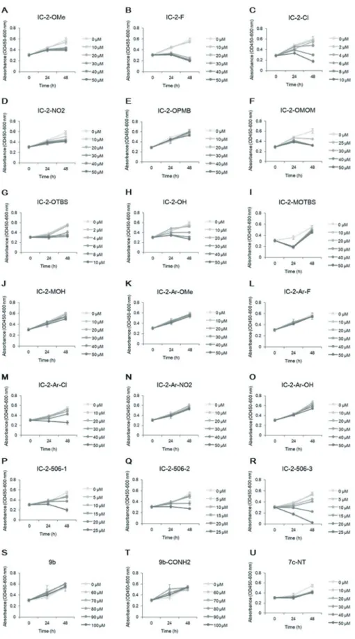
For screening of 21 derivatives of IC-2, cell viabilities were first assessed at 24 and 48 h after incubation with each compound at six different concentrations per compound (Fig. 2). The conditions that exhibited > 40% inhibition of the controls were excluded from subsequent screening. In the first step of screening, 9b and 9b-CONH2 were excluded, since we can not expect that these compounds suppress Wnt/β-catenin signaling at the concentrations where cell viability is hardly af-fected. The first screening selected 31 conditions from 19 small-molecule compounds.
Second-step screening that selected 11 small-molecule compounds
The TCF4 reporter assay was performed with 31 condi-tions from 19 compounds in addition to 20 μM IC-2 and 0.1% dimethyl sulfoxide (DMSO) as a control (Fig. 3A). Of the 31 conditions, 21 showed significant reduction of TCF4 activity in the TCF4 reporter assay relative to that for 0.1% DMSO (P < 0.01, each). The 21 conditions included 20 and 50 μM 2-OMe, 20 and 30 μM IC-2-F, 6 μM IC-2-Cl, 30 and 50 μM IC-2-NO2, 50 μM
IC-2-OPMB, 25 and 30 μM IC-2-OMOM, 6 and 10 μM IC-2-OTBS, 30 μM IC-2-OH, 50 μM IC-2-Ar-OMe, 50
μM IC-2-Ar-F, 50 μM IC-2-Ar-NO2, 50 μM
Ar-OH, 5 μM 506-1, 20 μM 506-2, 10 μM IC-2-506-3, and 10 μM 7c-NT. Twenty micromolar IC-2 gave about 50% TCF4 reporter activity relative to that of 0.1% DMSO as a positive control. However, 15 compounds including IC-2-OMe, IC-2-F, IC-2-NO2, IC-2-OPMB, 2-OMOM, 2-OTBS, 2-OH, 2-Ar-OMe, IC-2-Ar-F, IC-2-NO2, IC-2-Ar-OH, 1,
IC-2-506-2, IC-2-506-3, and 7c-NT exhibited <40% activity. In particular, 11 compounds such as IC-2-F, IC-2-NO2,
2-OPMB, 2-OMOM, 2-OTBS, 2-Ar-OMe, IC-2-Ar-F, IC-506-1, IC-2-506-2, IC-2-506-3, and 7c-NT exhibited < 30% activities. The second step screening selected 11 small-molecule compounds for further screening.
Third screening that identified IC-2-F as a potent deactivating small-molecule compound for fibro-genic hepatic stellate cells
Of 11 compounds that selected in the second screening, nine small-molecule compounds, i.e., 2-OMe, 2-F, 2-Cl, 2-OPMB, 2-OTBS, 2-506-1, IC-2-506-2, IC-2-506-3, and 7c-NT, were selected due to the intensity of suppression on the TCF4 reporter assay and the concentration < 20 μM, that is the concentra-tion of IC-2. The third screening included the effects of these compounds on mRNA expressions of α-SMA and COL1A1. Of note, α-SMA was used because a heterogeneous population of α-SMA-positive cells, defined as hepatic myofibroblasts, have been found to have a major pro-fibrogenic role in the process of liver fibrosis.19 IC-2 caused an approximately 60% reduction
in α-SMA mRNA relative to that of the 0.1% DMSO control. In addition to IC-2, six compounds, i.e., 2-OMe, 2-F, 2-Cl, 2-OPMB, 2-506-2, and IC-2-506-3, significantly suppressed expression of α-SMA mRNA relative to that of 0.1% DMSO (P < 0.01 for IC-2-OMe, IC-2-F, IC-2-Cl, and IC-2-506-2, P < 0.05 for IC-2-OPMB and IC-506-3) (Fig. 3B). IC-2-F showed the greatest suppression among the six compounds. Among all nine compounds, IC-2-OMe and IC-2-F significantly suppressed expression of COL1A1 mRNA (Fig. 3C). IC-2-F also caused the most potent suppression of COL1A1 mRNA among nine compounds (Fig. 3C). These results indicated that IC-2-F was the strongest inhibitor of α-SMA and COL1A1. Western blot analysis showed that IC-2-F inhibited the expressions of α-SMA and COL1A1 to a degree similar to that of IC-2 (Fig. 4). Taken together, these data suggested that IC-2-F is a novel deactivating small-molecule compound for HSCs
in vitro and is a promising medicine for suppressing
Fig. 2. Cell viability assay for various concentrations of small-molecule compounds. The effect of each small-molecule compound on cell viability was determined by a water-soluble tetrazolium salt assay at 24 and 48 h after addition of compound.
A, IC-2-OMe; B, IC-2-F; C, IC-2-Cl; D, IC-2-NO2; E, 2-OPMB; F, 2-OMOM; G, 2-OTBS; H, 2-OH; I, 2-MOTBS; J,
IC-2-MOH; K, IC-2-Ar-OMe; L, IC-2-Ar-F; M, IC-2-Ar-Cl; N, IC-2-Ar-NO2; O, IC-2-Ar-OH; P, IC-2-506-1; Q, IC-2-506-2; R, IC-2-506-3; S,
T. Sakaguchi et al.
Fig. 3. Small-molecule compounds that inhibit TCF4/β-catenin transcriptional activity and expressions of α-SMA and Col1A1. A. The effect of small-molecule compounds on Wnt/β-catenin signal activity in the human LX-2 hepatic stellate cell (HSC) line. TCF4/ β-catenin transcriptional activity is examined by dual-luciferase reporter assay in LX-2 cells treated with several concentrations for 48 h. The values of the 0.1% DMSO-treated cells are the controls, and the values obtained for each condition are expressed as a percentage of the control.
B. The effects of small-molecule compounds on HSC activation are shown. α-SMA mRNA expressions in LX-2 cells treated for 48 h as analyzed by qRT-PCR. Each expression level is normalized to GAPDH. All data are shown as the mean ± SD (n = 3). IC-2 is the positive control. One-way ANOVA followed by Tukey’s HSD test were performed. *P < 0.05, **P < 0.01 compared with 0.1% DMSO.
C. The effects of small-molecule compounds on collagen synthesis. Col1A1 mRNA expression in LX-2 cells treated for 48 hours as analyzed by qRT-PCR. Each expression level is normalized to GAPDH. All data are shown as the mean ± SD (n = 3). IC-2 is the positive control. One-way ANOVA followed by Tukey’s HSD test were performed. *P < 0.05, **P < 0.01 compared with 0.1% DMSO.
DISCUSSION
Liver fibrosis leads to decompensated LC, which is associated with the complications of functional liver failure, portal hypertension, and hepatic encephalopa-thy.1 In addition, HCC occurs in some patients with LC.
Since approved therapies targeting liver fibrosis and LC are lacking, development of new therapies to regress liver fibrosis are urgently needed.
Myofibroblasts are the driver cells of liver fibrosis.1, 4 HSCs are the major precursors of
myofi-broblasts, and activation of HSCs promotes conversion to myofibroblasts. Several external stimuli, including hepatocellular cell death due to immune reaction, viral infection, oxidative stress, apoptosis, and cytokines, cause activation of HSCs.1, 4, 20 These reports suggest
that anti-fibrotic therapies are supposed to include removal of external stimuli and/or direct deactivation of HSCs. Current treatments for chronic viral hepatitis are included in the former category, and those in the latter category await development.
Molecular dysregulation in activated HSCs/ myofibroblasts includes membrane signaling pathways, such as those involving TGF-β, PDGF, CTGF, Wnt/ β-catenin, and hedgehog signaling.1 Especially, TGF-β
signaling has a major role in promoting liver fibrosis. Recent studies have reported that Wnt/β-catenin signal-ing has an essential role in liver fibrosis.6 Crosstalk
between Wnt/β-catenin and TGF-β signaling has been previously demonstrated.7, 8 Wnt/β-catenin signaling
works in a combinatorial manner with TGF-β signaling. In contrast, suppression of Wnt/β-catenin signaling downregulates TGF-β signaling. Indeed, the molecular mechanism of the interactions between the β-catenin
and TGF-β signaling pathways has been clarified.21
Thus, suppression of Wnt/β-catenin signaling is a rea-sonable therapeutic approach for liver fibrosis.
In our previous study, we confirmed that addition of Wnt3a caused a slight increase of Wnt/β-catenin signaling, however inhibitors of this pathway caused relatively large decrease in this signal intensity in LX-2 cells.17 These data suggest that LX-2 cells displayed
higher activities of Wnt/β-catenin signaling, and may imply that LX-2 cells are suitable for selection of sup-pressors of Wnt/β-catenin signaling pathway.
In this study, although genetic controls were not employed, we previously confirmed Fzd8 siRNA potently suppressed the TCF4 activity.10 Therefore, to
minimize technical errors, we used the values of the 0.1% DMSO-treated cells as the controls. This assay was sup-ported by the character that LX-2 cells displayed higher activities of Wnt/β-catenin signaling.
Our screening was based on the concept that it is important to select small-molecule compounds that display higher suppressive activity on Wnt/β-catenin signaling within the concentrations exhibiting < 40% inhibition of the controls of cell viability. The small-molecule compounds selected in this way are expected to have higher deactivating activity on HSCs, but have lower toxic side effects. HSCs play important roles in maintenance of liver functions since HSCs secrete several hepatotrophic factors.5 In this study, the reason
why a cell viability assay on the basis of selecting the compounds exhibiting < 40% inhibition of the controls was due to this idea. Consequently, IC-2-F was selected as a promising medicine.
Activation or inactivation of Wnt/β-catenin signal
Fig. 4. Western blot analysis of α-SMA and Col1A1 protein expressions after IC-2-F treatment. The LX-2 cells were cultured with 2.5 ng TGF-β and 20 μM IC-2-F for the indicated hours. The α-SMA and Col1A1 protein levels determined by western blot analysis are shown. IC-2 is the positive control, and GAPDH is the internal control.
T. Sakaguchi et al.
is closely associated with subcellular distribution of β-catenin. β-catenin has dual roles, as an adhesion mol-ecule at the plasma membrane and as an intermediate molecule in the canonical Wnt pathway. In our previous study, on activation of the Wnt signal, β-catenin translo-cated into nucleus of UCBTERT-21 cells.10 On the other
hand, when the Wnt/β-catenin signal was suppressed by transfection of Fzd8 siRNA, β-catenin was located along the cell membrane and in the cytoplasm of the cells. In this study, although subcellular localization of β-catenin was not examined, β-catenin was supposed to be localized on the plasma membrane and in the cytoplasm.
The mechanism that these IC-2 derivatives exhibit suppressive activity on Wnt/β-catenin signals in HSCs remains to be clarified. It has been reported that ICG-001 suppresses Wnt/β-catenin signals by binding CREB-binding protein (CBP).12 Since IC-2 is
a derivative of ICG-001, IC-2 and IC-2 derivatives may interact with CBP. Further examination will be required to clarify the molecular mechanisms of IC-2 and IC-2 derivatives on Wnt/β-catenin signals. In previous study, we found that hexachlorophene suppressed Wnt/ β-catenin signals in UE7T-13 human mesenchymal stem cells.11 Treatment with hexachlorophene led to
reduction of β-catenin and cyclin D1, and the effect of hexachlorophene was abolished by MG-132, a protea-some inhibitor. These data suggest that hexachlorophene suppressed Wnt/β-catenin signals via intracellular β-catenin degradation in proteasome. Furthermore, we found that HC-1, the derivative of hexachlorophene, has a different mechanism suppressing Wnt/β-catenin signals from hexachlorophene (data not shown). Thus, the small-molecule compounds seem to have different mechanisms suppressing on Wnt/β-catenin signals even in their derivatives that have the similar structure with the original compounds.
We tried to explain the difference of biological activity from the viewpoint of the chemical structure of the compounds. For example, 2-OMe and IC-2-F, whose substitution groups are OMe and F, had similar suppressive activities on Wnt/β-catenin signals. However, the electronegativity of OMe is the opposite of F. Furthermore, OMe and F also have different molecular size. It seemed to be difficult to explain the difference of suppressive effects on Wnt/β-catenin signals from a viewpoint of chemical structure of the compounds. Therefore, novel molecular designs may be required to clarify the structure-activity relationship.
In conclusion, we screened newly developed derivatives of IC-2 in three steps: a cell viability assay, a TCF4 reporter assay, and induction of α-SMA and
Col1A1 expressions of human LX-2 HSCs in response to each compound. Consequently, we identified IC-2-F as a novel deactivating small-molecule compound for HSCs in vitro. In this study, since the effect of IC-2-F was examined at one dose, the dose-dependency and time-dependency on activation of HSCs and liver fibrosis are required in the near future. IC-2-F has more potent suppressive effects on a-SMA and Col1A1 expression than IC-2 at mRNA level. Although in vivo study about IC-2-F will be required, it is expected that IC-2-F will a promising medicine for treatment of liver fibrosis.
Acknowledgments: We thank all the members of the laboratory for their discussion and comments.
This work was partly supported by the project for realiza-tion of regenerative medicine and program for Creating Start-ups from Advanced Research and Technology of the Ministry of Education, Culture, Sports, Science, and Technology in Japan.
The authors thank Enago (www.enago.jp) for the English language review.
CONFLICT OF INTEREST
G.S. has licensed patents, holds more than 5% of the total shares of KanonCure Inc. and receives compensation as a member of KanonCure Inc. The other authors have declared that no compet-ing interests exist.
REFERENCES
1 Higashi T, Friedman SL, Hoshida Y. Hepatic stellate cells as key target in liver fibrosis. Adv Drug Deliv Rev. 2017;121:27-42. DOI: 10.1016/j.addr.2017.05.007, PMID: 28506744 2 Forouzanfar MH, Alexander L, Anderson HR, Bachman
VF, Biryukov S, Brauer M, et al.; GBD 2013 Risk Factors Collaborators. Global, regional, and national comparative risk assessment of 79 behavioural, environmental and occupational, and metabolic risks or clusters of risks in 188 countries, 1990–2013: a systematic analysis for the Global Burden of Disease Study 2013. Lancet. 2015;386:2287-323. DOI: 10.1016/S0140-6736(15)00128-2, PMID: 26364544 3 Tsochatzis EA, Bosch J, Burroughs AK. Liver cirrhosis.
Lan-cet. 2014;383:1749-61. DOI: 10.1016/S0140-6736(14)60121-5, PMID: 24480518
4 Friedman SL, Sheppard D, Duffield JS, Violette S. Therapy for fibrotic diseases: nearing the starting line. Sci Transl Med. 2013;5:167sr1. DOI: 10.1126/scitranslmed.3004700, PMID: 23303606
5 Friedman SL. Hepatic stellate cells: protean, multifunctional, and enigmatic cells of the liver. Physiol Rev. 2008;88:125-72. DOI: 10.1152/physrev.00013.2007, PMID: 18195085
6 Monga SP. β-catenin signaling and roles in liver homeostasis, and tumorigenesis. Gastroenterology. 2015;148:1294-310. DOI: 10.1053/j.gastro.2015.02.056, PMID: 25747274
7 Cheng JH, She H, Han YP, Wang J, Xiong S, Asahina K, et al. Wnt antagonism inhibits hepatic stellate cell activation and liver fibrosis. Am J Physiol Gastrointest Liver Physiol. 2008;294:G39-49. DOI: 10.1152/ajpgi.00263.2007, PMID: 18006602
8 Guo Y, Xiao L, Sun L, Liu F. Wnt/β-catenin signaling: a promising new target for fibrosis diseases. Physiol Res. 2012;61:337-46. PMID: 22670697
9 Shimomura T, Yoshida Y, Sakabe T, Ishii K, Gonda K, Murai R, et al. Hepatic differentiation of human bone marrow-derived UE7T-13 cells: Effects of cytokines and CCN family gene expression. Hepatol Res. 2007;37:1068-79. PMID: 17627621
10 Yoshida Y, Shimomura T, Sakabe T, Ishii K, Gonda K, Matsuoka S, et al. A role of Wnt/β-catenin signals in hepatic fate specification of human umbilical cord blood-derived mesenchymal stem cells. Am J Physiol Gastrointest Liver Physiol. 2007;293:G1089-98. DOI: 10.1152/ajpgi.00187.2007, PMID: 17884977
11 Itaba N, Matsumi Y, Okinaka K, Ashla AA, Kono Y, Osaki M, et al. Human mesenchymal stem cell-engineered hepatic cell sheets accelerate liver regeneration in mice. Sci Rep. 2015;5:16169. DOI: 10.1038/srep16169, PMID: 26553591 12 Itaba N, Sakabe T, Kanki K, Azumi J, Shimizu H, Kono Y,
et al. Identification of the small molecule compound which induces hepatic differentiation of human mesenchymal stem cells. Regenerative Therapy. 2015;2:32-41. DOI: 10.1016/ j.reth.2015.10.001, PMID: 31245457
13 Itaba N, Noda I, Oka H, Kono Y, Okinaka K, Yokobata T, et al. Hepatic cell sheets engineered from human mesenchymal stem cells with a single small molecule compound IC-2 ameliorate acute liver injury in mice. Regenerative Therapy. 2018;9:45-57. DOI: 10.1016/j.reth.2018.07.001, PMID: 30525075
14 Itaba N, Kono Y, Watanabe K, Yokobata T, Oka H, Osaki M, et al. Reversal of established liver fibrosis by IC-2-engineered mesenchymal stem cell sheets. Sci Rep. 2019;9:6841. DOI: 10.1038/s41598-019-43298-0, PMID: 31048740
15 Seto K, Sakabe T, Itaba N, Azumi J, Oka H, Morimoto M, et al. A Novel Small-molecule WNT Inhibitor, IC-2, Has the Potential to Suppress Liver Cancer Stem Cells. Anticancer Res. 2017;37:3569-79. PMID: 28668848
16 Urushibara S, Tsubota T, Asai R, Azumi J, Ashida K, Fujiwara Y, et al. WNT/β-Catenin Signaling Inhibitor IC-2 Suppresses Sphere Formation and Sensitizes Colorectal Cancer Cells to 5-Fluorouracil. Anticancer Res. 2017;37:4085-91. PMID: 28739692
17 Shiota G, Itaba N, Kanki K, Seto K, Shimizu H, Kono Y, et al. Suppressive effect of small-molecule compounds for cancer and organ fibrosis (Japanese). Japanese Patent No. 6552005. 2019 July 12.
18 Xu L, Hui AY, Albanis E, Arthur MJ, O’Byrne SM, Blaner WS, et al. Human hepatic stellate cell lines, LX-1 and LX-2: new tools for analysis of hepatic fibrosis. Gut. 2005;54:142-51. DOI: 10.1136/gut.2004.042127, PMID: 15591520
19 Novo E, Cannito S, Morello E, Paternostro C, Bocca C, Miglietta A, et al. Hepatic myofibroblasts and fibrogenic progression of chronic liver diseases. Histol Histopathol. 2015;30:1011-32. PMID: 25896393
20 Friedman SL. Mechanisms of Disease: mechanisms of hepatic fibrosis and therapeutic implications. Nat Clin Pract Gastroenterol Hepatol. 2004;1:98-105. DOI: 10.1038/ncpgas-thep0055, PMID: 16265071
21 Zhou B, Liu Y, Kahn M, Ann DK, Han A, Wang H, et al. Interactions between β-catenin and transforming growth factor-β signaling pathways mediate epithelial-mesenchymal transition and are dependent on the transcriptional co-activator cAMP-response element-binding protein (CREB)-binding protein (CBP). J Biol Chem. 2012;287:7026-38. DOI: 10.1074/jbc.M111.276311, PMID: 22241478