は じ め に
脳静脈血栓症は年間 100 万人中 4-8 人が発症し,死亡率
はその 10-20%とされており,的確な診断,早期の治療が
要求される疾患である
7).原因疾患は感染症,経口避妊
薬,悪性腫瘍,プロテイン C,プロテイン S などの凝固系
異常によるものがある
21).今回われわれは,鉄欠乏性貧血
(iron deficiency anemia:IDA)に合併した脳皮質静脈血栓
症の 1 例を経験した.その臨床像,治療経過について報告
し,病態について考察する.
市立奈良病院 脳神経外科(受稿日 2018. 8. 31)(脱稿日 2019. 5. 29)〔連絡先:〒 630-8305 奈良県奈良市東紀寺町 1-50-1 市立奈良病院 脳神経外科 森本尭之〕[Address correspondence: Takayuki MORIMOTO, M.D., Department of Neurosurgery, Nara City Hospital, 1-50-1 Higashikidera-cho, Nara, Nara 630-8305, Japan]
脳卒中の外科 49: 145 〜 150,2021
症 例
症
例
患 者:51 歳,女性.
主 訴:頭痛.
既往歴:過多月経.
家族歴:特記事項なし.
現病歴:数日間継続する頭痛があり,近医受診したとこ
ろ脳出血を指摘されたために当科紹介となった.
入院時現症:血圧 140/61 mmHg,脈拍 112 回/分.頭痛
はあったが意識清明であり,明らかな神経症状は認めな
鉄欠乏性貧血が原因と考えられた脳皮質静脈血栓症の 1 例
森本 尭之,永田 清,徳永 英守
出口 潤,小谷有希子,二階堂雄次
A Case of Isolated Cortical Venous Thrombosis Caused by Iron
Deficiency Anemia
Takayuki M
ORIMOTO, M.D., Kiyoshi N
AGATA, M.D., Hidemori T
OKUNAGA, M.D.,
Jun D
EGUCHI, M.D., Yukiko K
OTANI, M.D., and Yuji N
IKAIDO, M.D.
Department of Neurosurgery, Nara City Hospital, Nara, Japan
Summary: A 51-year-old woman was brought to our hospital with a major complaint of headache. Computed tomography (CT) showed a right temporal subcortical hemorrhage. Magnetic resonance imaging (MRI) and magnetic resonance angiography (MRA) did not show any abnormalities. Labora-tory evaluation on admission showed a severe iron deficiency anemia (IDA) due to myoma uteri. Therefore, the patient was admitted to our hospital, and a conservative treatment was administered to lower the blood pressure. However, 4 hours after admission, she became comatose, and CT revealed an increased hematoma size and signs of herniation. External decompression was performed and white thrombosis was observed in the superficial middle cerebral vein (SMCV). The patient was treat-ed with heparin and warfarin, and she was dischargtreat-ed with only left spatial neglect on hospital day 119. IDA induces thrombopoiesis, which is thought to be associated with a hypercoagulable state and microcytosis causing reduced red cell deformability and increased viscosity. This situation induced cerebral venous thrombosis.
Key words:
・ isolated cortical venous thrombosis
・ iron deficiency anemia ・ external decompression Surg Cereb Stroke (Jpn) 49: 145-150, 2021
かった.
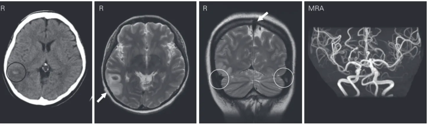
頭部単純 CT:右側頭葉皮質下に高吸収域を認めた(Fig.
1A
).
頭部 MRI:右側頭葉皮質下に脳出血を認め,上矢状静
脈 洞, 横 静 脈 洞,S 状 静 脈 洞 に 血 栓 は な か っ た. 頭 部
MRA では動脈性奇形や腫瘍性病変を認めなかった(Fig.
1B-D).
入院時一般採血:赤血球 279 万/μl,Hb 3.6 g/dl,MCV
57.7%と高度貧血がみられた.血小板 49.3 万/μl と増加して
おり,その他のデータは白血球 11,530/μl,CRP 0.56 mg/dl,
D-dimer 8.7 μg/dl であった.
入院後経過:画像検査にて動脈性奇形や腫瘍性病変はな
く,おもな静脈洞も MRI 上は開存していたために静脈洞
血栓症も否定的であり,脳出血としてしばらく保存的治療
を行い,後日あらためて原因精査を行う方針とした.ま
た,腹部 CT で子宮筋腫を認め,過多月経,高度の鉄欠乏
性貧血の原因であると考えた.その他,貧血に対しては濃
厚赤血球 2 単位を投与した.
入院 4 時間後に意識レベルが低下〔Japan Coma Scale
(JCS)Ⅱ-20〕し,血圧 143/80 mmHg,脈拍 75 回/分,左半
身麻痺(MMT 3/5),両側の瞳孔拡大(右/左:4 mm/4 mm)
を認めた.頭部 CT を施行したところ,血腫が増大し,正
中偏位がみられ,中脳周囲槽は消失していた(Fig. 2).脳
ヘルニア徴候を示していると判断して,緊急開頭術を施行
した.
手術所見:右前頭側頭開頭を行ったところ,脳表は蒼白
であり,膨隆してきた.また,浅中大脳静脈(superficial
middle cerebral vein:SMCV)に白色の血栓を認めた(Fig.
3
).以上のような術中所見から,脳皮質静脈血栓症(iso-lated cortical venous thrombosis:ICVT)による脳皮質下
出血と診断し,血腫除去術は行わず,外減圧術のみとした.
術後経過:頭蓋内圧コントロール目的に,鎮静下で人工
Fig. 1 Head computed tomography (CT) on admission reveals intracerebral
hemor-rhage in the right temporal lobe (circle).
Magnetic resonance imaging and angiography (MRI and MRA, respectively) on admission demonstrates patency of the superior sagittal sinus (white arrow), transverse sinus (circles), and no arterial malformation or tumor.
A: Head CT, B, C: T2-weighted image, D: MRA.
MRA R
R R
A B C D
Fig. 2 Computed tomography
(CT) performed after the coma shows an increase in the hematoma size and signs of herniation. R
呼吸管理を行った.貧血に対しては,濃厚赤血球を術中に
4 単位,術後に 4 単位投与し,第 2 病日には Hb 8.3 g/dl
まで改善していた.入院後も月経は継続しており,不正性
器出血も継続していたが,静脈血栓症に対する治療が優先
と考え,入院後第 3 病日よりヘパリン(12,000 単位/日)に
よる抗凝固療法を開始し,第 4 病日より意識清明となっ
た.追加の血清学的検査でプロテイン C 抗原量およびプ
ロテイン S 抗原量は正常範囲内で,ループスアンチコア
グラントや抗核抗体,抗 DNA 抗体,抗 RNP 抗体,抗カ
ルジオリピン β
2グリコプロテイン抗体はいずれも陰性で
あった.第 7 病日に脳血管造影検査を行ったところ,静脈
洞は開通していたが,右 SMCV の描出は不良であり,vein
of Labbe の描出はなかった(Fig. 4).第 23 病日よりワー
ファリン内服に切り替えた.第 47 病日に左上肢より始ま
る全身性強直間代発作がみられ,症候性てんかんとしてレ
ベチラセタム内服開始した.第 46 病日に頭蓋形成術を施
行した.左半側空間無視は残存するものの,第 69 病日に
modified Rankin Scale (mRS) 1 で自宅退院となった(Fig.
5
).退院後,産婦人科に再入院し,子宮全摘術を受けた.
病理診断は子宮平滑筋腫であった.
考
察
脳静脈血栓症は,硬膜静脈洞や静脈が血栓により狭窄あ
るいは閉塞し,静脈の還流障害により静脈圧が上昇するこ
とで生じる疾患で,静脈洞血栓症と皮質静脈血栓症(ICVT)
に分けられる.静脈洞血栓症を伴わない ICVT は比較的ま
れであり,梗塞を起こした部位によりさまざまな臨床所見
を認めたり,典型的な画像所見を呈することが少ないため
Fig. 3 White thrombosis (arrows) are observed in the superficial middle cerebral vein
(SMCV) on external decompression.
Fig. 4 Angiography performed on day 7 reveals the patency of the venous sinus and
oc-culusion of the Vein of Labbe (circle) and poor filling of the superficial middle ce-rebral vein (SMCV) and cavenous sinus (arrows).
に診断に難渋することが多い.本症例では感染症や遺伝性
疾患(アンチトロンビンⅢ,プロテイン C・S といった抗
凝固タンパク欠乏症,第 5 凝固因子 Leiden 変異,プロト
ロンビン G20210 変異),抗リン脂質抗体症候群,悪性腫
瘍,経口避妊薬の内服,妊娠,産褥など
8)21)がなかったた
めに,高度の鉄欠乏性貧血(IDA)により血栓症を生じたと
考えられた.IDA は女性や小児にしばしば認める血液疾
患であり,乳幼児ではまれに静脈洞血栓症を合併すること
がある
4).IDA による血栓形成の機序には血小板増加と血
液の粘度上昇がかかわっていると考えられている.前者に
ついては,血小板新生の抑制因子である鉄が欠乏している
ことが原因であるとされている他,貧血に対してエリスロ
ポエチンの濃度が上昇していることが血小板新生を促進し
ているとの報告もある
3).また,後者では IDA により赤血
球が小球化し,結果として変形能が低下してしまうことに
より血液の粘度が上昇してしまうといわれている
11).実
際,われわれの症例についても血小板 49.3 万/μl と上昇し
ており,MCV 57.7%と赤血球は小球化していた.本症例
では上記の機序により血栓形成傾向を示し,SMCV から
vein of Labbe にかけて血栓症により閉塞したために,静
脈還流障害を引き起こし,右側頭葉皮質下に脳出血を発症
したと考えられる.しかし,IDA は月経のある女性の
30%にみられるといわれており
6),脳静脈血栓症の罹患数
と比較すると IDA が直接的に関与しているわけではな
く,他の関連因子が発症に関与している可能性もあると考
えることができる.
また,成人での IDA と静脈洞血栓症の合併はきわめ
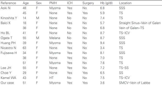
て ま れ で あ り, 数 編 の 報 告 が あ る の み で あ る(Table
1
)
1)2)5)10)12)-17)19).それぞれの報告の Hb の平均値は 7.1 g/dl
であり,本症例では 3.6 g/dl と貧血が高度であったことが
わかる.いずれの報告例も閉塞部位は静脈洞であったのに
対して,臨床的にも血液学的にもより重症の本症例では脳
皮質静脈のみに閉塞を認めていたことは,興味深い現象で
ある.
脳静脈血栓症に対する治療として,抗凝固療法の有用性
を検証したランダム化比較試験(randomized controlled
trial:RCT)は 2 つしかない.Einhaupl らは脳静脈血栓症
患者を対象に,ヘパリン静注群がプラセボ群に対して有意
に機能予後と生命転帰を改善したと報告した
18).また,de
Bruijin らは脳静脈血栓症患者を対象に,ヘパリン皮下注
後に経口抗凝固療法を施行した群はプラセボ群に対して有
意差はないものの良好な転帰をたどったと報告してい
る
18).さらに,脳出血により発症した例においても抗凝固
療法によって増悪はみられず,安全性が確認されたとい
う.以上の報告などから,出血症例に対しても,早期から
ヘパリン投与を行うことがよいとされている
18).また,脳
静脈血栓症の中でもヘルニアをきたすような mass effect
をもった静脈性梗塞や皮質下出血を伴う症例への開頭減圧
術の有効性も示唆されている
9)20).Ferro ら
8)が報告したメ
タ解析によると,脳ヘルニア徴候を示した 69 例の脳静脈
血栓症患者に対する開頭減圧術の有効性を検討してお
り,45 例に外減圧術,7 例に血腫除去術,17 例に両者が
施行され,いずれの開頭減圧術も良好な予後をもたらすと
されている.実際に,本症例では緊急開頭減圧術を施行
Fig. 5 Clinical course.
Discharge Resection of myoma Cranioplasty External decompression 119 102 46 1 0 Day Warfarin 2 mg Heparin 12,000 U/d Hb ICP 15 10 5 (g/dl) Hb Hemiplegia 20 15 10 5 0 (mmHg) ICP
後,抗凝固療法を施行し,良好な転帰をたどった.
本症例においては,来院時に右側頭葉に皮質下出血を認
めており,著明な IDA を伴っていたことから,静脈性梗
塞を除外するために脳血管造影検査を施行しておくべきで
あったかもしれない.その結果,vein of Labbe の閉塞な
どから静脈性梗塞と診断していたら,早期に抗凝固療法を
開始し,出血による急変にも備えることができただろ
う.側頭葉皮質下出血をみた場合,静脈性梗塞が鑑別に思
い浮かぶかどうかが明暗を分けると思われる.
結
語
IDA が原因と考えられた ICVT の 1 例を経験した.外
減圧術,ヘパリンによる抗凝固療法で良好な経過をたどっ
た.
著者全員は,日本脳神経外科学会への COI 自己申告を
完了している.本論文に関して,開示すべき COI はない.
文
献
1) Aoki N, Sakai T: Cerebral sinus thrombosis in patients with severe iron deficiency anaemia due to myoma uteri. Acta Neurochir(Wien)97: 131-134, 1989
2) Balci K, Utku U, Asil T, et al: Deep cerebral vein thrombosis associated with iron deficiency anaemia in adults. J Clin Neu-rosci 14: 181-184, 2007
3) Beguin Y: Erythropoietin and platelet production.
Haemato-logica 84: 541-547, 1999
4) Beri S, Khan A, Hussain N, et al: Severe anemia causing cere-bral venous sinus thrombosis in an infant. J Pediatr Neurosci 7: 30-32, 2012
5) Choe Y, Lee JB, Kim YJ, et al: Cerebral venous sinus throm-bosis and venous hemorrhagic infarction in a young woman. Ann Rehabil Med 38: 698-701, 2014
6) de Benoist B, McLean E, Egil I, et al(eds): Worldwide preva-lence of anaemia 1993-2005. WHO Global Database on Anae-mia, Geneva, World Health Organization, 2008
7) Dulli DA, Luzzio CC, Williams EC, et al: Cerebral venous thrombosis and activated protein C resistance. Stroke 27: 1731-1733, 1996
8) Ferro JM, Canhão P, Stam J, et al: Prognosis of cerebral vein and dural sinus thrombosis: results of the Internatinal Study on Cerebral Vein and Dural Sinus Thrombosis(ISCVT). Stoke 35: 664-670, 2004
9) Ferro JM, Crassard I, Coutinho JM, et al: Decompressive sur-gery in cerebrovenous thrombosis: a multicenter registry and a systematic review of individual patient data. Stroke 42: 2825-2831, 2011
10) 藤沢弘範,村松直樹,東馬康郎,ほか:鉄欠乏性貧血に関連 した脳静脈洞血栓症:成人 3 例の報告.脳卒中 35: 221-226, 2013
11) Hartfield DS, Lowry NJ, Keene DL, et al: Iron deficiency: a cause of stroke in infants and children. Pediatr Neurol 16: 50-53, 1997
12) Ho BL, Huang P, Khor GT, et al: Simultaneous thrombosis of cerebral artery and venous sinus. Acta Neurol Taiwan 17: 112-116, 2008
13) Huang PH, Su JJ, Lin PH: Iron deficiency anemia—a rare eti-ology of sinus thrombosis in adults. Acta Neurol Taiwan 19: 125-130, 2010
Table 1 Case reports on adult cerebral venous thrombosis associated with iron deficiency anemia. There was
no report on patients without venous sinus thrombosis
Reference Age Sex PMH ICH Surgery Hb (g/dl) Location Aoki N 48 F Myoma Yes No 6.8 SSS
45 F None Yes Yes 5.9 TS Kinoshita Y 14 M None No No 7.4 TS
Balci K 18 F None Yes No 5.7 Straight Sinus-Vein of Galen 38 F None No No 6.8 Vein of Galen-TS
Ho BL 41 F None No No 8.7 TS-ICV Ogata T 55 M Melana No No 8.7 SSS Huang PH 39 F Myoma Yes No 9.4 SSS Nicastro N 63 F None Yes No 3.4 TS Fujisawa H 34 F Myoma Yes No 8.1 SSS
38 F None Yes No 7.0 TS 51 F Myoma Yes No 7.6 TS Lee JH 55 F None Yes No 7.2 TS-SS Choe Y 29 F None Yes Yes 6.5 SS Kamel WA 43 F HT No No 7.5 TS-ICV
Our case 51 F Myoma Yes Yes 3.6 SMCV-Vein of Labbe
SSS: superior sagittal sinus, TS: transverse sinus, IJV: internal jugular vein, SMCV: superficial middle cerebral vein, F: female, M: male, PMH: past medical history, ICH: intracerebral hemorrhage
14) Kamel WA, AI-Hashel JY, Alexander KJ: Cerebral venous thrombosis in a patient with iron deficiency anemia and thrombocytopenia: a case report. Open Access Maced J Med Sci 5: 967-969, 2017
15) Kinoshita Y, Taniura S, Shishido H, et al: Cerebral venous si-nus thrombosis associated with iron deficiency: two case re-ports. Neurol Med Chir(Tokyo) 46: 589-593, 2006
16) Lee JH, Park KJ, Chung YG, et al: Isolated lateral sinus throm-bosis presenting as cerebellar infarction in a patient with iron deficiency anemia. J Korean Neurosurg Soc 54: 47-49, 2013
17) Nicastro N, Schnider A, Leemann B: Iron-deficiency anemia as a rare cause of cerebral venous thrombosis and pulmo-nary embolism. Case Rep Med 2012: 497814, 2012
18) 日本脳卒中学会,脳卒中ガイドライン委員会(編):脳卒中治 療ガイドライン 2015.東京,協和企画,2015
19) Ogata T, Kamouchi M, Kitazono T, et al: Cerebral venous thrombosis associated with iron deficiency anemia. J Stroke Cerebrovasc Dis 17: 426-428, 2008
20) Raza E, Shamim MS, Wadiwala MF, et al; Decompressive surgery for malignant cerebral venous sinus thrombosis: a retrospective case series from Pakistan and comparative lit-erature review. J Stroke Cerebrovasc Dis 23: e13-22, 2014 21) Saposnik G, Barinagarrementeria F, Brown RD Jr, et al:
Diag-nosis and management of cerebral venous thrombosis: a statement for healthcare professionals from the American Heart Association/American Stroke Association. Stroke 42: 1158-1192, 2011