1 1
Viscoelastic properties of the central region of porcine temporomandibular joint 2
disc in shear stress-relaxation. 3
4
Eva Barrientosa*, Fernandez Pelayoa, Eiji Tanakab, María Jesús Lamela-Reya, Alfonso
5
Fernández-Cantelia
6 7
a Department of Construction and Manufacturing Engineering, University of Oviedo,
8
Gijón, Spain 9
b Department of Orthodontics and Dentofacial Orthopedics, Institute of Biomedical
10
Sciences, Tokushima University Graduate School, Tokushima, Japan 11 12 *Corresponding Author: 13 Eva Barrientos 14
Department of Construction and Manufacturing Engineering 15
University of Oviedo, Gijón, Spain. 16
E-mail: [email protected]
17 18
Manuscript Click here to view linked References
© 2019. This manuscript version is made available under the CC-BY-NC-ND 4.0 license http://creativecommons.org/licenses/by-nc-nd/4.0/ The published version is available via https://doi.org/10.1016/j.jbiomech.2019.06.023.
2 Abstract
1
In this study, shear relaxation properties of the porcine temporomandibular joint (TMJ) 2
disc are investigated. Previous studies have shown that, in fatigue failure and damage 3
of cartilage and fibrocartilage, shear loads could be one of the biggest contributors to 4
the failure. The aim of the present study is to develop an evaluation method to study 5
shear properties of the disc and to do a mathematical characterization of it. For the 6
experiments, twelve porcine discs were used. Each disc was dissected from the TMJ 7
and, then, static strain control tests were carried out to obtain the shear relaxation 8
modulus for the central region of the discs. From the results, it was found that the disc 9
presents a viscoelastic behavior under shear loads. Relaxation modulus decreased 10
with time. Shear relaxation was 10% of the instantaneous stress, which implies that 11
the viscous properties of the disc cannot be neglected. The present results lead to a 12
better understanding of the discs mechanical behavior under realistic TMJ working 13
conditions. 14
15
Keywords: Temporomandibular Joint; Soft Tissues; Viscoelasticity; Biomechanical 16
Characterization; Experimental Techniques; Shear. 17
3 1. Introduction
1
Synovial joints allow various degrees of relative motion among the bones to be 2
regulated by muscles attached to the latter (Widegren et al., 2000). Daily activity 3
accompanies joint motion resulting in joint loads. The temporomandibular joint (TMJ), 4
a diarthrodial synovial joint, enables large relative movements between the temporal 5
bone and the mandibular condyle (Rees, 1954; Scapino et al., 2006). Within the joint, 6
both the articular surfaces of the condyle and temporal bone are covered by a thin 7
fibro-cartilaginous layer showing a very low coefficient of friction (Tanaka et al., 2004b). 8
A dense fibrocartilaginous articular disc is located between the bones in each TMJ. 9
The disc provides a largely passive movable articular surface accommodating the 10
traslatory movement made by the condyle (Koolstra and Tanaka, 2009). 11
The TMJ disc has an important load-bearing, stress absorbing and joint stabilizing 12
function (Barrientos et al., 2016; Fernández et al., 2013; Tanaka et al., 2008; Tanaka 13
and Eijden, 2003). The disc is subject to various types of loading, such as sustained 14
loading during clenching and intermittent loading during mastication (Hattori-Hara et 15
al., 2014; Hirose et al., 2006; Tanaka et al., 2007). Stresses are divided into 16
compression, tension and shear components. During every type of loading the disc 17
undergoes a deformation while internal forces arise within the tissue. The 18
viscoelasticity of such a material, as that of the disc, is the principal factor of energy 19
dissipation (Fung, 1969). These types of tissues show different mechanism of energy 20
dissipation that are result of the different phases in their structure: interstitial fluid flow 21
within and through the matrix and relaxation of the solid matrix (collagen fibers and 22
proteoglycans). Without strain energy dissipation, storage of the exceeding strain 23
energy can lead to breakage of the articular disc and other components of the TMJ 24
(Tanaka et al., 1999). 25
4 Since shear stress can result in fatigue, damage and deformation of cartilage, 1
investigation of shear properties in synovial joints is of particular interest (Spirt et al., 2
2005; Zhu et al., 1993, 1994). Gallo et al. (2000) suggest that, during mastication, 3
fatigue failure of the TMJ disc could result from shear stresses caused by medio-lateral 4
translation of stress location. Therefore, data on the shear modulus might contribute to 5
a better understanding of secondary tissue damage, such as perforation or thinning of 6
the disc due to long-term exposure to severe loadings. It has been reported that the 7
shear stress in cartilage is very sensitive to the frequency and direction of the loading 8
and to the amount of compressive strain (Mow et al., 1992). However, in the literature 9
few studies are available in which the viscoelastic properties of the TMJ disc are 10
measured in shear stress-relaxation. 11
This paper may provide better insight about the possible mechanism leading to tissue 12
fatigue and failure due to shear. Therefore, in this study the viscoelastic properties of 13
porcine TMJ disc are investigated under shear stress relaxation, aiming at advancing 14
in the design of biomimetic disc substitutes and in the understanding of the pathological 15
conditions of the TMJ disc. 16
17
2. Materials and Methods 18
In this study, twelve healthy-looking TMJ discs from 6 pigs (age: approx. 6–7 months, 19
gender not specified) were obtained at a local slaughterhouse (Noreña, Asturias, 20
Spain). The protocol of the experiment was approved by the Animal Care and Use 21
Committee at the University of Oviedo, Spain. The discs were carefully dissected 22
immediately after the sacrifice, introduced in hermetic containers immersed in a 23
physiologic saline solution (NaCl 0.09 g/100 ml), and frozen at -25 ºC for 3 days until 24
the experiment was initiated for testing (Allen and Athanasiou, 2005; Calvo-Gallego et 25
5 al., 2017). The discs were completely unfrozen in a refrigerator at 3-4 ºC and, then, 1
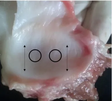
allow to reach room temperature (20 ºC) before testing. Using a cylindrical 4.0 mm 2
diameter tissue punch, two experimental specimens were dissected from the central 3
region of each disc (see Figure 1). 4
Although previous studies have shown region-dependent mechanical properties 5
(Fernández et al., 2013), this study is only focused on the central region, mainly due 6
to the complexity of extracting two specimens with the necessary dimensions of the 7
rest of regions. 8
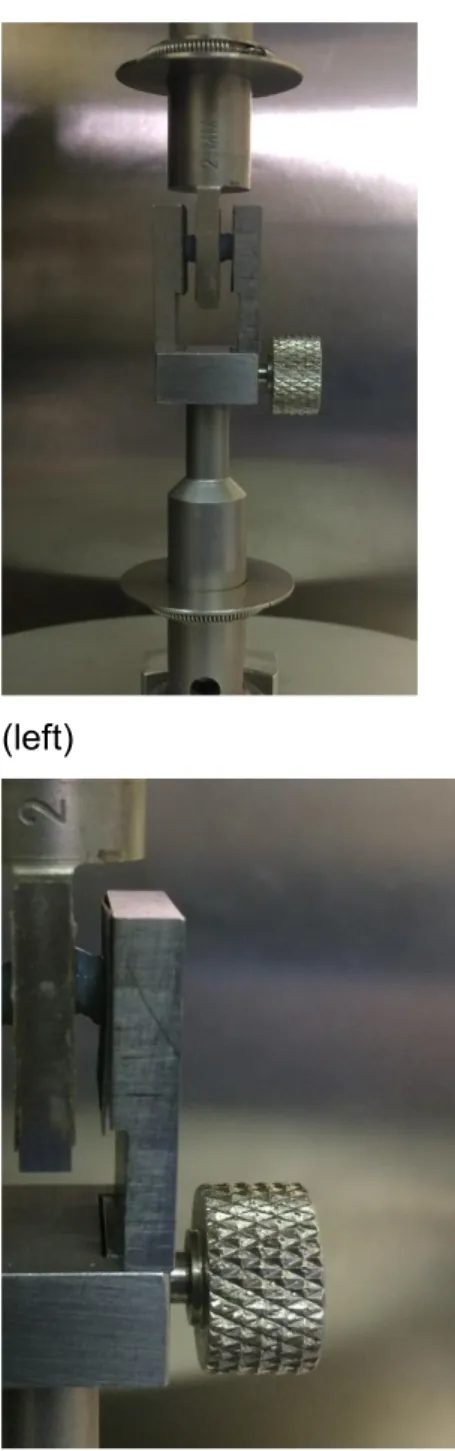
All the specimens were tested in a DMA Instrument (RSA3, T.A. Instruments, USA) in 9
unconfined shear using a shear tool (see Figure 2) at room temperature (20 ºC). The 10
loading was applied in the antero-posterior direction, since mechanical properties of 11
the disc, due to fiber distribution, will also be direction-dependent. 12
As mentioned before, two specimens of each disc were cut. In Figure 2, it can be seen 13
that the shear-tool has a sandwich configuration and samples need to be placed at 14
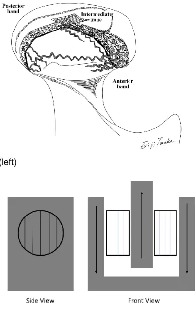
both sides of the tool. In order to test shear in antero-posterior direction, the fibers of 15
the specimens need to be aligned with the movement of the tool (vertical direction), 16
according to Figure 3. 17
To avoid the specimens’ slippage during shear loading, 600 grit sandpaper was glued 18
to the surfaces of the shear tool. Additionally, the selected inner part of the shear tool 19
would allow testing 2 mm thick specimens. Taking into account the average thickness 20
value for the discs, 1.84±0.11 mm, and the real gap for testing, 1.750 mm (subtracting 21
the sandpaper sheet thickness), an average initial value of 5% pre-strain in the 22
compression direction was applied before testing. After previous step, a 3-min 23
preconditioning test was performed with 1% sinusoidal strain before the subsequent 24
shear stress relaxation test. The shear strain was applied to the specimens moving the 25
6 lower part of the tool in the axial direction of the machine (vertical direction in Figure 2 1
and 3). Shear strain levels of the TMJ disc produced under ordinary mandibular 2
movement have not been reported. Previous studies do not show consensus for shear 3
strain (Lai et al., 1998; Tanaka et al., 2004a). Due to the limitations of testing the 4
specimens under shear conditions, i.e. very low loads for strain values lower than 5% 5
or problems of slippage for strain values larger than 10%, tests were carried out at 6
strain levels of 5% and 8% in order to obtain the corresponding relaxation modulus. 7
The specific level of shear strain was produced under an instantaneous strain step and 8
kept constant during 120 seconds for each stress relaxation test keeping the same test 9
procedure used in previous studies (Barrientos et al., 2016). 10
To apply and maintain the initial value of strain during the relaxation test, the DMTA 11
machine is equipped with a motor driven by an air bearing system, which applies the 12
corresponding displacement at a very high rate once the strain is commanded before 13
testing (T.A.Instruments, 2001). Loads were measured simultaneously under the 14
specified constant strain. 15
16
3. Results 17
3.1 Viscoelastic properties of porcine TMJ disc in shear stress relaxation 18
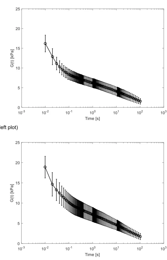
From the experimental tests, the mean and standard deviation of the shear modulus 19
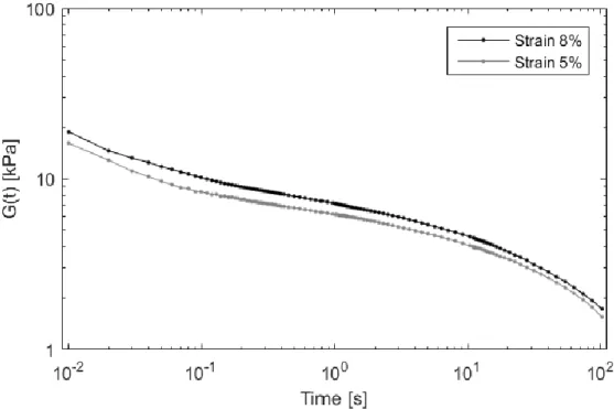
of the TMJ disc at convenient times were calculated. The resulting curves for the 5 and 20
8 % strain levels are presented in Figure 4 (left and right plots, respectively). 21
For comparison proposals both averaged curves are plotted in Figure 5. From Figure 22
5, a higher shear modulus is observed for the 8 % strain level. From the results (Figure 23
5), a dependence of the relaxation modulus, 𝐺(𝑡), with applied strain can be observed, 24
which is in agreement with the TMJ disc behaviour previously observed (Lamela et al., 25
7 2011).
1
The shear modulus obtained for both strain levels (see Figure 5) presents a large 2
relaxation ratio. For 1 s, the shear modulus decreases about 70% while a 90 % 3
reduction is observed for 100 s. 4
5
3.2 TMJ shear relaxation model 6
Due to its simplicity, even though other models could be used, generalized Maxwell 7
model was used to fit the experimental data to the viscoelastic model represented in 8
Figure 6, as a combination of spring and dashpot elements (Tschoegl, 2012), which 9
can be modelled using the Prony´s series model given by the equation: 10 G(t) = G0[1 − ∑ gi nt i=1 (1 − exp (− t τi))] (1) ( (1)
where 𝑔𝑖 and 𝜏𝑖 are the Prony parameters and 𝐺0 is the instantaneous shear 11
modulus. 12
To simplify the material model, as well as to take into account the dependence of the 13
𝐺(𝑡) with the applied strain, a unique set of Prony parameters was used to fit both 14
shear modulus curves. This procedure profits from the fact that a simple vertical shift 15
is observed between both material curves (see Figure 5) which could be interpreted as 16
a proportional shift of 𝐺(𝑡) with the strain. 17
Two steps were used for fitting the material model. Firstly, the shear curves for the 18
TMJ are averaged and, next, the generalized Maxwell model was applied to fit the 19
averaged curve by means of the Prony series equation (1). 20
To fit adequately the experimental data, 8 Prony terms were necessary being the R-21
square 0.994. The parameters of the Prony series presented in Table 1 define the 22
normalized viscoelastic curve for the material, as a function of the instantaneous 23
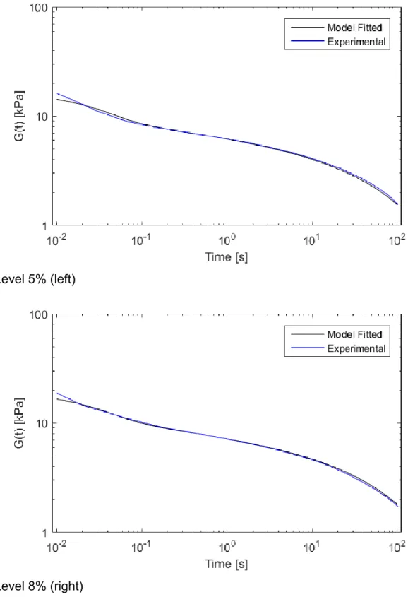
8 modulus of the material, G0. In this way, the curves for the 5% and the 8% strains are 1
gained from the fitted model, simply, by multiplying in each case equation (1), by the 2
corresponding instantaneous modulus. Accordingly, G05% = 1.6205e + 04 kPa and 3
G08%= 1.8883e + 04 kPa, for the 5 % and the 8 % shear modulus curves, respectively.
4
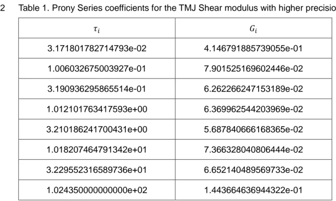
The Prony series parameters with higher precision are included in the appendix. 5
Table 1. Prony series parameters (𝑅2=0.994) for the normalized TMJ shear modulus
6 curve. 7 𝜏𝑖 𝐺𝑖 3.17e-02 4.14e-01 1.00e-01 7.90e-02 3.19e-01 6.26e-02 1.01e+00 6.36e-02 3.21e+00 5.68e-02 1.01e+01 7.36e-02 3.22e+01 6.65e-02 1.02e+02 1.44e-01
The experimental and the analytical curves (using equation (1)) are presented in Figure 8
7. The maximum error between the experimental results and the proposed model are 9
less than a 2% for both curves. 10
11
4. Discussion 12
Fatigue failure and damage of joint tissues, including both disc and cartilage, may be 13
more linked to repeated and prolonged extension and shear motions than to the joint 14
compression applied (Iatridis and ap Gwynn, 2004; Tanaka et al., 2003). Even when 15
the disc slides along smooth temporal cartilage during jaw movements, shear loading 16
9 of the disc and cartilage has been considered to be negligible due to almost zero 1
friction. However, several authors support the evidence that the disc and cartilage are 2
subjected to shear stress. For example, after prolonged clenching and grinding, only 3
solid contact may exist between the disc and cartilages, without boundary lubrication 4
between them, resulting in considerable shear stress (Forster and Fisher, 1999, 1996; 5
Tanaka et al., 2001). Few studies of the behaviour of the TMJ disc under dynamic 6
shear loads were performed in the past (Juran et al., 2013; Koolstra et al., 2007; 7
Tanaka et al., 2004a, 2003) to evaluate the mechanical properties of the disc at 8
different strain rates and frequencies. The present study is, as far as we know, the first, 9
in which the shear relaxation properties of the TMJ disc in shear stress relaxation were 10
examined. Wu et al. (2015) investigated the intrinsic viscoelastic shear properties in 11
porcine TMJ disc, but in contrast to the present study, they applied a rotational shear 12
loading. The present design might reproduce the actual environment in the TMJ disc. 13
Previous studies have shown that due to morphology, function and diet, pig discs are 14
the closest to human discs making them an appropriate model for TMJ studies 15
(Bermejo et al., 1993; Kalpakci et al., 2011). In this study, relaxation viscoelastic 16
behaviour of cut porcine specimens is evaluated in antero-posterior direction at 5 and 17
8% shear strain levels. As a result, the instantaneous shear moduli were increased 18
with increasing applied strain. This evidences a dependence with strain of the 19
behaviour of the disc which is in good agreement with the general mechanical 20
behaviour observed previously in the TMJ disc (Lamela et al., 2011; Tanaka and Eijden, 21
2003).The possible explanation for this increment is the stretching of collagen fibers in 22
antero-posterior direction (Barrientos et al., 2016; Lamela et al., 2011; Tanaka et al., 23
2003). Furthermore, present results show that the relaxed stress of the porcine TMJ 24
disc was approximately 10% of the instantaneous stress irrespective of shear strain 25
10 amplitude. This indicates that energy-dissipation function takes place in the TMJ disc. 1
Without the energy dissipation capacity of the disc, TMJ components including bony 2
components and soft tissue probably fail resulting in the tissue rupture. Thus far, it is 3
concluded that the TMJ disc plays an important role as a stress bumper during complex 4
mandibular movements. 5
When comparing the compression relaxation tests (Barrientos et al., 2016; Lamela et 6
al., 2011) with the shear relaxation tests, the present results clearly show that 7
compression relaxation modulus is 10 times higher than shear relaxation modulus. 8
Adam et al. (2015) investigated an image-based modelling study on the bovine caudal 9
disc, and concluded that shear resistance between lamellae confers disc mechanical 10
resistance to compression. This points out the relationship between shear and 11
compressive properties of the TMJ disc. Moreover, the present results reveal that the 12
porcine TMJ discs exhibited shorter relaxation times under shear stress relaxation than 13
under compressive stress relaxation. This may be due to the difference of an outflow 14
of interstitial fluid caused by pressurization of the compressed area. During shear 15
stress relaxation, the fluid within the disc is likely to move along the stretching collagen 16
fibers; however, during compressive stress relaxation, the disc maintains a fluid 17
pressure because of sustained interstitial fluids within the disc. Since the load bearing 18
functions of cartilaginous tissues are mainly provided by the viscoelastic property of 19
collagen fiber network and the osmotic pressure due to the presence of proteoglycans 20
(Hardingham and Fosang, 1992), the large proteoglycans and the related chondroitin 21
sulfate might be more important to counteract compression and shear, while the 22
collagen fibers are more important to counteract tension (Tanaka and Eijden, 2003). 23
Mow et al. (1980) reported about the biphasic theory, this theory is suitable for better 24
understanding of the mechanisms involved in energy dissipation. Due to the highly 25
11 heterogeneous structure of the TMJ disc, the viscoelastic approach used in this study 1
gives a global understanding of the mechanical properties of the disc rather than the 2
material constitutive law. 3
In literature, authors have used different models to characterize the viscoelastic 4
properties of the TMJ disc (Allen and Athanasiou, 2006; Tanaka and Eijden, 2003). For 5
large displacements, other models could be more appropriate (Fung, 1969). In this 6
study, a generalized Maxwell model, based on Prony´s series, was applied to 7
characterize the shear relaxation modulus of the material. Although the TMJ disc 8
presents a strain-dependent behavior, almost the same relaxation rate is observed for 9
the strain levels applied in the experiments (see Figure 5). This fact allows a unique 10
viscoelastic model to be fitted where the instantaneous modulus, 𝐺0 , at the 11
corresponding strain level must be used. The results obtained with the proposed Prony 12
series model can be considered adequate for the shear relaxation modulus of the TMJ 13
disc showing errors under 2%. 14
To be consistent with previous studies and allowed comparison (Barrientos et al., 2016; 15
Fernández et al., 2013), some testing conditions, such relaxation time and temperature, 16
and model parameters were chosen. Temperature affects mechanical results as higher 17
temperatures reduce stiffness and strength of the discs (Detamore and Athanasiou, 18
2003). 19
In conclusion, the relaxation properties of the porcine disc were determined under 20
shear in this study. A new methodology to test the disc under relaxation shear 21
conditions was proposed. The study shows that the viscoelastic properties of the disc 22
under shear loads cannot be neglected. Shear properties of the disc in antero-posterior 23
direction were characterized using a unique Maxwell model. Nevertheless, this study 24
is a first step in the shear characterization of the TMJ discs and further studies are 25
12 needed to conclude on the shear behavior of the disc in medio-lateral direction, cyclic 1
loads, pre-compression and region dependencies. 2
3 4 5
13 Acknowledgments
1
This research was supported in part by Grants-in-Aid 26293436 (E.T.) for Science 2
Research from the Ministry of Education, Culture, Sports, Science and Technology, 3
Japan. The funder had no role in study design, data collection and analysis, decision 4
to publish, or preparation of the manuscript. The authors would also like to 5
acknowledge the funds granted by CajAstur Fellowship-University of Oviedo 2011 6
programme. 7
8
Conflict of interest statement 9
We wish to confirm that there are no known conflicts of interest associated with this 10
publication and there has been no significant financial support for this work that could 11
have influenced its outcome. 12
14 5. References
1
Adam, C., Rouch, P., Skalli, W., 2015. Inter-lamellar shear resistance confers 2
compressive stiffness in the intervertebral disc: An image-based modelling 3
study on the bovine caudal disc. J. Biomech. 48, 4303–4308. 4
https://doi.org/10.1016/j.jbiomech.2015.10.041 5
Allen, K.D., Athanasiou, K.A., 2006. Viscoelastic characterization of the porcine 6
temporomandibular joint disc under unconfined compression. J. Biomech. 39, 7
312–322. https://doi.org/10.1016/j.jbiomech.2004.11.012 8
Allen, K.D., Athanasiou, K.A., 2005. A Surface–Regional and Freeze–Thaw 9
Characterization of the Porcine Temporomandibular Joint Disc. Ann. Biomed. 10
Eng. 33, 951–962. https://doi.org/10.1007/s10439-005-3872-6 11
Barrientos, E., Pelayo, F., Tanaka, E., Lamela-Rey, M.J., Fernández-Canteli, A., 2016. 12
Dynamic and stress relaxation properties of the whole porcine 13
temporomandibular joint disc under compression. J. Mech. Behav. Biomed. 14
Mater. 57, 109–115. https://doi.org/10.1016/j.jmbbm.2015.12.003 15
Bermejo, A., González, O., González, J.M., 1993. The pig as an animal model for 16
experimentation on the temporomandibular articular complex. Oral Surg. Oral 17
Med. Oral Pathol. 75, 18–23. 18
Calvo-Gallego, J.L., Commisso, M.S., Domínguez, J., Tanaka, E., Martínez-Reina, J., 19
2017. Effect of freezing storage time on the elastic and viscous properties of the 20
porcine TMJ disc. J. Mech. Behav. Biomed. Mater. 71, 314–319. 21
https://doi.org/10.1016/j.jmbbm.2017.03.035 22
Detamore, M.S., Athanasiou, K.A., 2003. Tensile Properties of the Porcine 23
Temporomandibular Joint Disc. J. Biomech. Eng. 125, 558–565. 24
https://doi.org/10.1115/1.1589778 25
Fernández, P., Lamela, M.J., Ramos, A., Fernández-Canteli, A., Tanaka, E., 2013. The 26
region-dependent dynamic properties of porcine temporomandibular joint disc 27
under unconfined compression. J. Biomech. 46, 845–848. 28
https://doi.org/10.1016/j.jbiomech.2012.11.035 29
Forster, H., Fisher, J., 1999. The influence of continuous sliding and subsequent 30
surface wear on the friction of articular cartilage. Proc. Inst. Mech. Eng. [H] 213, 31
329–345. https://doi.org/10.1243/0954411991535167 32
Forster, H., Fisher, J., 1996. The influence of loading time and lubricant on the friction 33
of articular cartilage. Proc. Inst. Mech. Eng. [H] 210, 109–119. 34
https://doi.org/10.1243/PIME_PROC_1996_210_399_02 35
Fung, Y., 1969. Biomechanics: Mechanical Properties of Living Tissues. Springer-36
Verlag. 37
15 Gallo, L.M., Nickel, J.C., Iwasaki, L.R., Palla, S., 2000. Stress-field Translation in the 1
Healthy Human Temporomandibular Joint. J. Dent. Res. 79, 1740–1746. 2
https://doi.org/10.1177/00220345000790100201 3
Hardingham, T.E., Fosang, A.J., 1992. Proteoglycans: many forms and many functions. 4
FASEB J. Off. Publ. Fed. Am. Soc. Exp. Biol. 6, 861–870. 5
Hattori-Hara, E., Mitsui, S.N., Mori, H., Arafurue, K., Kawaoka, T., Ueda, K., Yasue, A., 6
Kuroda, S., Koolstra, J.H., Tanaka, E., 2014. The influence of unilateral disc 7
displacement on stress in the contralateral joint with a normally positioned disc 8
in a human temporomandibular joint: An analytic approach using the finite 9
element method. J. Craniomaxillofac. Surg. 42, 2018–2024. 10
https://doi.org/10.1016/j.jcms.2014.09.008 11
Hirose, M., Tanaka, E., Tanaka, M., Fujita, R., Kuroda, Y., Yamano, E., Van Eijden, 12
T.M.G.J., Tanne, K., 2006. Three-dimensional finite-element model of the 13
human temporomandibular joint disc during prolonged clenching. Eur. J. Oral 14
Sci. 114, 441–448. https://doi.org/10.1111/j.1600-0722.2006.00389.x 15
Iatridis, J.C.J.C., ap Gwynn, I., 2004. Mechanisms for mechanical damage in the 16
intervertebral disc annulus fibrosus. J. Biomech. 37, 1165–1175. 17
https://doi.org/10.1016/j.jbiomech.2003.12.026 18
Juran, C.M., Dolwick, M.F., McFetridge, P.S., 2013. Shear Mechanics of the TMJ Disc: 19
Relationship to Common Clinical Observations. J. Dent. Res. 92, 193–198. 20
https://doi.org/10.1177/0022034512468749 21
Kalpakci, K.N., Willard, V.P., Wong, M.E., Athanasiou, K.A., 2011. An Interspecies 22
Comparison of the Temporomandibular Joint Disc. J. Dent. Res. 90, 193–198. 23
https://doi.org/10.1177/0022034510381501 24
Koolstra, J.H., Tanaka, E., 2009. Tensile stress patterns predicted in the articular disc 25
of the human temporomandibular joint. J. Anat. 215, 411–416. 26
https://doi.org/10.1111/j.1469-7580.2009.01127.x 27
Koolstra, J.H., Tanaka, E., Van Eijden, T.M.G.J., 2007. Viscoelastic material model for 28
the temporomandibular joint disc derived from dynamic shear tests or strain-29
relaxation tests. J. Biomech. 40, 2330–2334.
30
https://doi.org/10.1016/j.jbiomech.2006.10.019 31
Lai, W.F., Bowley, J., Burch, J.G., 1998. Evaluation of shear stress of the human 32
temporomandibular joint disc. J. Orofac. Pain 12, 153–159. 33
Lamela, M.J., Prado, Y., Fernández, P., Fernández-Canteli, A., Tanaka, E., 2011. Non-34
linear Viscoelastic Model for Behaviour Characterization of Temporomandibular 35
Joint Discs. Exp. Mech. 51, 1435–1440. https://doi.org/10.1007/s11340-011-36
9465-4 37
16 Mow, V.C., Kuei, S.C., Lai, W.M., Armstrong, C.G., 1980. Biphasic Creep and Stress 1
Relaxation of Articular Cartilage in Compression: Theory and Experiments. J. 2
Biomech. Eng. 102, 73–84. https://doi.org/10.1115/1.3138202 3
Mow, V.C., Ratcliffe, A., Robin Poole, A., 1992. Cartilage and diarthrodial joints as 4
paradigms for hierarchical materials and structures. Biomaterials 13, 67–97. 5
https://doi.org/10.1016/0142-9612(92)90001-5 6
Rees, LA., 1954. The structure and function of the mandibular joint. Br Dent J 96, 125– 7
133. 8
Scapino, R.P., Obrez, A., Greising, D., 2006. Organization and Function of the 9
Collagen Fiber System in the Human Temporomandibular Joint Disk and Its 10
Attachments. Cells Tissues Organs 182, 201–225. 11
https://doi.org/10.1159/000093969 12
Spirt, Mak Arthur F., Wassell Richard P., 2005. Nonlinear viscoelastic properties of 13
articular cartilage in shear. J. Orthop. Res. 7, 43–49. 14
https://doi.org/10.1002/jor.1100070107 15
T.A.Instruments, 2001. RSA3 UserManual. T.A. Instruments, USA. 16
Tanaka, E., Eijden, T. van, 2003. Biomechanical Behavior of the Temporomandibular 17
Joint Disc. Crit. Rev. Oral Biol. Med. 14, 138–150. 18
https://doi.org/10.1177/154411130301400207 19
Tanaka, E., Hanaoka, K., van Eijden, T., Tanaka, M., Watanabe, M., Nishi, M., Kawai, 20
N., Murata, H., Hamada, T., Tanne, K., 2003. Dynamic shear properties of the 21
temporomandibular joint disc. J. Dent. Res. 82, 228–231. 22
https://doi.org/10.1177/154405910308200315 23
Tanaka, E., Hirose, M., Inubushi, T., Koolstra, J.H., van Eijden, T.M., Suekawa, Y., 24
Fujita, R., Tanaka, M., Tanne, K., 2007. Effect of Hyperactivity of the Lateral 25
Pterygoid Muscle on the Temporomandibular Joint Disk. J. Biomech. Eng. 129, 26
890–897. https://doi.org/10.1115/1.2800825 27
Tanaka, E., Hirose, M., Koolstra, J.H., Eijden, T.M.G.J. van, Iwabuchi, Y., Fujita, R., 28
Tanaka, M., Tanne, K., 2008. Modeling of the Effect of Friction in the 29
Temporomandibular Joint on Displacement of Its Disc During Prolonged 30
Clenching. J. Oral Maxillofac. Surg. 66, 462–468. 31
https://doi.org/10.1016/j.joms.2007.06.640 32
Tanaka, E., Kawai, N., Hanaoka, K., Van Eijden, T., Sasaki, A., Aoyama, J., Tanaka, 33
M., Tanne, K., 2004a. Shear properties of the temporomandibular joint disc in 34
relation to compressive and shear strain. J. Dent. Res. 83, 476–479. 35
https://doi.org/10.1177/154405910408300608 36
Tanaka, E., Kawai, N., Tanaka, M., Todoh, M., Eijden, T. van, Hanaoka, K., Dalla-Bona, 37
17 D.A., Takata, T., Tanne, K., 2004b. The Frictional Coefficient of the 1
Temporomandibular Joint and Its Dependency on the Magnitude and Duration 2
of Joint Loading. J. Dent. Res. 83, 404–407. 3
https://doi.org/10.1177/154405910408300510 4
Tanaka, E., Rodrigo, D.P., Tanaka, M., Kawaguchi, A., Shibazaki, T., Tanne, K., 2001. 5
Stress analysis in the TMJ during jaw opening by use of a three-dimensional 6
finite element model based on magnetic resonance images. Int. J. Oral 7
Maxillofac. Surg. 30, 421–430. https://doi.org/10.1054/ijom.2001.0132 8
Tanaka, E., Tanaka, M., Miyawaki, Y., Tanne, K., 1999. Viscoelastic properties of 9
canine temporomandibular joint disc in compressive load-relaxation. Arch. Oral 10
Biol. 44, 1021–1026. https://doi.org/10.1016/S0003-9969(99)00097-7 11
Tschoegl, N.W., 2012. The Phenomenological Theory of Linear Viscoelastic Behavior: 12
An Introduction. Springer Science & Business Media, Berlin. 13
Widegren, U., Wretman, C., Lionikas, A., Hedin, G., Henriksson, J., 2000. Influence of 14
exercise intensity on ERK/MAP kinase signalling in human skeletal muscle. 15
Pflüg. Arch. 441, 317–322. https://doi.org/10.1007/s004240000417 16
Wu, Y., Kuo, J., Wright, G.J., Cisewski, S.E., Wei, F., Kern, M.J., Yao, H., 2015. 17
Viscoelastic shear properties of porcine temporomandibular joint disc. Orthod. 18
Craniofac. Res. 18, 156–163. https://doi.org/10.1111/ocr.12088 19
Zhu, Mow Van C., Koob Thomas J., Eyre David R., 1993. Viscoelastic shear properties 20
of articular cartilage and the effects of glycosidase treatments. J. Orthop. Res. 21
11, 771–781. https://doi.org/10.1002/jor.1100110602 22
Zhu, W., Chern, K.Y., Mow, V.C., 1994. Anisotropic viscoelastic shear properties of 23
bovine meniscus. Clin. Orthop. 34–45. 24
18 6. Appendix A
1
Table 1. Prony Series coefficients for the TMJ Shear modulus with higher precision 2 𝜏𝑖 𝐺𝑖 3.171801782714793e-02 4.146791885739055e-01 1.006032675003927e-01 7.901525169602446e-02 3.190936295865514e-01 6.262266247153189e-02 1.012101763417593e+00 6.369962544203969e-02 3.210186241700431e+00 5.687840666168365e-02 1.018207464791342e+01 7.366328040806444e-02 3.229552316589736e+01 6.652140489569733e-02 1.024350000000000e+02 1.443664636944322e-01 3
Figure 1. Area where the specimens were cut and fiber direction.
Figure 2. Specimens inside the test tool before test (left) and detail of a specimen after strain was applied (right).
(left)
Figure 3. Fiber distribution of discs (left) and direction of fibers in the tool during antero-posterior testing (right).
(left)
Figure 4. Shear relaxation modulus for the TMJ disc at 𝜀 = 5% (left) and 𝜀 = 8% (right).
(left plot)
Figure 5. Average shear relaxation modulus for the TMJ.
Figure 7. Experimental and analytical (using Eq. (1)) curves for the TMJ shear modulus for 5% (left) and 8% (right).
Level 5% (left)
10-3 10-2 10-1 100 101 102 103
Time [s]
0 5 10 15 20 25G(t) [kPa]
10
-310
-210
-110
010
110
210
3Time [s]
0
5
10
15
20
25
G(t) [kPa]
10
-210
-110
010
110
2Time [s]
1
10
100
G(t) [kPa]
Strain 8%
Strain 5%
10
-210
-110
010
110
2Time [s]
1
10
100
G(t) [kPa]
Model Fitted
Experimental
10
-210
-110
010
110
2Time [s]
1
10
100
G(t) [kPa]
Model Fitted
Experimental
3.17e-02 4.14e-01 1.00e-01 7.90e-02 3.19e-01 6.26e-02 1.01e+00 6.36e-02 3.21e+00 5.68e-02 1.01e+01 7.36e-02 3.22e+01 6.65e-02 1.02e+02 1.44e-01 Table1
3.171801782714793e-02 4.146791885739055e-01 1.006032675003927e-01 7.901525169602446e-02 3.190936295865514e-01 6.262266247153189e-02 1.012101763417593e+00 6.369962544203969e-02 3.210186241700431e+00 5.687840666168365e-02 1.018207464791342e+01 7.366328040806444e-02 3.229552316589736e+01 6.652140489569733e-02 1.024350000000000e+02 1.443664636944322e-01 Table1 appendix
1 1
Viscoelastic properties of the central region of porcine temporomandibular joint 2
disc in shear stress-relaxation. 3
4
Eva Barrientosa*, Fernandez Pelayoa, Eiji Tanakab, María Jesús Lamela-Reya, Alfonso
5
Fernández-Cantelia
6 7
a Department of Construction and Manufacturing Engineering, University of Oviedo,
8
Gijón, Spain 9
b Department of Orthodontics and Dentofacial Orthopedics, Institute of Biomedical
10
Sciences, Tokushima University Graduate School, Tokushima, Japan 11 12 *Corresponding Author: 13 Eva Barrientos 14
Department of Construction and Manufacturing Engineering 15
University of Oviedo, Gijón, Spain. 16
E-mail: [email protected]
17 18
2 Abstract
1
In this study, shear relaxation properties of the porcine temporomandibular joint (TMJ) 2
disc are investigated. Previous studies have shown that, in fatigue failure and damage 3
of cartilage and fibrocartilage, shear loads could be one of the biggest contributors to 4
the failure. The aim of the present study is to develop an evaluation method to study 5
shear properties of the disc and to do a mathematical characterization of it. For the 6
experiments, twelve porcine discs were used. Each disc was dissected from the TMJ 7
and, then, static strain control tests were carried out to obtain the shear relaxation 8
modulus for the central region of the discs. From the results, it was found that the disc 9
presents a viscoelastic behavior under shear loads. Relaxation modulus decreased 10
with time. Shear relaxation was 10% of the instantaneous stress, which implies that 11
the viscous properties of the disc cannot be neglected. The present results lead to a 12
better understanding of the discs mechanical behavior under realistic TMJ working 13
conditions. 14
15
Keywords: Temporomandibular Joint; Soft Tissues; Viscoelasticity; Biomechanical 16
Characterization; Experimental Techniques; Shear. 17
3 1. Introduction
1
Synovial joints allow various degrees of relative motion among the bones to be 2
regulated by muscles attached to the latter (Widegren et al., 2000). Daily activity 3
accompanies joint motion resulting in joint loads. The temporomandibular joint (TMJ), 4
a diarthrodial synovial joint, enables large relative movements between the temporal 5
bone and the mandibular condyle (Rees, 1954; Scapino et al., 2006). Within the joint, 6
both the articular surfaces of the condyle and temporal bone are covered by a thin 7
fibro-cartilaginous layer showing a very low coefficient of friction (Tanaka et al., 2004b). 8
A dense fibrocartilaginous articular disc is located between the bones in each TMJ. 9
The disc provides a largely passive movable articular surface accommodating the 10
traslatory movement made by the condyle (Koolstra and Tanaka, 2009). 11
The TMJ disc has an important load-bearing, stress absorbing and joint stabilizing 12
function (Barrientos et al., 2016; Fernández et al., 2013; Tanaka et al., 2008; Tanaka 13
and Eijden, 2003). The disc is subject to various types of loading, such as sustained 14
loading during clenching and intermittent loading during mastication (Hattori-Hara et 15
al., 2014; Hirose et al., 2006; Tanaka et al., 2007). Stresses are divided into 16
compression, tension and shear components. During every type of loading the disc 17
undergoes a deformation while internal forces arise within the tissue. The 18
viscoelasticity of such a material, as that of the disc, is the principal factor of energy 19
dissipation (Fung, 1969). These types of tissues show different mechanism of energy 20
dissipation that are result of the different phases in their structure: interstitial fluid flow 21
within and through the matrix and relaxation of the solid matrix (collagen fibers and 22
proteoglycans). Without strain energy dissipation, storage of the exceeding strain 23
energy can lead to breakage of the articular disc and other components of the TMJ 24
(Tanaka et al., 1999). 25
4 Since shear stress can result in fatigue, damage and deformation of cartilage, 1
investigation of shear properties in synovial joints is of particular interest (Spirt et al., 2
2005; Zhu et al., 1993, 1994). Gallo et al. (2000) suggest that, during mastication, 3
fatigue failure of the TMJ disc could result from shear stresses caused by medio-lateral 4
translation of stress location. Therefore, data on the shear modulus might contribute to 5
a better understanding of secondary tissue damage, such as perforation or thinning of 6
the disc due to long-term exposure to severe loadings. It has been reported that the 7
shear stress in cartilage is very sensitive to the frequency and direction of the loading 8
and to the amount of compressive strain (Mow et al., 1992). However, in the literature 9
few studies are available in which the viscoelastic properties of the TMJ disc are 10
measured in shear stress-relaxation. 11
This paper may provide better insight about the possible mechanism leading to tissue 12
fatigue and failure due to shear. Therefore, in this study the viscoelastic properties of 13
porcine TMJ disc are investigated under shear stress relaxation, aiming at advancing 14
in the design of biomimetic disc substitutes and in the understanding of the pathological 15
conditions of the TMJ disc. 16
17
2. Materials and Methods 18
In this study, twelve healthy-looking TMJ discs from 6 pigs (age: approx. 6–7 months, 19
gender not specified) were obtained at a local slaughterhouse (Noreña, Asturias, 20
Spain). The protocol of the experiment was approved by the Animal Care and Use 21
Committee at the University of Oviedo, Spain. The discs were carefully dissected 22
immediately after the sacrifice, introduced in hermetic containers immersed in a 23
physiologic saline solution (NaCl 0.09 g/100 ml), and frozen at -25 ºC for 3 days until 24
the experiment was initiated for testing (Allen and Athanasiou, 2005; Calvo-Gallego et 25
5 al., 2017). The discs were completely unfrozen in a refrigerator at 3-4 ºC and, then, 1
allow to reach room temperature (20 ºC) before testing. Using a cylindrical 4.0 mm 2
diameter tissue punch, two experimental specimens were dissected from the central 3
region of each disc (see Figure 1). 4
Although previous studies have shown region-dependent mechanical properties 5
(Fernández et al., 2013), this study is only focused on the central region, mainly due 6
to the complexity of extracting two specimens with the necessary dimensions of the 7
rest of regions. 8
All the specimens were tested in a DMA Instrument (RSA3, T.A. Instruments, USA) in 9
unconfined shear using a shear tool (see Figure 2) at room temperature (20 ºC). The 10
loading was applied in the antero-posterior direction, since mechanical properties of 11
the disc, due to fiber distribution, will also be direction-dependent. 12
As mentioned before, two specimens of each disc were cut. In Figure 2, it can be seen 13
that the shear-tool has a sandwich configuration and samples need to be placed at 14
both sides of the tool. In order to test shear in antero-posterior direction, the fibers of 15
the specimens need to be aligned with the movement of the tool (vertical direction), 16
according to Figure 3. 17
To avoid the specimens’ slippage during shear loading, 600 grit sandpaper was glued 18
to the surfaces of the shear tool. Additionally, the selected inner part of the shear tool 19
would allow testing 2 mm thick specimens. Taking into account the average thickness 20
value for the discs, 1.84±0.11 mm, and the real gap for testing, 1.750 mm (subtracting 21
the sandpaper sheet thickness), an average initial value of 5% pre-strain in the 22
compression direction was applied before testing. After previous step, a 3-min 23
preconditioning test was performed with 1% sinusoidal strain before the subsequent 24
shear stress relaxation test. The shear strain was applied to the specimens moving the 25
6 lower part of the tool in the axial direction of the machine (vertical direction in Figure 2 1
and 3). Shear strain levels of the TMJ disc produced under ordinary mandibular 2
movement have not been reported. Previous studies do not show consensus for shear 3
strain (Lai et al., 1998; Tanaka et al., 2004a). Due to the limitations of testing the 4
specimens under shear conditions, i.e. very low loads for strain values lower than 5% 5
or problems of slippage for strain values larger than 10%, tests were carried out at 6
strain levels of 5% and 8% in order to obtain the corresponding relaxation modulus. 7
The specific level of shear strain was produced under an instantaneous strain step and 8
kept constant during 120 seconds for each stress relaxation test keeping the same test 9
procedure used in previous studies (Barrientos et al., 2016). 10
To apply and maintain the initial value of strain during the relaxation test, the DMTA 11
machine is equipped with a motor driven by an air bearing system, which applies the 12
corresponding displacement at a very high rate once the strain is commanded before 13
testing (T.A.Instruments, 2001). Loads were measured simultaneously under the 14
specified constant strain. 15
16
3. Results 17
3.1 Viscoelastic properties of porcine TMJ disc in shear stress relaxation 18
From the experimental tests, the mean and standard deviation of the shear modulus 19
of the TMJ disc at convenient times were calculated. The resulting curves for the 5 and 20
8 % strain levels are presented in Figure 4 (left and right plots, respectively). 21
For comparison proposals both averaged curves are plotted in Figure 5. From Figure 22
5, a higher shear modulus is observed for the 8 % strain level. From the results (Figure 23
5), a dependence of the relaxation modulus, 𝐺(𝑡), with applied strain can be observed, 24
which is in agreement with the TMJ disc behaviour previously observed (Lamela et al., 25
7 2011).
1
The shear modulus obtained for both strain levels (see Figure 5) presents a large 2
relaxation ratio. For 1 s, the shear modulus decreases about 70% while a 90 % 3
reduction is observed for 100 s. 4
5
3.2 TMJ shear relaxation model 6
Due to its simplicity, even though other models could be used, generalized Maxwell 7
model was used to fit the experimental data to the viscoelastic model represented in 8
Figure 6, as a combination of spring and dashpot elements (Tschoegl, 2012), which 9
can be modelled using the Prony´s series model given by the equation: 10 G(t) = G0[1 − ∑ gi nt i=1 (1 − exp (− t τi))] (1) ( (1)
where 𝑔𝑖 and 𝜏𝑖 are the Prony parameters and 𝐺0 is the instantaneous shear 11
modulus. 12
To simplify the material model, as well as to take into account the dependence of the 13
𝐺(𝑡) with the applied strain, a unique set of Prony parameters was used to fit both 14
shear modulus curves. This procedure profits from the fact that a simple vertical shift 15
is observed between both material curves (see Figure 5) which could be interpreted as 16
a proportional shift of 𝐺(𝑡) with the strain. 17
Two steps were used for fitting the material model. Firstly, the shear curves for the 18
TMJ are averaged and, next, the generalized Maxwell model was applied to fit the 19
averaged curve by means of the Prony series equation (1). 20
To fit adequately the experimental data, 8 Prony terms were necessary being the R-21
square 0.994. The parameters of the Prony series presented in Table 1 define the 22
normalized viscoelastic curve for the material, as a function of the instantaneous 23
8 modulus of the material, G0. In this way, the curves for the 5% and the 8% strains are 1
gained from the fitted model, simply, by multiplying in each case equation (1), by the 2
corresponding instantaneous modulus. Accordingly, G05% = 1.6205e + 04 kPa and 3
G08%= 1.8883e + 04 kPa, for the 5 % and the 8 % shear modulus curves, respectively.
4
The Prony series parameters with higher precision are included in the appendix. 5
Table 1. Prony series parameters (𝑅2=0.994) for the normalized TMJ shear modulus
6 curve. 7 𝜏𝑖 𝐺𝑖 3.17e-02 4.14e-01 1.00e-01 7.90e-02 3.19e-01 6.26e-02 1.01e+00 6.36e-02 3.21e+00 5.68e-02 1.01e+01 7.36e-02 3.22e+01 6.65e-02 1.02e+02 1.44e-01
The experimental and the analytical curves (using equation (1)) are presented in Figure 8
7. The maximum error between the experimental results and the proposed model are 9
less than a 2% for both curves. 10
11
4. Discussion 12
Fatigue failure and damage of joint tissues, including both disc and cartilage, may be 13
more linked to repeated and prolonged extension and shear motions than to the joint 14
compression applied (Iatridis and ap Gwynn, 2004; Tanaka et al., 2003). Even when 15
the disc slides along smooth temporal cartilage during jaw movements, shear loading 16
9 of the disc and cartilage has been considered to be negligible due to almost zero 1
friction. However, several authors support the evidence that the disc and cartilage are 2
subjected to shear stress. For example, after prolonged clenching and grinding, only 3
solid contact may exist between the disc and cartilages, without boundary lubrication 4
between them, resulting in considerable shear stress (Forster and Fisher, 1999, 1996; 5
Tanaka et al., 2001). Few studies of the behaviour of the TMJ disc under dynamic 6
shear loads were performed in the past (Juran et al., 2013; Koolstra et al., 2007; 7
Tanaka et al., 2004a, 2003) to evaluate the mechanical properties of the disc at 8
different strain rates and frequencies. The present study is, as far as we know, the first, 9
in which the shear relaxation properties of the TMJ disc in shear stress relaxation were 10
examined. Wu et al. (2015) investigated the intrinsic viscoelastic shear properties in 11
porcine TMJ disc, but in contrast to the present study, they applied a rotational shear 12
loading. The present design might reproduce the actual environment in the TMJ disc. 13
Previous studies have shown that due to morphology, function and diet, pig discs are 14
the closest to human discs making them an appropriate model for TMJ studies 15
(Bermejo et al., 1993; Kalpakci et al., 2011). In this study, relaxation viscoelastic 16
behaviour of cut porcine specimens is evaluated in antero-posterior direction at 5 and 17
8% shear strain levels. As a result, the instantaneous shear moduli were increased 18
with increasing applied strain. This evidences a dependence with strain of the 19
behaviour of the disc which is in good agreement with the general mechanical 20
behaviour observed previously in the TMJ disc (Lamela et al., 2011; Tanaka and Eijden, 21
2003).The possible explanation for this increment is the stretching of collagen fibers in 22
antero-posterior direction (Barrientos et al., 2016; Lamela et al., 2011; Tanaka et al., 23
2003). Furthermore, present results show that the relaxed stress of the porcine TMJ 24
disc was approximately 10% of the instantaneous stress irrespective of shear strain 25
10 amplitude. This indicates that energy-dissipation function takes place in the TMJ disc. 1
Without the energy dissipation capacity of the disc, TMJ components including bony 2
components and soft tissue probably fail resulting in the tissue rupture. Thus far, it is 3
concluded that the TMJ disc plays an important role as a stress bumper during complex 4
mandibular movements. 5
When comparing the compression relaxation tests (Barrientos et al., 2016; Lamela et 6
al., 2011) with the shear relaxation tests, the present results clearly show that 7
compression relaxation modulus is 10 times higher than shear relaxation modulus. 8
Adam et al. (2015) investigated an image-based modelling study on the bovine caudal 9
disc, and concluded that shear resistance between lamellae confers disc mechanical 10
resistance to compression. This points out the relationship between shear and 11
compressive properties of the TMJ disc. Moreover, the present results reveal that the 12
porcine TMJ discs exhibited shorter relaxation times under shear stress relaxation than 13
under compressive stress relaxation. This may be due to the difference of an outflow 14
of interstitial fluid caused by pressurization of the compressed area. During shear 15
stress relaxation, the fluid within the disc is likely to move along the stretching collagen 16
fibers; however, during compressive stress relaxation, the disc maintains a fluid 17
pressure because of sustained interstitial fluids within the disc. Since the load bearing 18
functions of cartilaginous tissues are mainly provided by the viscoelastic property of 19
collagen fiber network and the osmotic pressure due to the presence of proteoglycans 20
(Hardingham and Fosang, 1992), the large proteoglycans and the related chondroitin 21
sulfate might be more important to counteract compression and shear, while the 22
collagen fibers are more important to counteract tension (Tanaka and Eijden, 2003). 23
Mow et al., (1980) reported about the biphasic theory, this theory is suitable for better 24
understanding of the mechanisms involved in energy dissipation. Due to the highly 25
11 heterogeneous structure of the TMJ disc, the viscoelastic approach used in this study 1
gives a global understanding of the mechanical properties of the disc rather than the 2
material constitutive law. 3
In literature, authors have used different models to characterize the viscoelastic 4
properties of the TMJ disc (Allen and Athanasiou, 2006; Tanaka and Eijden, 2003). For 5
large displacements, other models could be more appropriate (Fung, 1969). In this 6
study, a generalized Maxwell model, based on Prony´s series, was applied to 7
characterize the shear relaxation modulus of the material. Although the TMJ disc 8
presents a strain-dependent behavior, almost the same relaxation rate is observed for 9
the strain levels applied in the experiments (see Figure 5). This fact allows a unique 10
viscoelastic model to be fitted where the instantaneous modulus, 𝐺0 , at the 11
corresponding strain level must be used. The results obtained with the proposed Prony 12
series model can be considered adequate for the shear relaxation modulus of the TMJ 13
disc showing errors under 2%. 14
To be consistent with previous studies and allowed comparison (Barrientos et al., 2016; 15
Fernández et al., 2013), some testing conditions, such relaxation time and temperature, 16
and model parameters were chosen. Temperature affects mechanical results as higher 17
temperatures reduce stiffness and strength of the discs (Detamore and Athanasiou, 18
2003). 19
In conclusion, the relaxation properties of the porcine disc were determined under 20
shear in this study. A new methodology to test the disc under relaxation shear 21
conditions was proposed. The study shows that the viscoelastic properties of the disc 22
under shear loads cannot be neglected. Shear properties of the disc in antero-posterior 23
direction were characterized using a unique Maxwell model. Nevertheless, this study 24
is a first step in the shear characterization of the TMJ discs and further studies are 25
12 needed to conclude on the shear behavior of the disc in medio-lateral direction, cyclic 1
loads, pre-compression and region dependencies. 2
3 4 5
13 5. Acknowledgments
1
This research was supported in part by Grants-in-Aid 26293436 (E.T.) for Science 2
Research from the Ministry of Education, Culture, Sports, Science and Technology, 3
Japan. The funder had no role in study design, data collection and analysis, decision 4
to publish, or preparation of the manuscript. The authors would also like to 5
acknowledge the funds granted by CajAstur Fellowship-University of Oviedo 2011 6
programme. 7
14 6. References
1
Adam, C., Rouch, P., Skalli, W., 2015. Inter-lamellar shear resistance confers 2
compressive stiffness in the intervertebral disc: An image-based modelling 3
study on the bovine caudal disc. J. Biomech. 48, 4303–4308. 4
https://doi.org/10.1016/j.jbiomech.2015.10.041 5
Allen, K.D., Athanasiou, K.A., 2006. Viscoelastic characterization of the porcine 6
temporomandibular joint disc under unconfined compression. J. Biomech. 39, 7
312–322. https://doi.org/10.1016/j.jbiomech.2004.11.012 8
Allen, K.D., Athanasiou, K.A., 2005. A Surface–Regional and Freeze–Thaw 9
Characterization of the Porcine Temporomandibular Joint Disc. Ann. Biomed. 10
Eng. 33, 951–962. https://doi.org/10.1007/s10439-005-3872-6 11
Barrientos, E., Pelayo, F., Tanaka, E., Lamela-Rey, M.J., Fernández-Canteli, A., 2016. 12
Dynamic and stress relaxation properties of the whole porcine 13
temporomandibular joint disc under compression. J. Mech. Behav. Biomed. 14
Mater. 57, 109–115. https://doi.org/10.1016/j.jmbbm.2015.12.003 15
Bermejo, A., González, O., González, J.M., 1993. The pig as an animal model for 16
experimentation on the temporomandibular articular complex. Oral Surg. Oral 17
Med. Oral Pathol. 75, 18–23. 18
Calvo-Gallego, J.L., Commisso, M.S., Domínguez, J., Tanaka, E., Martínez-Reina, J., 19
2017. Effect of freezing storage time on the elastic and viscous properties of the 20
porcine TMJ disc. J. Mech. Behav. Biomed. Mater. 71, 314–319. 21
https://doi.org/10.1016/j.jmbbm.2017.03.035 22
Detamore, M.S., Athanasiou, K.A., 2003. Tensile Properties of the Porcine 23
Temporomandibular Joint Disc. J. Biomech. Eng. 125, 558–565. 24
https://doi.org/10.1115/1.1589778 25
Fernández, P., Lamela, M.J., Ramos, A., Fernández-Canteli, A., Tanaka, E., 2013. The 26
region-dependent dynamic properties of porcine temporomandibular joint disc 27
under unconfined compression. J. Biomech. 46, 845–848. 28
https://doi.org/10.1016/j.jbiomech.2012.11.035 29
Forster, H., Fisher, J., 1999. The influence of continuous sliding and subsequent 30
surface wear on the friction of articular cartilage. Proc. Inst. Mech. Eng. [H] 213, 31
329–345. https://doi.org/10.1243/0954411991535167 32
Forster, H., Fisher, J., 1996. The influence of loading time and lubricant on the friction 33
of articular cartilage. Proc. Inst. Mech. Eng. [H] 210, 109–119. 34
https://doi.org/10.1243/PIME_PROC_1996_210_399_02 35
Fung, Y., 1969. Biomechanics: Mechanical Properties of Living Tissues. Springer-36
Verlag. 37
15 Gallo, L.M., Nickel, J.C., Iwasaki, L.R., Palla, S., 2000. Stress-field Translation in the 1
Healthy Human Temporomandibular Joint. J. Dent. Res. 79, 1740–1746. 2
https://doi.org/10.1177/00220345000790100201 3
Hardingham, T.E., Fosang, A.J., 1992. Proteoglycans: many forms and many functions. 4
FASEB J. Off. Publ. Fed. Am. Soc. Exp. Biol. 6, 861–870. 5
Hattori-Hara, E., Mitsui, S.N., Mori, H., Arafurue, K., Kawaoka, T., Ueda, K., Yasue, A., 6
Kuroda, S., Koolstra, J.H., Tanaka, E., 2014. The influence of unilateral disc 7
displacement on stress in the contralateral joint with a normally positioned disc 8
in a human temporomandibular joint: An analytic approach using the finite 9
element method. J. Craniomaxillofac. Surg. 42, 2018–2024. 10
https://doi.org/10.1016/j.jcms.2014.09.008 11
Hirose, M., Tanaka, E., Tanaka, M., Fujita, R., Kuroda, Y., Yamano, E., Van Eijden, 12
T.M.G.J., Tanne, K., 2006. Three-dimensional finite-element model of the 13
human temporomandibular joint disc during prolonged clenching. Eur. J. Oral 14
Sci. 114, 441–448. https://doi.org/10.1111/j.1600-0722.2006.00389.x 15
Iatridis, J.C.J.C., ap Gwynn, I., 2004. Mechanisms for mechanical damage in the 16
intervertebral disc annulus fibrosus. J. Biomech. 37, 1165–1175. 17
https://doi.org/10.1016/j.jbiomech.2003.12.026 18
Juran, C.M., Dolwick, M.F., McFetridge, P.S., 2013. Shear Mechanics of the TMJ Disc: 19
Relationship to Common Clinical Observations. J. Dent. Res. 92, 193–198. 20
https://doi.org/10.1177/0022034512468749 21
Kalpakci, K.N., Willard, V.P., Wong, M.E., Athanasiou, K.A., 2011. An Interspecies 22
Comparison of the Temporomandibular Joint Disc. J. Dent. Res. 90, 193–198. 23
https://doi.org/10.1177/0022034510381501 24
Koolstra, J.H., Tanaka, E., 2009. Tensile stress patterns predicted in the articular disc 25
of the human temporomandibular joint. J. Anat. 215, 411–416. 26
https://doi.org/10.1111/j.1469-7580.2009.01127.x 27
Koolstra, J.H., Tanaka, E., Van Eijden, T.M.G.J., 2007. Viscoelastic material model for 28
the temporomandibular joint disc derived from dynamic shear tests or strain-29
relaxation tests. J. Biomech. 40, 2330–2334.
30
https://doi.org/10.1016/j.jbiomech.2006.10.019 31
Lai, W.F., Bowley, J., Burch, J.G., 1998. Evaluation of shear stress of the human 32
temporomandibular joint disc. J. Orofac. Pain 12, 153–159. 33
Lamela, M.J., Prado, Y., Fernández, P., Fernández-Canteli, A., Tanaka, E., 2011. Non-34
linear Viscoelastic Model for Behaviour Characterization of Temporomandibular 35
Joint Discs. Exp. Mech. 51, 1435–1440. https://doi.org/10.1007/s11340-011-36
9465-4 37
16 Mow, V.C., Kuei, S.C., Lai, W.M., Armstrong, C.G., 1980. Biphasic Creep and Stress 1
Relaxation of Articular Cartilage in Compression: Theory and Experiments. J. 2
Biomech. Eng. 102, 73–84. https://doi.org/10.1115/1.3138202 3
Mow, V.C., Ratcliffe, A., Robin Poole, A., 1992. Cartilage and diarthrodial joints as 4
paradigms for hierarchical materials and structures. Biomaterials 13, 67–97. 5
https://doi.org/10.1016/0142-9612(92)90001-5 6
Rees, LA., 1954. The structure and function of the mandibular joint. Br Dent J 96, 125– 7
133. 8
Scapino, R.P., Obrez, A., Greising, D., 2006. Organization and Function of the 9
Collagen Fiber System in the Human Temporomandibular Joint Disk and Its 10
Attachments. Cells Tissues Organs 182, 201–225. 11
https://doi.org/10.1159/000093969 12
Spirt, Mak Arthur F., Wassell Richard P., 2005. Nonlinear viscoelastic properties of 13
articular cartilage in shear. J. Orthop. Res. 7, 43–49. 14
https://doi.org/10.1002/jor.1100070107 15
T.A.Instruments, 2001. RSA3 UserManual. T.A. Instruments, USA. 16
Tanaka, E., Eijden, T. van, 2003. Biomechanical Behavior of the Temporomandibular 17
Joint Disc. Crit. Rev. Oral Biol. Med. 14, 138–150. 18
https://doi.org/10.1177/154411130301400207 19
Tanaka, E., Hanaoka, K., van Eijden, T., Tanaka, M., Watanabe, M., Nishi, M., Kawai, 20
N., Murata, H., Hamada, T., Tanne, K., 2003. Dynamic shear properties of the 21
temporomandibular joint disc. J. Dent. Res. 82, 228–231. 22
https://doi.org/10.1177/154405910308200315 23
Tanaka, E., Hirose, M., Inubushi, T., Koolstra, J.H., van Eijden, T.M., Suekawa, Y., 24
Fujita, R., Tanaka, M., Tanne, K., 2007. Effect of Hyperactivity of the Lateral 25
Pterygoid Muscle on the Temporomandibular Joint Disk. J. Biomech. Eng. 129, 26
890–897. https://doi.org/10.1115/1.2800825 27
Tanaka, E., Hirose, M., Koolstra, J.H., Eijden, T.M.G.J. van, Iwabuchi, Y., Fujita, R., 28
Tanaka, M., Tanne, K., 2008. Modeling of the Effect of Friction in the 29
Temporomandibular Joint on Displacement of Its Disc During Prolonged 30
Clenching. J. Oral Maxillofac. Surg. 66, 462–468. 31
https://doi.org/10.1016/j.joms.2007.06.640 32
Tanaka, E., Kawai, N., Hanaoka, K., Van Eijden, T., Sasaki, A., Aoyama, J., Tanaka, 33
M., Tanne, K., 2004a. Shear properties of the temporomandibular joint disc in 34
relation to compressive and shear strain. J. Dent. Res. 83, 476–479. 35
https://doi.org/10.1177/154405910408300608 36
Tanaka, E., Kawai, N., Tanaka, M., Todoh, M., Eijden, T. van, Hanaoka, K., Dalla-Bona, 37
17 D.A., Takata, T., Tanne, K., 2004b. The Frictional Coefficient of the 1
Temporomandibular Joint and Its Dependency on the Magnitude and Duration 2
of Joint Loading. J. Dent. Res. 83, 404–407. 3
https://doi.org/10.1177/154405910408300510 4
Tanaka, E., Rodrigo, D.P., Tanaka, M., Kawaguchi, A., Shibazaki, T., Tanne, K., 2001. 5
Stress analysis in the TMJ during jaw opening by use of a three-dimensional 6
finite element model based on magnetic resonance images. Int. J. Oral 7
Maxillofac. Surg. 30, 421–430. https://doi.org/10.1054/ijom.2001.0132 8
Tanaka, E., Tanaka, M., Miyawaki, Y., Tanne, K., 1999. Viscoelastic properties of 9
canine temporomandibular joint disc in compressive load-relaxation. Arch. Oral 10
Biol. 44, 1021–1026. https://doi.org/10.1016/S0003-9969(99)00097-7 11
Tschoegl, N.W., 2012. The Phenomenological Theory of Linear Viscoelastic Behavior: 12
An Introduction. Springer Science & Business Media, Berlin. 13
Widegren, U., Wretman, C., Lionikas, A., Hedin, G., Henriksson, J., 2000. Influence of 14
exercise intensity on ERK/MAP kinase signalling in human skeletal muscle. 15
Pflüg. Arch. 441, 317–322. https://doi.org/10.1007/s004240000417 16
Wu, Y., Kuo, J., Wright, G.J., Cisewski, S.E., Wei, F., Kern, M.J., Yao, H., 2015. 17
Viscoelastic shear properties of porcine temporomandibular joint disc. Orthod. 18
Craniofac. Res. 18, 156–163. https://doi.org/10.1111/ocr.12088 19
Zhu, Mow Van C., Koob Thomas J., Eyre David R., 1993. Viscoelastic shear properties 20
of articular cartilage and the effects of glycosidase treatments. J. Orthop. Res. 21
11, 771–781. https://doi.org/10.1002/jor.1100110602 22
Zhu, W., Chern, K.Y., Mow, V.C., 1994. Anisotropic viscoelastic shear properties of 23
bovine meniscus. Clin. Orthop. 34–45. 24
18 7. Appendix A
1
Table 1. Prony Series coefficients for the TMJ Shear modulus with higher precision 2 𝜏𝑖 𝐺𝑖 3.171801782714793e-02 4.146791885739055e-01 1.006032675003927e-01 7.901525169602446e-02 3.190936295865514e-01 6.262266247153189e-02 1.012101763417593e+00 6.369962544203969e-02 3.210186241700431e+00 5.687840666168365e-02 1.018207464791342e+01 7.366328040806444e-02 3.229552316589736e+01 6.652140489569733e-02 1.024350000000000e+02 1.443664636944322e-01 3