不整脈と抗不整脈薬
著者 柳澤 輝行
不整脈と
抗不整脈薬
arrhythmias &
antiarrhythmic drugs
<イオンチャネル の分子薬理学>に続いて
• (ギリシャ語) A + rhythm
• 正常な心拍動から逸脱した心筋の脱分極
からなる。
• 心臓急死 cardiac sudden death の原因とな
る。
• 薬物療法で問題となるのは主に頻拍性不
整脈
分子薬理学 柳澤輝行 20130519
Distribution of the molecular functions of 26,383 human genes. Science (2001) Fig. 15.
Science, Vol 306, Issue 5693, 79-86 , 1 October 2004
Figure S4. (eukaryotic cluster of orthologous groups)
Functional classification of T. pseudonana proteins compared with those from 6 other eukaryotes.
酸素 供給 酸素 需要 収縮力 心拍数 壁張力 左心室圧 心室容積 冠動静脈酸素含有量の較差 心筋酸素分布 冠血流量 大動脈圧 (拡張期) 冠血管抵抗 虚血 狭心痛 自覚症状 心電図異常 代謝障害 心室機能不全 ST下降(労作性狭心症) ST上昇(安静時狭心症) 不整脈 乳酸・H の産生+ K の漏出 + アデノシン遊離 壁運動異常 拡張終期圧上昇 心拍出量低下 血圧(後負荷) 静脈還流(前負荷) 図4-6 心筋酸素需要・供給と狭心症の病態
small oxygen debt: extract about 75% of available oxygen
Lead V4
at rest
(top) and after 4.5 min ofexercise
(bottom).There is 0.3 mV of horizontal ST-segment depression, indicating a positive test for ischemia.
1 sec
Acute ischemia causes a current of injury
労作性狭心症
predominant
subendocardial
ischemia
myocardial infarction
ischemia involving
the outer ventricular
layer (transmural or
epicardial injury)
Acute ischemia causes ST changes
KATP チャネル開口 KATP チャネル:細胞内ATP減少で開く 心筋保護作用を持つ。 Preconditioningに関与。 Subendocardium Epicardium 伝導性ありKATP チャネル開口 Subendocardium Epicardium Reentry→Ventricular fibrillation 動脈血 ニコランジル
Ischemia-induced ST changes
Ischemia (SG-75)-induced VF
Acute ischemia causes a current of
injury????
虚血の際の心筋
K
ATP
チャネル開口
心筋活動電位への影響
(虚血性不整脈)
心筋K
ATPチャネル開口により、高用量の
SG-75による心室細動VFも説明できる。
心室中隔 乳頭筋
SG-75-induced VF
Reentry→Ventricular fibrillation
動脈血 SG-75 ニコランジル 心房 心室 ボラス動脈注射でSG-75の 組織濃度の不均一が生じ医学部3年生の女子学生が
レジデント医師Carterと交わした会話の中で、
でてきた、“PVC”という言葉を理解した人は?
4月20日(金)のER シリーズV
を見た人は?
心室性期外収縮
Premature
Ventricular
Contraction
心室細動 ventricular fibrillation
R on T型心室性期外収縮
から
心室頻拍
、
次いで
心室細動
が出現している。
1 sec
心電図E
lectro
C
ardio
G
ram
心室頻拍
不整脈と抗不整脈薬
正常拍動Normal cardiac rhythm
• site of origin of the impulse
SA node
• its rate or regularity
60 - 100 beat/min
変動が10%以下
• its conduction.
normal pathway
• normal rate (beats/min): SA node (100); AV
node (70); His bundle (60); Purkinje fiber (40)
正常伝導路 AV node 房室結節 Ectopic focus 異所性自動能 正常 ペースメーカ SA node 洞房結節
異所性自動能による心室性期外収縮
心室性期外収縮
Premature
Ventricular
Contraction/Beat
連結期先行するP波がない
その心拍のみ先行RR間隔が短い 幅広い異常な形をしたQRS波
心室性期外収縮 PVC
心室性期外収縮診断のポイント
"R on T"
心室の受攻期
P
Q
R
S
T
図4-22 4)
心室性不整脈
心室細動
心室頻拍
心室頻拍( )
short run
多源性心室性期外収縮
心室細動
心室性期外収縮が心室内の複数部位から発生すること。期外収縮の連結期と QRS波の形が発生部位ごとに異なる。 連結期 3個以上連発する心室性期外収縮をいう。心室細動
心房 心室
Ventricular
Fibrillation (Vf)
ECG
No Cardiac OutputDeath
No synchronization図4-22
5)
心室細動
心筋の応答性と反応性
B. 膜応答性曲線 C. 閾値曲線 最大立ち上り速度 (V/s) 膜 電 位 (mV) 閾 値 電 流 (mA) ERP 伝導 A. 早期刺激に対する反応様式 時間 (ms)心筋の応答性と反応性
細胞が一度興奮した後の再分極相には、次の刺激に対 して反応しなくなる不応期refractory periodが存在す る。 A. 活動電位での早期刺激
に対する反応様式 B.膜電位と第0相の最大立ち上り速度(Vmax, Na電
流の指標)
との関係(膜応答性曲線) C.再分極相における閾値曲線
絶対不応期ARP
、全不応期TRP、有効不応期ERP
、 完全回復時間FRP、相対不応期RRP、および過常
期SNP.
図5-140 新臨床内科学 8th不応性refractoriness
二つの刺激の連結期と閾値電流の関係をみたの
が閾値曲線(強さ一期間曲線)である。
•絶対的不応期absolute refractory periodはどん
なに強い電気刺激にも反応しない期間である。
•相対不応期relative refractory periodは大きな刺
激にのみ反応する期間で、絶対的不応期から過
常期supernormal period(閾値電流が完全回復期
のそれよりも小さくなる時期)が始まるまでである。
•臨床的には有効不応期effective refractory
period(興奮伝播できる活動電位が発生するもっ
とも早期の刺激までの時間)がよく用いられる。
Mechanisms of arrhythmias
Disturbances of impulse formation
興奮生成の異常
正常自動能の亢進あるいは低下
異常自動能の発生
Disturbances of impulse conduction
興奮伝導の異常
0.5sec -80 -100 -45 -65 ① ② ③ ④ +20 0 -20 -40 -60 -80 -100 膜電位( mV ) Mem bra ne pote nti al (m V) 徐拍(ペースメーカ活動を遅くする)の決定因子 図4-20 ①最大拡張期電位の過分極、②ペースメーカ電位の脱分極速度の遅延、 ③活動電位閾値の上昇、④活動電位持続時間APDの延長
徐拍
Disturbances of impulse formation
興奮生成の異常 i)
increased
pacemaker rate
図4-20の逆1) less negative maximum diastolic potential 2) increase of the slope of diastolic (phase 4)
depolarization
3) less positive threshold potential
4) shortening of the action potential duration (APD)
例 sympathetic and parasympathetic effects on SA node (参照、標準生理学)
Disturbances of impulse formation
興奮生成の異常 ii)
Hypokalemia
; ectopic pacemaker generation
due to the decrease in g
K(inherent in K
+channels)
Hypokalemia
results in an increased risk of early
and delayed afterdepolarization (EAD, DAD),
and ectopic beat
Hyperkalemia
stabilizes the membrane close to
the E
K(due to the increase in g
K)
Disturbances of impulse formation
興奮生成の異常 iii)
Triggered pacemaker activity
due to intracellularCa
overload
図4-19C)遅延後脱分極 (DAD:
delayed afterdepolarization
)低カリウム血症ではジギタリス中毒digitalis toxicityが発
生しやすい。
Transient inward current一過性内向き電流
oscillation of [Ca2+]i; Ca-induced Ca release
spontaneous Ca release from SR through ryanodine R.
トリガー活動
Triggered activity 活動電位 によるCICR 自発的 CICR (Caオーバーロード) Ca overload 活動電位 電気刺激[Ca
2+]
iDADによるトリガー活動,
Ryanodine 受容体DAD
Disturbances of impulse formation
興奮生成の異常 iv)
EAD: early afterdepolarization
(arises from the plateau)早期後脱分極
図4-19D)
EAD at various membrane potentials APDの延長がある。多くの場合、除拍。
a) inhibition of K+ current (Cs+, Ba2+, low K+e)
b) modulation of Na+ current (aconitine, veratridine)
c)enhancement of Ca2+ current (isoproterenol, Bay K 8644)
プラトー
活動電位
EAD
トリガー活動
Early AfterDepolarization
EADによるトリガー活動
伝導
APD延長
心室筋活動電位(B)と心電図(A)との関係
Relationship of the ventricular action potential (B) to the electrocardiogram (ECG) (A)
EAD
Early afterdepolarization
Prolonged
APD
→ Long QT syndrome
torsade de pointes (TdP)
仏語縒り房
(よりふさ)QT prolongation
延長
ventricular tachycardiaECG
しめ縄
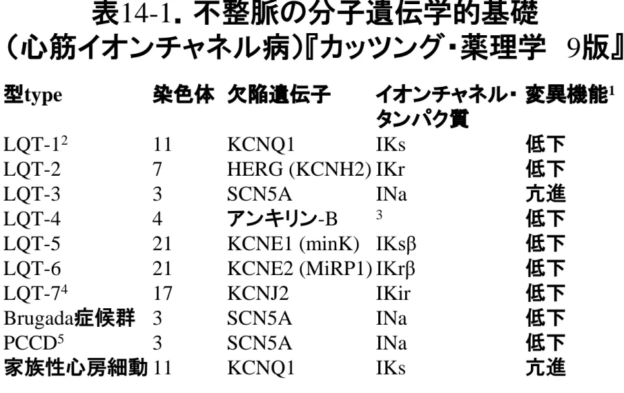
表14-1.不整脈の分子遺伝学的基礎 (心筋イオンチャネル病)『カッツング・薬理学 9版』 型type 染色体 欠陥遺伝子 イオンチャネル・ 変異機能1 タンパク質 LQT-12 11 KCNQ1 IKs 低下 LQT-2 7 HERG (KCNH2) IKr 低下 LQT-3 3 SCN5A INa 亢進 LQT-4 4 アンキリン-B 3 低下
LQT-5 21 KCNE1 (minK) IKsβ 低下 LQT-6 21 KCNE2 (MiRP1) IKrβ 低下 LQT-74 17 KCNJ2 IKir 低下
Brugada症候群 3 SCN5A INa 低下 PCCD5 3 SCN5A INa 低下
家族性心房細動 11 KCNQ1 IKs 亢進
1変異による機能変化,低下loss of function; 亢進gain of function.
2LQT: QT延長症候群long QT syndrome(LQT-1: Ward症候群タイプ1; QT-3: Romano-Ward症候群タイプ3,Na+チャネル不活性化機構の障害;Jervell and Lange-Nielsen症候群,先天性
難聴も伴うタイプ),β:βサブユニット
3アンキリンankyrinはNa+チャネル,Na+/K+ ATPアーゼ,Na+/Ca2+交換体,Ca2+遊離チャネル(リアノ
Disturbances of impulse conduction
興奮伝導の異常 i)
AV block; SA block (sick sinus syndrome)
Bradycardia 徐拍
"Bradyarrhythmia"
isoproterenol or atropine
Pacemaker ペースメーカ療法
Disturbances of impulse conduction
興奮伝導の異常 ii)
Reentry リエントリ(ー)
p.150
•興奮伝導の異常の一種、不整脈の大きな原因。
リエントリとは1つの刺激が旋回路を介して再びも
との部位に戻り、興奮を起こさせる現象である。心
臓のあらゆる部位で起こりうる。
•連結期の短い期外収縮では興奮伝導が不均一と
なるとランダム・リエントリが生じやすい。このほか
に渦巻き状の興奮旋回の存在もある。
1 sec
プルキンエ線維の三角形、
プルキンエ線維の2辺と心室筋の底辺の三角形
リエントリ(2)
(3)
1
1
2
2
1
2
(二段脈bigeminy )一方向性
伝導部
unidirectional block
リエントリ reentry の条件
p.151
1) obstacle to homogeneous conduction,
establishing a circuit 均一伝導の障害、旋回路
の形成。
2)
unidirectional block
in the circuit (depressed
conduction)
一方向性ブロック
。
3) conduction time around the circuit
>
effective
refractory period
旋回路の伝導時間が
有効不
応期
よりも長い。(距離一定なら,伝導速度が遅
Therapeutic strategy against reentry
1) Lengthening of the refractory period or APD (I
Kblock)
2) Conduction around the circuit a
slow,
bweek,
clate
bydepression of INa or ICa (esp. AV node)
a bidirectional block; b failed conduction;
c collision with the next regular beat
3) if conduction is too rapid: bidirectional conduction, refractory
Disturbances of impulse conduction
興奮伝導の異常iii)
WPW syndrome
(Wolff-Parkinson-White) 症候群 •Kent束に代表される心房と心室間の副伝導路を通じて の心室への早期興奮伝導により、頻拍発作paroxysmal supraventricular tachycardiaを伴う疾患. •WPW症候群に伴うPSVTに対する治療には薬物療法(Ic 群、ベラパミル)と非薬物療法がある。後者では手術療法 が行われていたが、現在は心臓電気生理学的検査により 副伝導路の位置を推定し、カテーテルアブレーション
catheter ablation
が主になっている。WPW症候群の心電図所見(WPW型心電図)
1)デルタ波出現 2)PR間隔短縮 3)QRS間隔延長 4)しばしばSTーT変化。
⊿
デルタ波I II III aVR aVL aVF V1 V2 V3 V4 V5 V6 ★ 仙台市立病院 石田明彦先生 ★ ⊿デルタ波
Atrial Flutter心房粗動
I II III aVR aVL aVF V1 V2 V3 V4 V5 V6 仙台市立病院 石田明彦先生冠状静脈洞
左心房
肺
静
脈
心臓の静脈 ①冠状静脈洞 ②中心臓静脈 ③大心臓静脈 ④左心室後静脈 ⑤左心房斜静脈radiofrequency (RF) linear ablation
心房細動(粗動) のラジオ波焼灼
AF termination by radiofrequency (RF) linear ablation I II III aVR aVL aVF V1 V2 V3 V4 V5 V6 ★ ★ ★ 仙台市立病院 石田明彦先生
Therapeutic mechanisms of
action against arrhythmias
A. Na
+channel blockade (Class 1)
-
modulated receptor theory
- dynamism of blockade
B. β Blockade (Class 2)
C. K
+channel blockade(Class 3)
prolongation of refractory period (APD)
D. Ca
2+channel blockade (Class 4)
E. Miscellaneous
局所麻酔薬
と
TTX
のNa
+チャネル遮断様式
(
LA:local anesthetic)
(図3-21) 細胞外 ゲート Na + HN + LA (中性) 細胞内 Na+チャネル (非特異的・低親和性遮断) Na + 電流 細胞膜 LA+ ←→ LA + H + pKa 細胞膜透過形 (細胞外より栓をす るように遮断する) TTX結合部位 フグ毒TTX LA + 高親和性 開チャネル遮断形H+ 細胞内より 電離LA (lidocaine) 細胞外 脂質二重層 中性LA 高親和性 1対1関係 低親和性 非特異的
細胞膜
Na
+channel
A. Na channel blocking drugs
(Class 1)
1) Quinidine (Class 1A)
• アンデス山脈原産のアカネ科キナノキ(Cinchona) a. Cardiac effects
• Blocks Na
+channel in the activated state
• Slow kinetics recovery from block• Prolong APD → prolong QT (block of KV channel ) • Ca2+ channel blocking action
Quinidine -- Side effects
1. Quinidine syncope 失神:
torsade de pointesトル
サード
: a drug-induced rapid, disorganized
ventricular tachycardia resembling ventricular
fibrillation, which usually terminates spontaneously,
associated with a striking prolongation of QT
interval
2. Sinus depression esp. in
elderly with sick sinus
syndrome
[due to Ca channel blocking action]
Quinidine -- Side effects
4. Side effects due to
anitmuscarinic
effects
(
atropine
-like, inhibition of vagus n.)
a. Sinus tachycardia
b. Facilitation of AV nodal conduction;
• increased ventricular rate in accompanying with atrial fibrillation or flutter (paradoxical tachycardia)
• to prevent: preadministration of digitalis, β-blocker or verapamil
5. GI tract; diarrhea, nausea, vomiting
• Like quinidine; Slow kinetics recovery from block
• anitmuscarinic
effects (more marked than
quinidine),
• structurally likes
isopropamide
; all of the side
effects of
atropine
参考 図3-14 抗コリン薬
ジソピラミド
anitmuscarinic
3) Procainamide (Class 1A)
• procaine (LA) 'amide' (KdI = 370
m
M)
• Like quinidine; Slow kinetics recovery from block
• without anitmuscarinic effects
• Side effects; heart failure; lupus
erythematosus-like syndrome。slow acetylator に多い。
• 肝臓で N-acetylation 代謝される。
N-アセチルプロカインアミド
アセチルトランスフェラーゼ
(N-アセチル転移酵素)
アセチルCoA
3群抗不整脈作用, prolongation of APD
K
+チャネル遮断作用
4) Propafenone#, Flecainide,
Pilsicainide* (Class 1C)
• Blocks Na channel in the activated state (like quinidine); no effect on APD
• #incidence of heart failure (9.3%)
• *Very slow kinetics recovery from block, l→R = 0.05 /msec
• premature ventricular contraction; very effective • without anitmuscarinic effects
• Myocardial infarction with arrhythmias (long term
administration) higher mortality than placebo
CAST
study
: New Engl. J. Med. 321: 406-412, 1989. (新薬理学入門 p.156) Risk-benefit ratio生存率(%) プラセボ(n=725) エンカイニドまたは フレカイニド(n=730) P=0.0006 期間(日) エンカイニドあるいはフレカイニド群とプラセボ群の生存率。 心室性不整脈は抑制されたものの、死亡原因は不整脈も しくは心停止であった。 CAST study (N Engl J Med 321: 406-412, 1989)
5) (Class 1B) Lidocaine
(i.v. administration);
Mexiletine
(p.o. administration)• Blocks both activated and inactivated Na channels • Fast kinetics recovery from block; l→R = 1 /msec • Shorten APD
• no suppression of normal conduction,
• inhibition of the ectopic beat with short coupling intervals (< 500 ms)
• selective inhibition of the ectopic beat at high basal frequency
• very effective for suppressing arrhythmias associated with depolarization (ischemia, myocardial infarction, digitalis toxicity)
Toxicity of class 1 antiarrhythmic
drugs
1) suppression of normal conduction with
use-dependency
2) prolongation of QRS (proarrhythmic,
drug-induced arrhythmias)
hyperkalemia
increases
the depressant effect of class 1
対策
by increasing serum pH (reduction of [K
+]
o)
to reduce the depressant effect of class 1 at
normal or high [K
+]
o3) Convulsion けいれん
Modulated receptor theory
p.152
Channel blocking drugs have high affinity for
activated or inactivated channels but low affinity
for rested channels.
多くの電位依存性チャネル (K
+チャネルを祖先と
して進化、分化, p.25) に作用する薬物の作用動態
を理解する上で参考になる
“
modulated receptor theory” とは、 「チャネル遮断
薬は活性化 (A) あるいは不活性化状態 (I) のチャ
ネルに高親和性を有し、静止状態のチャネルには
低い親和性を示す。」と言うもの。
Vh
Test 保持電位
Dependence of
Na channel
function on the membrane potential preceding the stimulus (Vh).The shift in the curve illustrates the effect of a typical
local
anesthetic antiarrhythmic drug
.Resting membrane potential
Drug
Control
Channels available, percent of maximum
Drug
Control
Resting membrane potential
Recovery time constant (ms) 保持電位 Vh C t1 t2 tn “recovery”
The time constant for recovery from inactivation
after repolarization also depends on the resting
potential.
‘state-dependent block`
活性化 (A)、不活性化 (I)状態
• 'voltage-dependent block' esp.
I
• 'frequency-dependent block'
esp.
A
• 'use-dependent block'
esp.
A
• fast tachycardia (many channels in
A & I
)
• loss of resting potential (many channels in
I
)
• longer APD (many channels in
A & I
,
Ventricle > Atrium)
Na
+channel
D D D R A I RD AD ID H-H H-H H-H H-H' H-H' H-H' k R l R k A l A k I l I Unblocked: Blocked: k R l R k A l A k I l I k R l R k A l A k I l I Lidocaine Quinidine (msec M ) -1 -1 (msec ) -1 (msec M ) -1 -1 (msec ) -1 =0.4 =1.0 =0 =0.05 =5X10 =2.6X10 =2X10 =6X10 =50 =1.5 =2.7 =0 4 4 -5 -3B L I J K L GNa Vmax B Q E F G H GNa Vmax
心筋のV
maxに対するLidocaineとQuinidineの使用
依存性抑制の差違
心筋V
max
に対するquinidineの使用依存
性抑制
Q:quinidine (1.5×10⁻⁵M) 投与下では、興奮開始と共に 活性化状態のNaチャンネルに薬物が結合し、それが 0.25Hzと言った緩くりした興奮頻度でも、静止状態の チャンネルからの薬物の解離速度が遅いため、次の興 奮が生じるまでに完全に薬物が解離しない。従って興 奮回数と共に薬物に結合したチャンネル(B)が蓄積性に 増加するので利用可能なチャンネル数(GNa)が暫減し、 それによりVmaxが減少する。これをVmaxの使用依存 性抑制と呼ぶ。この抑制効果は頻度依存性に増強する。心筋のV
max
に対するlidocaine
の使用依存性抑制
L:lidocaine存在下では、興奮頻度が低い(0.25〜
1Hz)場合には興奮時に活性化状態のNaチャンネ
ルと薬物が結合しても、静止状態の間に薬物は完
全にチャネルから解離するので、GNaとVmaxは減
少せぬ。しかし高い興奮頻度(2Hz)では薬物と結合
したチャンネルが生じ、従ってGNaとVmaxは減少
す る 。 こ の 場 合 の 使 用 依 存 性 の Vmax抑 制 は 、
lidocaineとチャネルの結合解離度が速いので1〜2
拍以内に平衡に達する。(Hondeghem LM et al:
Circulation 61:1217,1980)
1sec 0.3sec
Ex
quinidine
Vmax Na channellidocaine
Vmax Na channel 連結期使用依存性抑制の差違によるquinidineと
lidocaineの抗不整脈作用の比較
左図:心拍数60回/分で興奮する心室に短い連結期の 期外収縮(Ex)が生じた際のquinidineとlidocaineの作用 を、Vmax(Na+電流の指標)に対する使用依存性抑制の 立場から検討した概略図。 Quinidineでは正常洞調律に相当する心拍数(60回/分) でも、それによる心室興奮時、Vmaxに対するquinidine の使用依存性抑制により遅延し、したがってQRS巾は延 長する。そして期外収縮によるVmaxに対しても、正常 興奮のそれに対する抑制作用を発揮した程度にしか有 効でない。この理由として、quinidineとNa+チャンネルと の相互作用が遅いため、1秒の興奮間隔では薬物と結 合して遮断されたNa+チャンネルの割合(BF)が蓄積性 に増加するためと考えられる。使用依存性抑制の差違によるquinidineと
lidocaineの抗不整脈作用の比較
左図:心拍数60回/分で興奮する心室に短い連結期の 期外収縮(Ex)が生じた際の概略図。 Lidocaineではチャンネルとの相互作用が速いため、1秒 の興奮間隔があれば、興奮の際にチャンネルと結合し た薬物は充分に解離できるので、BFの蓄積性の増加は 生じない。したがってQRS巾の延長は起らぬ。しかし短 い連結期の期外収縮に対しては充分に興奮抑制効果 を発揮する。 右図:180回/分程度の頻拍が生じた場合には、quinidine では無論のことlidocaine投与下でもBFが蓄積し、頻拍 による心室興奮を抑制する。したがって頻拍の抑制効 果についてはquinidineとlidocaineの間で本質的な差違 はない。Slow kinetic
(recovery from block )
drugs
•Slow kinetics (disopyramide); quinidine: intemed. •Very slow kinetics (flecainide, pilsicainide)
i. merits
•inhibition of the ectopic beat異所性興奮at low frequency of sinus rhythm
•inhibition of the ectopic beat with long coupling intervals 連結期
ii. demerits
•suppression of normal conduction with use-dependency使 用依存性に正常拍動を抑制する。prolongation of QRS
Fast kinetics (lidocaine, mexiletine)
i. merits
• no suppression of normal conduction,
• inhibition of the ectopic beat with short coupling intervals (< 500 ms)
• selective inhibition of the ectopic beat at high basal frequency 高頻度時の異所性興奮を抑制する。
ii. demerits
• no inhibition of the ectopic beat with long coupling intervals
B.
b
Adrenoceptor blocking drugs
(Class 2)
b
1-blockers (metoprolol, bisoprolol, carvedilol) are
better than propranolol
esmolol (
short acting, intraoperative, acute arrhythmias)
• prevent recurrent infarction and sudden death in
Pts recovering acute myocardial infarction
• Arrhythmias with chronic heart failure
• Thyrotoxicosis 甲状腺機能亢進症;老人の症状
は不整脈 (特に、Af) のみの場合あり。
β遮断薬
←イソプロテレノール
β遮断薬の開発と発展 in 「休み時間の薬物治療学」講談社、2009、p21-23 ノルアドレナリン; アドレナリン
β遮断薬の分化・進化
部分作動薬 partial agonist エステラーゼ加水分解 t1/2:4分 t1/2:3.6分 Tmax:4時間後 t1/2:4.8時間(α相)、19.6時間(β相) Tmax:1.5~2時間後 t1/2:錠3.9±0.5時間C. Class 3 antiarrhythmic drugs
• Amiodarone,
Sotalol, Nifekalant, Vernakalant • Prolongation of APD and refractory period; prolong QTinterval
• Amiodarone (all actions of Class 1A, 2,
3
, 4)noncompetitive blockade of b-receptor • Very effective against both supraventricular and
ventricular arrhythmias
• Wolff-Parkinson-White syndrome (WPW)
– accessory conduction pathway (Kent bundle)
Pharmacokinetics of amiodarone
(Melmon & Morreli's Clinical Pharmacology 3rd Ed.)
• Oral bioavailability (F) 0.22 - 0.86 • Time to maximum conc. (Tmax) 3 - 7 hr
• Volume of distribution (Vd) 18 - 148 L/kg
• Protein binding 96%
• Clearance (% renal < 1) 100 - 175 ml/min • half-life t1/2a (redistribution) NR
t1/2b (clearance) 2.5 - 10 days (initial)
40 - 55 days (terminal)
左 CAMIAT Studyの成績.1202人の心筋梗塞後の心室性期外収縮を認め る患者でのアミオダロン投与は、心室細動・不整脈に関してアミオダロン群が良 好であった。 (Lancet 1997;349: 675) 右 CHF-STATの成績. うっ血性心不全と無症候性心室性不整脈を有する 患者に対するアミオダロン投与は、生命予後に関して全体ではプラセボ群との差 はなかったが、非虚血性の心筋症患者に関して検討したところ、有意にアミオダ ロン群の生命予後は良好であった。 (N Engl J Med 1995;333: 77)
Toxicity of amiodarone
• Its toxicity limits its clinical usefulness 他の抗不整脈薬 が無効で、危険な不整脈の時に用いる。
• Deposition in every tissue in all cases with long term administration
• Corneal deposition (yellowish-brown microcrystals) • Photodermatitis
• Paresthesias 異常感覚, tremor 振戦, ataxia 失調, headache
• Thyroid dysfunction; hypo- or hyper-
化学構造を見るとI ヨウ素を含むため
アミオダロンに関して
• 長期的には専門医が用いるべきものであ
る。
• 逆に短期的には、静脈内投与で高用量を
用いても、副作用を気にせずともよく、重
症の心室頻拍や急性心房細動に対して、
安全で大変有効な抗不整脈薬として、研
修医レベルでも用いられるようになってき
た。
逆頻度依存性活動電位延長
Reverse use-dependent APD prolongation
APD 延長度 (m sec) N-アセチルプロカインアミド APD 延長度 (m sec ) キニジン ソタロール アミオ ダロン 理想 サイクル長(msec) サイクル長(msec) 頻度依存性一抗不整脈薬による活動電位持続時間 APD 延長効果と刺激間隔の関係 左図のソタロールではAPDの延長効果は刺激間隔の増加(刺激頻度の減少)に伴って増 加を示している(逆頻度依存性)が、右図のアミオダロンではAPD延長効果は刺激間 隔に関わらずほぼ一定で、頻度依存性を示さない。右図の点線はHondeghemらのクラ スIII群薬の理想的な頻度依存性を示している。 (Circulation 81: 686, 1990)
D. Class 4 antiarrhythmic drugs
• Non-vasoselective Ca
2+channel blockers
– Cf. vascular selective Ca2+ channel blockers (DHP Ca
antagonists: nifedipine, amlodipine, …)
• Verapamil (anti-anginal drug)
• Diltiazem (anti-anginal, antihypertensive drug)
• Bepridil (+ 1, 3 actions)
• Paroxysmal supraventricular tachycardia
E. Miscellaneous antiarrhythmic drugs
1)
Adenosine, ATP
• supraventricular tachycardia
• increased gK (Gi protein-coupled A
1R →K
GIRchannel (i.e. K
ACh) 開)
• inhibition of cAMP-induced Ca
2+influx
2)Magnesium
• digitalis-induced arrhythmias
(hypomagnesemia)
• torsade de pointes
E. Miscellaneous antiarrhythmic drugs
3)
Potassium
• Hypokalemia results in an increased risk of EAD,
DAD, and ectopic pacemaker activity, especially in
the presence of digitalis.
• normalizing potassium gradients and pools in the
body
4)
Digitalis
as an Antiarrhythmic (via vagal n. st.)
• 心房粗動atrial flutter →心房細動atrial fibrillation
• 洞結節性および房室結節性リエントリ
Trigger
Modulating factors
図4-23 不整脈の病態生理学的見方
(Triad)
Substrate
VT, VF
Sudden death
頻拍PVC
伝導遅延 slow conduction 交感神経、血中CAs 電解質異常 壁張力wall tensionDrugs
虚血 ischemia 再分極異常 abnormal repolarizationHypertension
Heart failure
A.経験的治療 不整脈の診断 治療 臨床帰結 B.病態生理学的治療 不整脈の診断 房室結節リエントリ頻拍 発生機序の想定 不整脈の成立条件 受攻性因子 標的分子 治療 臨床帰結 房室接合部リエントリ 心房の解剖学的経路
(fast pathway, slow pathway) 房室結節(緩徐伝導) 房室結節の活動電位 L型Caチャネル 洞調律 bブロッカー Caチャネルブロッカー 不整脈の経験的治療(A)と病態生理学的治療(B)
不整脈の機序 受攻性因子 治療の選択 治療の標的 使用しうる薬剤 広い興奮間隔を有するリエントリ 房室結節あるいはケント束の伝導 a) 房室結節内伝導(Caチャネル依存性) あるいは b) ケント束伝導(Naチャネル依存性)の抑制 a) Caチャネル b) Naチャネル a) Ca拮抗薬 b) Naチャネル抑制薬
発作性上室性頻拍症例の薬剤選択の流れ
調節因子の考慮心室細動に対する除細動
心房細動は認知症の独立した
危険因子
次の図を見て、各自その病因を考察
して下さい。
心房細動; 心房内血栓; 脳塞栓症
心房細動は認知症の独立した危険因子
教科書・参考書
イオンチャネル
不整脈 arrhythmiaと抗不整脈薬
新薬理学入門(第3版)(2008)
Katzung (12th ): Basic & Clinical Pharmacology (2011) 柳澤他監訳:カッツング・薬理学,丸善(2009) 大地陸男:生理学テキスト(第7版)(2013) 倉智嘉久:心筋細胞イオンチャネル(2000) (参考)唐木英明 編著:イオンシグナルの謎−カルシウ ムの40億年を渉猟する(1999) 柳澤輝行、今野俊宏:カリウムチャネルの分子薬理学. 臨床麻酔 30(4): 659-667 (2006) 2011.5.25 薬理学II
抗不整脈薬
アンケート課題4)
Ventricular cellの活動電位に対す
る
抗不整脈薬IB, III, IV群薬
の作用をコントロールと比較して
それぞれ図示して下さい。
Ib
Ia, III
IV
100 msec