Fukushima Medical University
福島県立医科大学 学術機関リポジトリ
This document is downloaded at: 2021-11-08T00:39:05Z
Title Two cases of pancreatic abscess associated with penetration to the gastrointestinal tract during treatment using endoscopic ultrasound-guided drainage
Author(s)
Imamura, Hidemichi; Irisawa, Atsushi; Takagi, Tadayuki;
Shibukawa, Goro; Wakatsuki, Takeru; Takahashi, Yuta; Sato, Ai; Sato, Masaki; Hikichi, Takuto; Obara, Katsutoshi; Ohira, Hiromasa
Citation Fukushima Journal of Medical Science. 53(1): 39-49
Issue Date 2007-06
URL http://ir.fmu.ac.jp/dspace/handle/123456789/207
Rights © 2007 The Fukushima Society of Medical Science
DOI
Text Version publisher
Fukushima J. Med. Sci., Vol. 53, No.1, 2007
[Case Report]
TWO CASES OF PANCREATIC ABSCESS ASSOCIATED WITH PENETRATION TO THE GASTROINTESTINAL TRACT
DURING TREATMENT USING ENDOSCOPIC ULTRASOUND-GUIDED DRAINAGE
HIDEMICHI IMAMURN), ATSUSHI IRISAWN), TADAYUKI TAKAGP), GORO SHIBUKAWN), TAKERU WAKATSUKP), YUTA TAKAHASHP),
AI SAT01), MASAKI SAT01l, T AKUTO HIKICHP), KA TSUTOSHI OBARN) and HIROMASA OHIRN)
')Department of Internal Medicine 2, Fukushima Medical University School of Medicine 2)Department of Endoscopy, Fukushima Medical University Hospital
(Received July 13, 2006, accepted December 20, 2006)
Abstract: Of pancreatic pseudocysts, approximately 30% are complicated by ab- scess formation, perforation into the abdominal cavity, penetration to the gastro- intestinal tract, or bleeding. We report two cases of pancreatic abscess complicat- ing severe acute pancreatitis in which the abscess penetrated to the gastrointestinal tract during the course of treatment with endoscopic ultrasound (EUS)-guided pseudocyst drainage. In these cases, neither aggravation nor recurrence of the pancreatic abscess has been identified since the event occurred. The EUS-guided treatment was effective for improvement of severe inflammation of the pseudocyst as an initial treatment. However, drainage tube placement limitations pertained because the pseudocyst was present with multilocular infection. Penetrations eventually contributed to their resolution because the fistulas were used as wide drainage routes.
Itis important to understand the courses of these cases for preparation of therapeutic strategies to treat pancreatic pseudocyst/abscess.
Key words: pancreatic pseudocyst, penetration, EUS-guided drainage
INTRODUCTION
Pancreatic pseudocysts, which contain pancreatic juice and effusion, are formed by pancreatic inflammation. A secondary pseudocyst develops in 10-15% of
4-ff~~, A~.;!;,
il1Ii**z.,
~}lIm:~,;f5j; ~,il1Ii:flltim:;t, fti:jJj ~,fti:jJjg~, i3ljtgni.A., Ij\Jjj{)jffj&:,*
lIZ~lECorrespondence to: Atsushi Irisawa, MD, PhD Department of Internal Medicine 2 Fukushima Medical University School of Medicine Hikarigaoka I, Fukushima City, 960-1295, Japan.
E-mail: [email protected]
39
40 H. IMAMURA et at.
patients with acute pancreatitis and chronic pancreatitis
l ).Of those, approximately 30% are complicated by abscess formation, perforation into the abdominal cavity, penetration to the gastrointestinal tract, or bleeding (gastrointestinal bleeding, intra-abdominal bleeding, intracystic bleeding)2,3). About 20-60% of pancreatic pseudocysts will disappear spontaneously. However, that rate falls to less than 10% for pseudocysts of 6 cm diameter or greater. Therefore, if a cyst is not reduced, even after the sixth week when the cyst wall starts to organize and adhere to surrounding organs, cyst drainage is indicated
4,5).On the other hand, a pancreatic pseudocyst associated with infection or pancreatic abscess might cause sepsis and multi-organ failure.
It might penetrate into the gastrointestinal tract, cause bleed-ing, and form a pseudo-aneurysm
ifinflammation spreads to surrounding organs.
Consequently, for conservative treatment of uncontrolled intracystic infection, immediate drainage is required, even if in the early phase of pseudocyst formation.
We have recently experienced two cases of pancreatic abscess complicating severe acute pancreatitis in which the abscess penetrated to the gastrointestinal tract during treatment with endoscopic ultrasound (EUS)-guided pseudocyst drain- age. In these cases, penetrations eventually contributed to their resolution because the fistulas were used as wide drainage routes. The courses of these cases should be understood for preparation of therapeutic strategies of pancreatic pseudocyst/abs- cess.
CASE REPORT
Casel
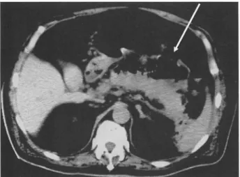
A 65-year-old man was admitted to another hospital because of severe acute pancreatitis. Although the pancreatitis was relieved using various treatments, pancreatic pseudocyst developed around the pancreas. After eating was restarted, he displayed a high fever (39°C); infection of the pseudocyst was suspected. He was then referred to our hospital, to which he was admitted for treatment. Laboratory tests performed on admission indicated a severe inflammatory reaction with WBC of 21,200/ pI and CRP of 12.7 mg/dl. Mild anemia, mild thrombocytopenia, liver dysfunction, and marked hypoalbuminemia were also indicated. Renal functions, blood glucose, serum AMY, and tumor markers were normal (Table 1). Abdominal CT showed a 12-cm-diameter abscess around the pancreatic tail and another abscess around the pancreatic head (Fig. 1).
Itwas considered that immediate drainage for abscess should be done. Therefore, EUS-guided drainage was perfor- med using a 19-9auge puncture needle (Echotip19G; Wilson-Hirata, Osaka, Japan) guided by ultrasound using a convex type echoendoscope (GF-UCT240AL-5;
Olympus Medical Systems. Co., Tokyo, Japan) (Fig.2a) with a transgastric
approach. After puncture of the abscess, the needle stylet was removed. The
aspirated fluid, viscous, white, and slightly bloody, was collected. A 0.025-inch
guide wire (Jagwire; Boston Scientific Japan, Tokyo) was inserted into the needle
PANCREATIC ABSCESS ASSOCIATED IIHH PENETRATION TO THE GASTROINTESTINAL TRACT
Table 1. Laboratory tests of Case 1 (on admission)
WBC 21.200//11 TP 6.2 g/dl RBC 349 X 10"//11 ALB 1.6 g/dl Hb 10.9 g/dl BUN 18 mg/dl Hct 32.9% CREA 0.6mg/dl PLT 13.8 X 10'//11 Na 137 mEq/1
K 4.3 mEq/1
TB 1.6 mg/dl CI 102 mEq/1
DB l.lmg/dl AMY 82 LU/I AST 162 lU/1
ALT 73LU/1 BS 128 mg/dl
LDI-I 391 IU/I
ALP 374 IU/I CEA 1.5 ng/ml
y-GTP 153 LU/I CA19-9 29.1 U/ml
Fig.1. Abdominal CT showed a 12-cm-diameter abscess around the pancreatic tail and another abscess around the pancreatic head in case 1.
41
and was advanced into the abscess. Then a 6 Fr endoscopic naso-biliary drainage (ENBD) tube (Flexima; Boston Scientific Japan. Tokyo) was placed in the abscess over the guide wire (Fig. 2b). The abscess cavity was washed every day with saline mixed with amikacin sulfate. After this treatment. although the inflammatory reaction decreased and the patient's general condition improved. follow-up CT revealed residual abscess cavities. Two weeks after the drainage. hematochezia was observed. Emergent colonoscopy revealed two ulcerative lesions with mucosal edema in the transverse colon; penetrations of pancreatic abscesses into the colon were suspected (Fig. 3). Images obtained by CT scanning revealed an abscess containing air near the pancreatic tail. Pseudo-aneurysm was not seen in the locations of the fistulas. Fortunately. apparent anemia and an inflammatory reac-
42 H. IMAMURA el al.
a
b
Fig. 2. a: EUS-guided drainage was performed using a 19-9auge puncture needle guided by ultrasound using a convex type echoendoscope with the transgastric approach.
b: An endoscopic naso-biliary drainage (ENBD) tube was placed in the abscess.
Fig. 3. Colonoscopy revealed two ulcerative lesions with mucosal edema in the transverse colon. Penetrations of pancreatic abscesses into the colon were suspected.
tion were not identified. Therefore, conservative treatments were performed. An abdominal CT scan performed 2 weeks after the hematochezia showed communica- tion between the abscesses and the transverse colon, as well as the reduced abscesses (Fig. 4). Since the event occurred, neither aggravation nor recurrence of the pancre- atic abscess has been identified.
PANCREATIC ABSCESS ASSOCIATED WITH PENETRATION TO THE GASTROINTESTINAL TRACT 43
Fig. 4. An abdominal CT scan performed 2 weeks after hematochezia showed com·
munication between the abscesses and the transverse colon (arrow), as well as the red uced abscesses.
Case 2
A 53-year-old man with a history of choledocholithiasis was admitted to another hospital with a diagnosis of severe acute alcoholic pancreatitis. The pancreatitis was relieved through treatment with protease inhibitors and antibiotics under a fasting condition. Nevertheless, the patient developed a fever of 39"C and abdominal pain. Abdominal CT images suggested infection of a pancreatic pseudocyst associated with acute pancreatitis. For treatment, he was referred to our hospitaL On admission, laboratory tests showed WBC of 9,900/,u1, CRP of 11.8 mg/dl, mild anemia, thrombocytosis, and mild liver dysfunction. Renal function
Table 2. Laboratory tests of Case 2 (on admission)
'NBC 9,900/11 1 TP 7.2 g/dl
RBC 376 X 10'/111
Hb 11.9 g/dl BUN 12 mg/dl
Hct 33.7% CREA 0.6 mg/dl
PLT 51.7 X 10"/111 Na 133 mEq/1 K 4.7 mEq/1
TB 0.5 mg/dl CI 97 mEq/1
DB 0.3 mg/dl AMY l11IU/1
AST 231U/1
ALT 25lU/1 BS 140 mg/dl
LDH 2511U/1
ALP 4791U/1
44 H. IMAMURA el al.
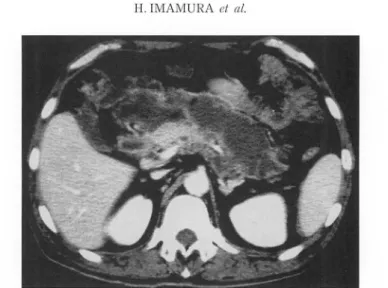
Fig. 5. Abdominal CT revealed extensively developed abscesses in case 2.
a
b
Fig. 6. a: EUS-guided drainage was done using the same procedure as that used in
"case I".
b: ENBD tube was indwelled in the abscess.
and serum AMY were normal; a marked increase of blood glucose was not ob- served (Table 2). Abdominal CT revealed extensively developed abscesses around the pancreas (Fig. 5). It was considered that immediate drainage should be perfor- med. Therefore, EUS-guided drainage was performed using the same procedure as that used in "case 1" (Fig.6a). Yellow and white pus was aspirated from the abscess. At that time, a 6 Fr ENBD tube (Flexima; Boston Scientific Japan, Tokyo) was indwelled in the abscess (Fig. 6b). The abscess cavity was washed through the tube daily with saline mixed with amikacin sulfate. Improvement of fever and the inflammatory reaction were apparent after the drainage. Two weeks later, X-ray fluoroscopic examination was done by injection of contrast medium via
PANCREATIC ABSCE S ASSOCIATED WITH PENETRATION TO THE GASTROINTESTINAL TRACT 45
Fig. 7. On the fluoroscopic image, the small intestine was visualized in images taken inside of the abscess cavity (arrow), suggesting that the pancreatic abscess had penetrated into the small intestine.
Fig. 8. Abdominal CT showed the reduced pancreatic abscess. Contrast medium was recognized in the small intestine and the abscess cavity.
an ENBD tube to decrease the pancreatic abscess. Then, the small intestine was visualized in images taken inside the abscess cavity, which suggested that the pancreatic abscess had penetrated into the small intestine (Fig. 7). Subsequently, CT showed the reduced pancreatic abscess (Fig. 8). During the course of the case, abdominal pain, melena, inflammatory reaction, and anemia were not seen. Since that event occurred, neither aggravation nor recurrence of the pancreatic abscess has been identified.
46 H. IMAMURA et at.
DISCUSSION
Pancreatic pseudocysts are classifiable into three types depending on their etiology: type I, pseudocyst complicating acute pancreatitis; type II, pseudocyst complicating the aggravation of chronic pancreatitis; or type III, chronic pseudocyst [retention cystJ6). Both type I and type II described above are condi- tions of post-necrotizing pseudocyst. The space of the omental sac between the front of the pancreas and the stomach becomes a cyst cavity when the inflammation spreads from the pancreas. The exudate is retained in that space, and a pseudocyst is formed because Winslow's foramen is closed by severe inflammation. Type III is a cyst within the pancreas. Although the cyst wall might adhere to the gastro- intestinal tract because of repeated inflammation in chronic pancreatitis, the cyst wall is usually separated from the wall of the gastrointestinal tract. Generally, type I and type II cysts, which develop secondary to acute pancreatitis and acute aggrava- tion of chronic pancreatitis, tend to be complicated by bleeding, infection, and penetration into the gastrointestinal tract. In both cases described in this report, pancreatic pseudocysts complicating severe acute pancreatitis (above-mentioned type I) were infected, consequently forming pancreatic abscesses. They were at high risk of sepsis and might have bled severely by prolonged inflammation.
Therefore, immediate drainage was necessary. As noted previously, the cyst wall might include the gastric wall itself, and it was considered that endoscopic retro- grade pancreatography-guided transpapillary drainage might aggravate the acute pancreatitis. For those reasons, EUS-guided trans-gastrointestinal drainage was chosen. In both cases, inflammatory reactions improved gradually after drainage, showing the effect of washing through the drainage tube. Our treatment was inferred to be effective as an initial treatment.
Pancreatic pseudocyst is known to perforate into the abdominal cavity and the
retroperitoneum, and to penetrate into the gastrointestinal tract. The frequency of
penetration into the gastrointestinal tract is approximately
15-20%among pancre-
atic pseudocyst cases
3,7).In the case of a pseudocyst complicating acute pan-
creatitis, the partial wall of the pseudocyst was composed of the wall of the
gastrointestinal tract, and adhered to surrounding gastrointestinal tract because of
severe inflammation. The pancreatic enzymes in the pseudocyst weaken the gastro-
intestinal tract wall. Consequently, the pseudocyst might penetrate into the gastro-
intestinal tract and naturally generate an internal fistula. Moreover, internal
pressure of the pseudocyst caused by hemorrhage might involve penetration into the
gastrointestinal tract8). To our knowledge, there are some such cases of penetra-
tion into other organs in the literature. Of those, the most frequently penetrated
organ was the stomach
(9cases)12-16.19); other organs including the duodenum (4
cases)l°,16), transverse colon (4 cases)9.1l), spleen (4 cases), aorta
(1case)l8), and bile
duct
(1case) were also penetrated (two or more organs were penetrated in some
PANCREATIC ABSCESS ASSOCIATED WITH PENETRATION TO THE GASTROINTESTINAL TRACT 47
cases). No penetration has ever been recorded into the small intestine, as occurred in case 2. The fistula which was formed by penetration into the gastrointestinal tract imparted a drainage effect that might have eliminated some pseudocysts naturally. In both cases reported here, naturally generated internal fistulas as a complication eventually contributed to elimination of the pseudocyst.
Pseudocysts complicating acute pancreatitis often form multilocular cysts because of widely expanded inflammation. In recent years, EUS-guided drainage has been performed widely as a treatment for pancreatic pseudocyst including abscess and infectious pancreatic necrosis. Many studies have examined the useful- ness of the technique
2o,21).As an initial treatment for advanced inflammation, EUS- guided drainage is effective. On the other hand, limitation of the drainage tube placement in the case of multilocular infection is known. In such cases, multiple drainage tubes might have been required to be punctured from other sites.
Additionally, because of the large amount of necrotic debris contained in the abscess, debris removal might be distressful, even with larger drainage tubes, and sufficient treatment might be difficult. In such cases, an endoscopic approach into the pseudocyst through the gastrointestinal tract wall (endoscopic necrosectomy) should be performed
22,23).In our case 1, pancreatic abscesses penetrated into the transverse colon and caused bleeding. Further massive bleeding in the cyst and the abdominal cavity might occur if inflammation involves major vessels near the pseudocyst and forms a pseudo-aneurysm
24 ,25).Therefore, as noted above, trans-gastrointestinal endos- copy-guided necrosectomy should be considered early when EUS-guided drainage is not effective.
In conclusion, EUS-guided treatment was effective for improvement of severe inflammation of a pseudocyst as an initial treatment. However, limitations of the drainage tube placement pertained in these cases of pseudocyst with multilocular infection. For treatment of pseudocyst/abscess complicating acute pancreatitis, a therapeutic strategy should include consideration of the pathogenic mechanism and configuration, with particular attention to the potential secondary complications.
REFERENCES
1. Simpson A, Srivastava VK. Pseudocyst of pancreas. Br J Surg, 60: 45-49, 1973.
2. Stabile BE, Wilson SE, Debas HT. Reduced mortality from bleeding pseudocyst and pseudoaneurysm caused by pancreatitis. Arch Surg, 118: 45-51, 1983.
3. Sankaran S, Walt AJ. The natural and unnatural history of pancreatic pseudocysts.
Surgery, 62: 37-44, 1975.
4. Vitas GJ, Sarr MG. Selected management of pancreatic pseudocysts: Operative versus expectant management. Surgery, 111: 123-130, 1992.
5. Yeo CJ, Bastidas JA, Lynch-Nyhan A, Fishman EK, Zinner MJ, Cameron JL. The natural history of pancreatic pseudocysts documented by computed tomography. Surg Gynecol Obstet, 170: 411-417, 1990.
6. D'Egidio A, Schein M. Pancreatic pseudocysts: a proposed classification and its
48 H. IMAMURA et al.
management implications. Br J Surg, 78: 981-984, 1991.
7. Bradley EL, Clements JL, Gonzalez AC. The natural history of pancreatic pseudocysts.
A united concept of management. Am J Surg, 137: 135, 1979.
8. Greenstein A, DeMaio EF, N abseth DC. Acute hemorrhage associated with pancreatic pseudocyst. Surgery, 69: 56-62, 1972.
9. Goda M, Yukawa N, Fujisawa J, Matsukawa H, Shimizu S, Tomita Y. A case of serious melena and shock associated with a pancreatic pseudocyst. (in Japanese, English abstract) Journal of Japan Surgical Association, 65: 2194-2199, 2004.
10. Yajima S, Yamasaki S, Shimada K, Sano T, Esaki M, Koide N, Kosuge T. A Case of Pancreatic Abscess Mimicking Mucinous Cystic Tumor of the Pancreas. (in Japanese, English abstract) Jpn J Gastroenterol Surg, 37: 1428-1432, 2004.
11. Ando T, Sakakibara K, Nishiwaki N. A Case of Hemorrhagic Pseudocyst Fenestrating into the Transverse Colon. (in Japanese, English abstract) Jpn J Gastroenterol Surg, 35:
63-67, 2002.
12. Shida A, Yamadera H, Nakabayashi Y, Ishida Y, Anazawa S, Yamazaki Y. Total gastrectomy for gastric penetration of pseudocyst of pancreas with upper gastro- intestinal bleeding - report of a case -. (in Japanese, English abstract) Journal of Japan Surgical Association, 62: 2900-2904, 2001.
13. Takahashi T, Shimada K, Kobayashi N, Kakita A. Migration of Steel-Wire Coils into the Stomach after Transcatheter Arterial Embolization for a Bleeding Splenic Artery Pseudo aneurysm : Report of a case. Surgery Today, 31: 458-462, 2001.
14. Ohmura Y, Kashiwa T. Pancreatic carcinoma coexistent with pancreatic pseudocyst which penetrated into the stomach. International Journal of Clinical Oncology, 5: 195- 199, 2000.
15. Ishihara K, Yamada T, Suzuki N, Eirai M, Aki A, Nishikawa M, Hujii H. A Case Report of Hemorrhagic Pancreatic Pseudocyst Fenestrated into the Stomach. (in Japanese, English abstract) Jpn J Gastroenterol Surg, 32: 870-874, 1999.
16. Shimoyama T, Ishikawa H, Shimizu T, Hisano H, Honjo S, Matsuo S, Sumida Y, Matsuo S, Nishikawa K, Terada R, Kusano H, Nakagoe T, Miura T, Tomita M, Kawaguchi A, Ashizawa K. Pancreatic Pseudocysts - Proposal for Diagnosis and Surgical Management -. Acta Medica Nagasakiensia, 40: 18-21, 1995.
17. Yokota T, Naruse M, Oohira Y, Abe S, Aoki H, Nimura H, Tsutsumi J, Moriya Y, Tokita I, Soji K, Aoki T. A case of pancreatic pseudocyst with colonic penetration. (in Japanese, English abstract) Journal of Japanese Society for Clinical Surgery, 56: 605- 609, 1995.
18. Tsubono T, Fukushige H, Muto T. Pancreatic pseudocyst penetrated into the abdomi- nal aorta - a case report -. (in Japanese, English abstract) Journal of Japanese Society for Clinical Surgery, 53: 1698-1701, 1992.
19. Hirotsuji N, Takeda Y, Ohshiba S, Tsukiyama J. Pancreatic Pseudocyst Resulting in a Submucosal Hematoma, Report of a Case. (in Japanese, English abstract) I to Cho (Stomach and Intestine), 25: 211-215, 1990.
20. Imamura H, Irisawa A, Bhutani MS, Hikichi T, Shibukawa G, Takagi TJ, Katakura K, Wakatsuki T, Sato A, Obara K, Sato Y. A New Device for Dilation of the Fistula Tract between Gastric and Cystic Wall in Performing EUS-Guided Pancreatic Pseudocyst Drainage. Gastrointestinal Endoscopy, 61: AB283, 2005.
21. Irisawa A, Hikichi T, Obara K, Sato Y. Current State of EUS-Guided Therapy. (in Japanese, English abstract) Endoscopia Digestiva, 16: 1247-1252, 2004.
22. Seewald S, Groth S, Omar S, Imazu H, Seitz U, Weerth AD, Soetikno R, Zhong Y, Sriram VJS, Ponnudurai R, Sikka S, Thonke F, Soehendra Nib. Aggressive endoscopic therapy for pancreatic necrosis and pancreatic abscess: a new safe and effective treatment algorithm (videos). Gastrointestinal Endoscopy, 62: 92-100, 2005.
23. Matthes H, Janiszewski C, Schenk G. Endoscopic Transgastric EUS Guided Drainage and Necrosectomy of Infected Pancreas Pseudocysts. Gastrointestinal Endoscopy, 61:
PANCREATIC ABSCESS ASSOCIATED WITH PENETRATION TO THE GASTROINTESTINAL TRACT 49 AB292, 2005.
24. EI Hamael A, Parc R, Adda G, Bouteloup P.Y, Huguet C, Malafosse M. Bleeding pseudocysts and pseudo aneurysms in chronic pancreatitis. Br
J
Surg, 8: 1059-1063, 1991.25. Adams DB, Zellner JL, Anderson MC. Arterial hemorrhage associated with pancreatic pseudocysts: role of angiography. J Surg Res, 54: 150-156, 1993.