INTRODUCTION
Bone is one of the most important oral tissues. It is maintained by the balance between bone formation and resorption
1,2
. It is remodeled continuously by two specialized cell types: bone-forming osteoblasts and bone-resorbing osteo- clasts
2. Osteoclasts are tar- trate-resistant acid phosphatase (TRAP)-positive, multinucleated giant cells that originate from the mono- cyte/macrophage lineage of hematopoi- etic stem cells
3. Stromal cells and os- teoblasts express macrophage col-
ony-stimulating factor (M-CSF) and re- ceptor activator of nuclear factor-κB ligand (RANKL), which are essential for the induction of osteoclasts
1-3. In terms of tissue engineering, osteoclasts and macrophages resorb scaffolds, making space for bone matrix formation
4. Con- sequently, osteoclasts play an important role in bone regeneration.
We reported previously microRNAs (miRNAs) expression profiles during osteoclastogenesis using microarrays
5. miRNAs are small, endogenous, non- coding RNAs of approximately 22 nu-
Expression of MicroRNAs in the Extracellular Microvesicles of
Murine Osteoclasts
Tadayoshi KAGIYA
1and Masayuki TAIRA
21
Division of Functional Morphology, Department of Anatomy,
2
Department of Biomedical Engineering, Iwate Medical University, Iwate, Japan
SYNOPSIS
Bone is one of the most important oral tissues. Osteoclasts play important roles in bone tissue engineering. Previously, we profiled microRNA (miRNA) expression during osteoclastogenesis using microarrays. miRNAs are small, noncoding RNAs that are involved in various biological processes, including cellular differentiation, proliferation, apoptosis, and organ development. Recently, the presence of miRNAs in extracellular microvesicles was reported. It is not known whether os- teoclasts secrete extracellular microvesicles containing miRNAs. We investigated miRNA expression in extracellular microvesicles in conditioned medium of cultured osteoclasts using RT-PCR. Specifically, we investigated eight miRNAs deemed important for osteoclastogenesis in our previous study: let-7e, miR-21, miR-33, miR-155, miR-210, miR-223, miR-378, and miR-1224. Of these, the expression levels of miR-378, miR-210, and miR-21 were very high, while no significant miR-33 or miR-1224 expression was detected. These results suggest that osteo- clasts secrete extracellular microvesicles containing specific miRNAs, but that they do not contain the entire set of intracellular miRNAs.
Key words: MicroRNA, Extracellular Microvesicle, Exosome, Osteoclast
ORIGINAL ARTICLE
143 cleotides
1,5. miRNAs are incorporated into an RNA-induced silencing complex that contains argonaute-family proteins.
The argonaute proteins recruit miRNAs specific for target mRNAs
1,5, and the RNA-induced silencing complex either inhibits the translation of the target mRNAs or degrades the target mRNAs
1,5
. Consequently, miRNAs are involved in the post-transcriptional regulation of mRNA function and participate in regu- lation of cellular differentiation, prolifera- tion, apoptosis, and organ development
5
.
Recently, miRNAs were reported to be present extracellular microvesicles and to function in other cells
6-9. Ex- tracellular microvesicles are small parti- cles derived from the endosomal com- partment
7,9-11, and 40–100-nm-diameter microvesicles are defined as exosomes
11-13
. Extracellular microvesicles have an important role in cell-to-cell communica- tion via the transfer of miRNAs, mRNAs, proteins, and bioactive lipids to target cells
6-13. The secretion of microvesicles containing miRNAs depends on the cell type, biological conditions, and what kinds of miRNAs the cells contain
12,13. Although osteoclasts play important roles in bone remodeling and regenera- tion, it is not known whether osteoclasts secrete extracellular microvesicles containing miRNAs. Here, hypothesizing that osteoclasts secrete extracellular microvesicles containing miRNAs, we investigated the expression of miRNAs in extracellular microvesicles in condi- tioned medium of cultured osteoclasts using RT-PCR.
MATERIALS AND METHODS 1. Cell culture
All animal experiments were reviewed and approved by the Animal Use and Care Committee of Iwate Medical Uni- versity (registration number 23-029).
Five-week-old male ddY mice were purchased from SLC Japan
(Hamamatsu, Japan). The mice were sacrificed, and their femurs and tibias were removed and dissected free of adherent soft tissue. The ends of the bones were cut, α-MEM (Invitrogen, Fredrick, MD, USA) was injected into one end of the bone, and the marrow cavity was slowly flushed to collect the marrow cells. Red blood cells were re- moved by treatment with phos- phate-buffered saline (pH 7.2) contain- ing 10 mM Tris and 0.83% NH
4Cl. The cells were seeded at a density of 2 x 10
5cells / cm
2and cultured in α-MEM con- taining 10% heat-inactivated fetal bo- vine serum (FBS) (Lot. 75300103; Mo- regate Biotech, Bulimba, Australia) and 10 ng / mL recombinant mouse macro- phage colony-stimulating factor ( M-CSF ) ( R&D Systems, Minneapolis, MN, USA ) without antibiotics. After 2 days, the medium was changed, and the adherent cells were cultured in the presence of 10 ng / mL M-CSF and 100 ng / mL RANKL for up to an additional 82 h. The medium was exchanged after 48 h of RANKL / M-CSF treatment.
2. Extracellular microvesicle precipi- tation
Extracellular microvesicles exist not only in conditioned medium, but also in FBS.
To measure the background expression
of miRNAs, we isolated the microvesi-
cles in FBS. After the culture period
(RANKL/M-CSF treatment for 82 h), 6
mL each of conditioned medium treated
with RANKL/M-CSF for 48 to 82 h and
α-MEM containing 10% FBS without
culturing for measurement of the back-
ground were collected. To pellet dead
cells and cellular debris, the samples
were centrifuged at 3,000 rpm for 5 min
and 5 mL of each supernatant was
transferred to a fresh tube. Next, 1 mL of
ExoQuick-TC
TMExosome Isolation Re-
agent (System Biosciences, Mountain
View, CA, USA) was added to each tube,
and the samples were incubated over-
night at 4C. The next day, they were centrifuged at 3,000 rpm for 30 min to pellet the microvesicles. The super- natant was removed, and the pellets containing microvesicles were isolated.
3. Quantitative RT-PCR (qRT-PCR) analysis of mRNA and miRNA ex- pression
Total RNA was harvested from bone marrow macrophages that had been treated with RANKL / M-CSF for 0, 24, or 82 h, and the RNA was isolated from the pellets of microvesicles as described above using a mirVana
™miRNA isola- tion kit (Ambion, Austin, TX, USA). To measure mRNA expression, the RNA was reverse-transcribed using PrimeScript RT reagent kit (Takara Bio, Otsu, Japan). The resulting cDNAs were then amplified using SYBR Premix EX Taq II (Takara Bio) with gene-specific primers (Takara Bio) (Table 1). The tar- get gene expression in each sample was normalized to the glyceralde- hyde-3-phosphate dehydrogenase (Gapdh) signal. The 2
–ΔΔCtmethod was used to calculate the relative mRNA ex- pression levels. To evaluate miRNA ex- pression, the RNA was re- verse-transcribed using a TaqMan Mi- croRNA Reverse Transcription Kit (Ap- plied Biosystems, Foster City, CA, USA) with miRNA-specific primers (Applied Biosystems), as described previously
5. The expression of mature miRNAs was analyzed using appropriate TaqMan
miRNA assays (Applied Biosystems).
The intracellular expression was quanti- fied using snoRNA 202 as an endoge- nous control
14,15. The 2
–ΔΔCtmethod was used to calculate the relative miRNA expression levels.
4. TRAP staining
After culture, the cells were fixed with formalin / acetone / citrate (10% / 65% / 25%) for 1 min at room temperature and stained for TRAP using an acid phos- phatase assay kit (Sigma-Aldrich, St.
Louis, MO, USA), as described previ- ously
5.
5. Statistical analysis
Data are presented as means ± stan- dard deviation (SD). For multiple-group comparisons, one-way analysis of vari- ance (ANOVA) was performed. For two independent groups, unpaired two-tailed Student’s t-tests were used, assuming unequal variance, to identify statistically significant differences (* P < 0.05; ** P <
0.01).
RESULTS
1. Bone marrow macrophages dif- ferentiated into osteoclasts
We cultured murine bone marrow cells treated with M-CSF for 2 d. Of the M-CSF treated cells, the adherent cells are defined as bone marrow macro- phages (BMMs). The BMMs differenti- ated into TRAP-positive multinucleated giant cells; i.e., osteoclasts, after treat-
Primer GenBank Acc. Forward (5’ - 3’) Reverse (5’ - 3’)
CtsK NM_007802.3 CAGCAGAACGGAGGCATTGA
CTTTGCCGTGGCGTTATACATACA
Gapdh NM_008084.2 TGTGTCCGTCGTGGATCTGA
TTGCTGTTGAAGTCGCAGGAG
Nfatc1 NM_198429.1 AGGTGGCAATGAGCAGTCTGTG
TCCAGAGGTAGACCCTGATGGAAG Table 1 Primer pairs used in qRT-PCR analyses.
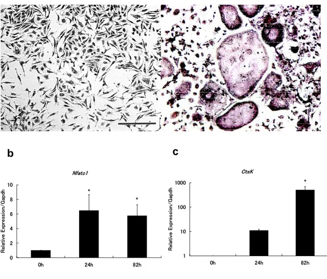
145 ment with RANKL and M-CSF (RANKL/M-CSF) for 82 h (Fig. 1 a). Nu- clear factor of activated T cells, cyto- plasmic 1 (Nfatc1), which is a master regulator of osteoclastogenesis, was upregulated after RANKL/M-CSF treatment for 82 h (Fig. 1 b). The os-
osteoclast-specific gene Cathepsin K (CtsK) strongly expressed after treat- ment with RANKL/M-CSF for 82 h (Fig.
1 c). Therefore, we used this culture system to analyze the intra- and ex- tracellular miRNA expression in os- teoclasts.
a
Nfatc1
0 2 4 6 8 10
0h 24h 82h
Relative Expression/Gapdh *
*
CtsK
1 10 100 1000
0h 24h 82h
Relative Expression/Gapdh
*
b c
Figure 1 Differentiation of osteoclasts from murine bone marrow macrophages.
Murine bone marrow macrophages (BMMs) were incubated for 82 h with RANKL (100 ng/mL) and M-CSF (10 ng/mL); osteoclasts formed after this treatment.
a; Representative microscopic images of BMMs before (left panel) and after (right panel) 82 h stimulation with RANKL and M-CSF and stained for TRAP. Bar = 50 μm.
b and c; Relative expression of the osteoclast genes Nuclear factor of activated T cells, cytoplasmic 1 (Nfatc1) (b) and Cathepsin K (CtsK) (c) in BMMs treated with RANKL and M-CSF for 0, 24, or 82 h, as measured by qRT-PCR. The values are relative to 1 at 0 h (before stimulation) for each mRNA. The target gene expression in each sample was normalized to the Gapdhsignal. The experiments were performed in three times. The data are presented as means ± SD. One-way ANOVA was performed (* P < 0.05 vs. 0 h).
0 0.5 1 1.5 2
0h 82h
Relative Expression/snoRNA202
0 0.5 1 1.5
0h 82h
Relative Expression/snoRNA202
miR-33
0 1 2 3 4
0h 82h
Relative Expression/snoRNA202
**
miR-155
0 0.5 1 1.5
0h 82h
Relative Expression/snoRNA202
miR-210
0 1 2 3 4
0h 82h
Relative Expression/snoRNA202
miR-223
0 0.5 1 1.5
0h 82h
Relative Expression/snoRNA202 **
miR-378
0 10 20 30 40
0h 82h
Relative Expression/snoRNA202
*
miR-1224
0 1 2 3 4 5
0h 82h
Relative Expression/snoRNA202
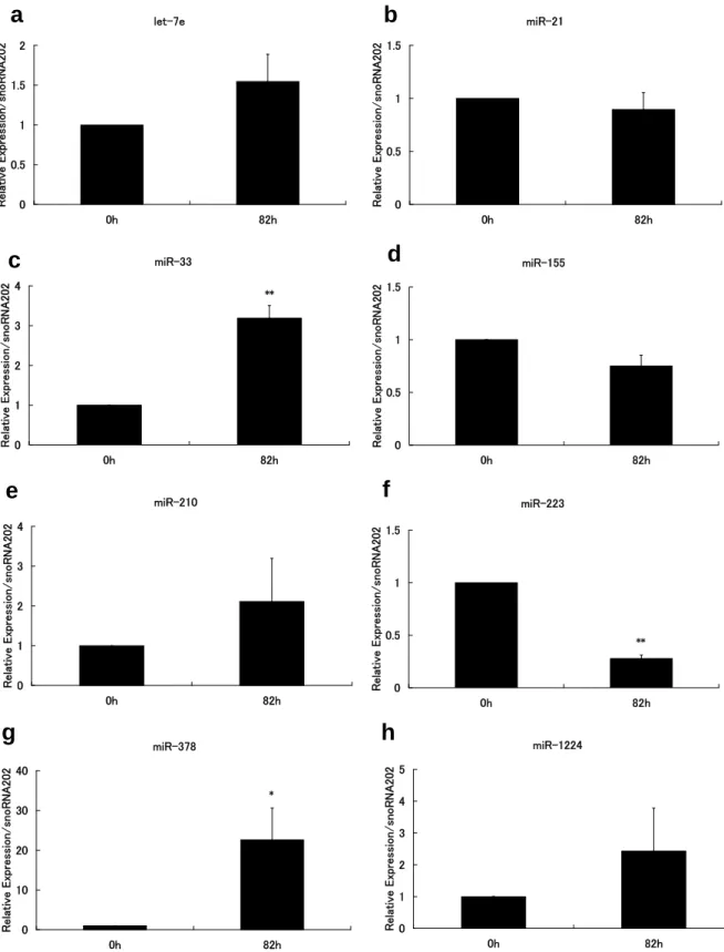
Figure 2 Intracellular microRNA expression in BMMs during osteoclastogenesis.
Murine bone marrow macrophages (BMMs) were cultured in the presence of RANKL (100 ng/mL) and M-CSF (10ng/mL) for 0 or 82 h, and then subjected to qRT-PCR. The expression levels of let-7e (a), miR-21 (b), miR-33 (c), miR-155 (d), miR-210 (e), miR-223 (f), miR-378 (g), and miR-1224 (h) are shown.
The values are relative to 1 at 0 h (before stimulation) for each microRNA. Quantification was performed using snoRNA 202 as an endogenous control. The experiments were performed in three times. The data are presented as means ± SD. Unpaired two-tailed Student’s t-tests were used, assuming unequal vari- ance, to identify statistically significant differences (* P < 0.05 vs. 0 h, ** P < 0.01 vs. 0 h).
c d
e f
g h
147 2. The expression of intracellular miRNAs in osteoclasts
We reported global intracellular miRNA expression profiling during osteoclast differentiation
5. Based on that study, we selected eight miRNAs that we consider important for osteoclastogenesis: let-7e, miR-33, miR-155, miR-210, miR-223, miR378, and miR-1224. The expression levels of let-7e, miR-33, miR-210, miR378, and miR-1224 were upregu- lated compared with before RANKL/
M-CSF treatment (Fig. 2 a, c, e, g, and h), while those of miR-155 and miR-223 were downregulated compared with before RANKL/M-CSF treatment (Fig. 2 d and f). Despite the downregulation of miR-155 and miR-223, the expression baselines of these miRNAs were rela- tively high (data not shown). The level of miR-21 expression did not change sig- nificantly in RANKL/M-CSF-treated BMMs (Fig. 2 b); the baseline expres- sion was relatively high (data not shown).
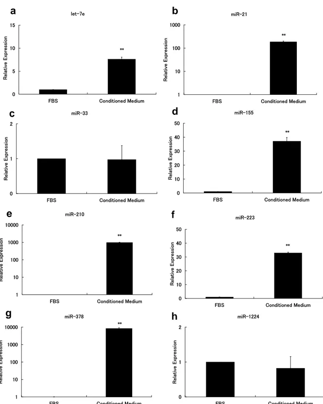
3. The expression of miRNAs in the extracellular microvesicles of osteo- clasts
We next evaluated the expression of miRNAs in the extracellular microvesi- cles of osteoclasts. Of the eight miRNAs we investigated, miR-378 was ex- pressed to the greatest degree in the extracellular microvesicles (Fig. 3 g), followed by miR-210 and miR-21 (Fig. 3 b and e). miR-155 and miR-223 were highly expressed in the extracellular microvesicles (Fig. 3 d and f), and let-7e expression was detected (Fig. 3 a). No significant miR-33 or miR-1224 expres- sion was detected in the extracellular microvesicles (Fig. 3 c and h). These results are similar to those obtained from qRT-PCR amplification curves after elimination of the background miRNA expression in FBS (data not shown).
DISCUSSION
For the first time, we demonstrated that osteoclasts secrete extracellular mi- crovesicles containing specific miRNAs.
Of the eight miRNAs we investigated, miR-378 was expressed at the highest level in the extracellular microvesicles.
We previously postulated that miR-378 plays an important role in osteoclasto- genesis and its intracellular expression increased during osteoclast differentia- tion, with a 21-fold increase in RANKL/
M-CSF-treated BMMs
5. It is reported that miR-378 promotes cell survival and participates in blood vessel formation
16. Since bone tissue is highly vascular, miR-378 in the microvesicles might promote cell survival and blood vessel formation around osteoclasts.
In microvesicles, miR-210 showed the second-highest expression; its in- tracellular expression in BMMs also in- creased after RANKL/M-CSF treatment.
miR-210 regulates various genes in- volved in the cell cycle, differentiation, development, membrane trafficking, and migration / adhesion
17,18. In microvesi- cles, miR-210 might play various roles in cell-to-cell communication. The level of miR-21 expression in the microvesicles was higher than that of let-7e, miR-155, or miR-223. miR-21 is one of the most abundant miRNAs detected in microve- sicles circulating in the blood of cancer patients
19. Considering both our results and these reports, both cancer cells and normal cells, such as osteoclasts, might secrete miR-21-containing microvesi- cles into the circulation.
We detected high-level miR-223 expression in the microvesicles.
Although the intracellular expression of
miR-223 decreased in RANKL / M-CSF-
treated murine BMMs, the expression
baseline was relatively high. miR-223 is
highly expressed in myeloid and bone
marrow cells
20. miR-223 plays a key role
in osteoclastogenesis
1, 5,21, and the
expression of intracellular miR-223
let-7e
0 5 10 15
FBS Conditioned Medium
Relative Expression
**
miR-21
1 10 100 1000
FBS Conditioned Medium
Relative Expression
**
miR-33
0 1 2
FBS Conditioned Medium
Relative Expression
miR-155
0 10 20 30 40 50
FBS Conditioned Medium
Relative Expression
**
miR-210
1 10 100 1000 10000
FBS Conditioned Medium
Relative Expression
**
miR-223
0 10 20 30 40 50
FBS Conditioned Medium
Relative Expression
**
**
miR-378
1 10 100 1000 10000
FBS Conditioned Medium
Relative Expression
*
**
miR-1224
0 1 2
FBS Conditioned Medium
Relative Expression
Figure 3 MicroRNA expression in the extracellular microvesicles in osteoclasts.
Murine bone marrow macrophages (BMMs) were cultured in the presence of RANKL (100 ng/mL) and M-CSF (10 ng/mL) for 82 h. RNA was isolated from extracellular microvesicles in the RANKL/M-CSF-treated conditioned medium after 48 to 82 h, and then subjected to qRT-PCR. The ex- pression levels of let-7e (a), miR-21 (b), miR-33 (c), miR-155 (d), miR-210 (e), miR-223 (f), miR-378 (g), and miR-1224 (h) are shown. The values are relative to 1 for α-MEM containing 10% FBS without culturing for each microRNA. The experiments were performed in three times. The data are presented as means ± SD. Unpaired two-tailed Student’s t-tests were used, assuming unequal variance, to identify statistically significant differences (** P < 0.01 vs. FBS).
a b
c d
e f
g h
149 decreases during osteoclast differentia- tion
5. The overexpression of miR-223 blocks osteoclast differentiation, whereas the inhibition of miR-223 ex- pression has the reverse effect
21. Since the expression of miR-223 increases incrementally as granulocytes mature
20, it might affect granulocyte maturation around osteoclasts in the bone marrow.
miR-155 expression was downregulated in RANKL/M-CSF-treated murine BMMs during osteoclast differentiation. Active macrophage differentiation involves the rapid upregulation of miR-155 expres- sion, which leads to the suppression of RANKL-induced osteoclastogenesis
22. If the miR-155 in the extracellular mi- crovesicles functions similarly, it might inhibit osteoclast differentiation via negative feedback. Let-7e expression was also detected in the microvesicles.
Identified let-7e targets include cell cycle regulators such as CDC25A and CDK6
23. Thus let-7e within microvesi- cles might be involved in regulation of the cell cycle of other cells.
No significant miR-33 or miR-1224 expression was detected in the ex- tracellular microvesicles. However, the intracellular expression of these miRNAs was upregulated during osteo- clast differentiation. Our results suggest that the entire set of intracellular miRNAs is not present in the extracel- lular microvesicles of osteoclasts.
Here, we demonstrated that osteo- clasts secrete extracellular microvesi- cles containing let-7e, miR-21, miR-155, miR-210, miR-223, and miR-378. It is widely known that growth factors play important roles in tissue engineering
24. In future, extracellular microvesicles might be useful as new growth factors in oral tissue engineering.
ACKNOWLEDGMENTS
We thank Prof. Akira Fujimura, Iwate Medical University, for reading the manuscript and giving helpful advice.
This study was supported, in part, by grants from the Scientific Research (C) (No. 23593042), and a Grant-in-Aid for Strategic Medical Science Research Center (2010–2014) from the Ministry of Education, Culture, Sports, Science and Technology, Japan.
REFERENCES
1)
Sugatani T, Hruska KA. Impaired mi- cro-RNA pathways diminish osteoclast differentiation and function. J Biol Chem 2009; 284: 4667-4678.
2)
Kim K, Kim JH, Lee J, et al. MafB nega- tively regulates RANKL-mediated os- teoclast differentiation. Blood 2007; 109:
3253-3259.
3)
Takeshita S, Kaji K, Kudo A. Identifica- tion and characterization of the new osteoclast progenitor with macrophage phenotypes being able to differentiate into mature osteoclasts. J Bone Miner Res 2000; 15: 1477-1488.
4)
Taira M, Nezu T, Sasaki K, et al.
Preparation and in vivo evaluation of apatite/collagen packed composite by alternate immersion method and New- ton press. J Biomed Mater Res B Appl Biomater 2009; 90: 566-573.
5)
Kagiya T, Nakamura S. Expression pro- filing of microRNAs in RAW264.7 cells treated with a combination of tumor ne- crosis factor alpha and RANKL during osteoclast differentiation. J Periodontal Res 2012. [Epub ahead of print]
6)
Valadi H, Ekstrom K, Bossios A, Sjostrand M, Lee JJ, Lotvall JO.
Exosome-mediated transfer of mRNAs and microRNAs is a novel mechanism of genetic exchange between cells. Nat Cell Biol 2007; 9: 654-659.
7)
Skog J, Wurdinger T, van Rijn S, et al.
Glioblastoma microvesicles transport RNA and proteins that promote tumour growth and provide diagnostic bio- markers. Nat Cell Biol 2008; 10: 1470- 1476.
8)
Kosaka N, Iguchi H, Yoshioka Y, Take- shita F, Matsuki Y, Ochiya T. Secretory mechanisms and intercellular transfer of microRNAs in living cells. J Biol Chem 2010; 285: 17442-17452.
9)
Cantaluppi V, Gatti S, Medica D, et al.
Microvesicles derived from endothelial
progenitor cells protect the kidney from
ischemia-reperfusion injury by mi- croRNA-dependent reprogramming of resident renal cells. Kidney Int 2012; 82:
412-427.
10) Record M, Subra C, Silvente-Poirot S,
Poirot M. Exosomes as intercellular signalosomes and pharmacological ef- fectors. Biochem Pharmacol 2011; 81:
1171-1182.
11) Bobrie A, Colombo M, Raposo G, Thery
C. Exosome secretion: molecular mechanisms and roles in immune re- sponses. Traffic 2011; 12: 1659-1668.
12) Mathivanan S, Fahner CJ, Reid GE,
Simpson RJ. ExoCarta 2012: database of exosomal proteins, RNA and lipids.
Nucleic Acids Res 2012; 40: D1241- 1244.
13) Simons M, Raposo G. Exosomes-
-vesicular carriers for intercellular communication. Curr Opin Cell Biol 2009; 21: 575-581.
14) Bak M, Silahtaroglu A, Moller M, et al.
MicroRNA expression in the adult mouse central nervous system. RNA 2008; 14: 432-444.
15) Lin EA, Kong L, Bai XH, Luan Y, Liu CJ.
miR-199a, a bone morphogenic protein 2-responsive MicroRNA, regulates chondrogenesis via direct targeting to Smad1. J Biol Chem 2009; 284: 11326- 11335.
16) Lee DY, Deng Z, Wang CH, Yang BB.
MicroRNA-378 promotes cell survival, tumor growth, and angiogenesis by targeting SuFu and Fus-1 expression.
Proc Natl Acad Sci U S A 2007; 104:
20350-20355.
17) Fasanaro P, Greco S, Lorenzi M, et al.
An integrated approach for experimental target identification of hypoxia-induced miR-210. J Biol Chem 2009; 284:
35134-35143.
18) Tsuchiya S, Fujiwara T, Sato F, et al.
MicroRNA-210 regulates cancer cell proliferation through targeting fibroblast growth factor receptor-like 1 (FGFRL1).
J Biol Chem 2011; 286: 420-428.
19) Krichevsky AM, Gabriely G. miR-21: a
small multi-faceted RNA. J Cell Mol Med 2009; 13: 39-53.
20) O'Connell RM, Zhao JL, Rao DS. Mi-
croRNA function in myeloid biology.
Blood 2011; 118: 2960-2969.
21) Sugatani T, Hruska KA. MicroRNA-223
is a key factor in osteoclast differentia- tion. J Cell Biochem 2007; 101: 996- 999.
22) Mann M, Barad O, Agami R, Geiger B,
Hornstein E. miRNA-based mechanism for the commitment of multipotent pro- genitors to a single cellular fate. Proc
Natl Acad Sci U S A 2010; 107: 15804- 15809.
23) Peter ME. Let-7 and miR-200 microR-
NAs: guardians against pluripotency and cancer progression. Cell Cycle 2009; 8: 843-852.
24) Zaky SH, Cancedda R. Engineering
craniofacial structures: facing the chal- lenge. J Dent Res 2009; 88: 1077-1091.
(Received, December 30, 2012/
Accepted, March 18, 2013)
Corresponding author: