THE SCIENCE AND ENGINEERING REVIEW OF DOSHISHA UNIVERSITY, VOL. 49, NO.4January2009
Development of Capillary Electrophoresis Equipped with a Novel PTFE Cell for Chemiluminescence Detection
Misa YANAGIUCHI, Kosuke SUZUKI, Masahiko HASHIMOTO, and Kazuhiko TSUKAGOSHI* (Received September 19, 2008)
We developed a capillary electrophoresis–chemiluminescence detection system, using a novel PTFE detection cell.
The detection cell is comprised of a T-type micro-flow line (0.5 mm i.d.), in which the reaction of luminol-hydrogen peroxide-metal ion catalyst is used to generate chemiluminescence. The two reagent solutions, hydrogen peroxide and metal ion catalyst solution, are delivered toward the T-junction from the two ends of the T-type line positioned on a straight line. A fused silica capillary is inserted through the T-junction into the flow line where the sample eluted from the capillary outlet is mixed with the reagent solutions. In the direct detection method, luminal was determined based on the positive chemiluminescence peak at concentrations over the ranges of 110-5–110-3 M and 110-6–310-5 M for Fe(II) and Cu(II) catalysts, respectively. In the indirect detection method, antioxidants were detected based on negative chemiluminescence response; catechin mixture was determined over the concentration range of 310-5–110-3 M. The indirect detection method using the present system was applied to analysis of catechin in commercial green tea beverages.
Key Words: Capillary electrophoresis, chemiluminescence, antioxidant.
*Department of Chemical Engineering and Materials Science, Faculty of Engineering, Doshisha University, Kyotanabe, Kyoto 610-0321. Tel. +81-774-65-6595, Fax. +81-774-65-6595, E-mail: [email protected]
1. Introduction
Chemiluminescence (CL) is one of the most useful detection methods in addition to absorption and fluorescence detection. CL is characterized by high sensitivity, wide determinable range, simple and rapid operation, inexpensive reagents and apparatus, and ease of setup for flow analysis. Such CL detection methods have been combined with HPLC, FIA, and capillary electrophoresis (CE) [1,2]. Studies regarding CE with CL detection were first reported in the early 1990s.
Various CL detection cells for CE, i.e., interfaces between CE and CL detection, were developed, including flow-type, batch-type, and optical fiber setting-type systems [3-5]. Generally, flow-type systems are complicated with a number of joints and flow lines,
batch-type systems lack repeatability for continuous sample injection, and optical fiber setting-type systems suffer from light loss inside the fiber. Thus, a novel detection CL cell for CE is required for the wider use of CE-CL detection systems.
PTFE (polytetrafluoroethylene) is an attractive material for analytical use, because it is chemically inert, easy to process, inexpensive, and non-conductive.
However, PTEF lacks light penetration properties, a characteristic that has prevented its use in optical detection methods. To our knowledge, only very thin PTFE tubing has been applied to detection cells in HPLC, FIA, and CE [6,7].
In this study, we developed a novel PTFE CL detection cell for CE. The PTFE cell has a T-type micro-flow line (0.5 mm i.d.). Part of the flow line for CL detection has a thickness of 0.5 mm between the
inner wall of the line and the surface of the PTFE cell (PTEF block). The PTFE detection cell was confirmed to be applicable to a CE with CL detection system, including direct detection (luminal sample) and indirect detection (antioxidant sample).
2. Experimental
2.1 Reagents
Water was purified using a Millipore Elix 3 UV water purification system (Millipore, Tokyo, Japan). All reagents used were of commercially available special grade and were purchased from the following sources:
luminol, copper(II) sulfate, Mohr’s salt (ammonium iron (II) sulfate hexahydrate), ascorbic acid, 2-propanol, sodium azide, and nitroblue tetrazolium were from Nacalai Tesque (Kyoto, Japan); catechin (catechin mixture from green tea) and hydrogen peroxide (30 wt%) were from Wako Pure Chemical Industries (Osaka, Japan).
2.2 PTFE CL detection cell
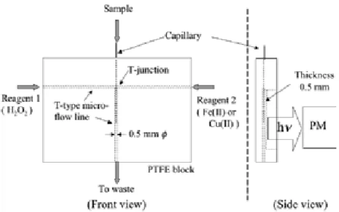
A schematic diagram of the PTFE CL detection cell is shown in Fig. 1. The detection cell is comprised of a T-type micro-flow line (0.5 mm i.d.). The two reagent solutions are delivered at a defined flow rate toward the T-junction from the two ends of the T-type line positioned on a straight line, with the reagents mixed in the other flow line. The fused silica capillary is inserted through the T-junction into the flow line as shown in Fig.
1. The part of the flow line for CL detection is 0.5 mm in thickness between the inner wall of the line and the surface of the PTFE cell (PTEF block).
2.3 Analytical conditions and procedures
Direct detection of luminal. The reaction of luminal-hydrogen peroxide-metal ion catalyst was used to generate CL in the micro-flow line. The two reagent solutions, 10 mM hydrogen peroxide (10 mM carbonate buffer; pH 10.8) and 0.1 mM Fe(II) (as ammonium iron (II) sulfate hexahydrate) (water solvent) or 10 mM Cu(II) (as copper (II) sulfate) (10 mM carbonate buffer
Fig. 1. Schematic diagram of PTFE CL detection cell.
including 200 mM potassium tartrate; pH 10.8), were delivered at a defined flow rate toward the T-junction from both ends of the T-type line positioned on a straight line, with the reagents mixed in the other flow line. The fused silica capillary (75 mm i.d., 150 mm o.d., and 70 cm in length) was inserted through the T-junction into the flow line. The distance from the junction point to the capillary outlet was set as 0, 1, or 2 cm. Luminol solved with migration buffer (10 mM carbonate buffer; pH 10.8) was injected into the capillary by the gravity method (20 cm, 15 s). The applied voltage was 12 kV. The CL occurring at the tip of the capillary outlet was detected with a photomultiplier tube (PM) (H6780; Hamamatsu Photonics Co. Ltd, Shizuoka, Japan).
Indirect detection of antioxidants. CL reaction of luminol-hydrogen peroxide-Cu(II) catalyst was used for the indirect detection of antioxidants in the CE with CL detection system. Similar analytical conditions and procedures were carried out for the indirect detection system except for the migration buffer and sample solution. The migration buffer was prepared by dissolving luminal in 20 mM carbonate buffer (pH 10.8), yielding a final solution of 1.0 mM luminol.
Antioxidants dissolved in 20 mM carbonate buffer (pH 10.8) were injected into the capillary as samples by the gravity method (20 cm height, 30 s). The baseline CL was determined by the reaction of luminal, hydrogen peroxide, and Cu(II) catalyst at the tip of the capillary outlet. Antioxidants migrated in the capillary were mixed with active oxygen species generated by the reaction Misa YANAGIUCHI, Kosuke SUZUKI, Masahiko HASHIMOTO and Kazuhiko TSUKAGOSHI
188
between Cu(II) and hydrogen peroxide, such as superoxide radical anions, at the capillary outlet, providing a negative peak from the baseline CL.
3. Results and Discussion
3.1 Design of PTFE CL detection cell
As described in the Introduction, PTFE has a number of characteristics that make it an attractive material for analytical use. However, its light-penetration characteristics are inappropriate for use in devices for use in optical CL detection methods. To our knowledge, only PTFE tubing (0.5 mm i.d and 1.6 or 2.0 mm o.d.) has been used successfully for flow-type CL detection cells in CE with CL detection systems [2,8]. Here, micro-flow lines or channels were prepared mechanically in the PTFE block, and the thickness between the inner wall of the flow line and the surface of the PTFE block was narrow at 0.5 mm. This thickness was sufficient to allow the CL detected with the PM to pass through. The PTFE CL detection cell could be easily combined with CE and then used for CE analysis.
3.2 Direct detection of luminol
Luminol-hydrogen peroxide-Cu(II) or Fe(II) catalyst CL reaction was adapted for direct luminol detection in the CE with CL detection system. The effects of the flow rates of the two reagents as well as the distance from the junction point to the tip of the capillary for Fe(II) and Cu(II) catalysts were examined for detection of luminol.
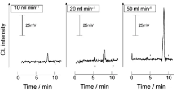
As examples, the electropherograms of luminol obtained with all flow rates and lengths for the Fe(II) catalyst are shown in Figs. 2 and 3, respectively. Although the lengths of 0 and 1.0 cm sometimes gave large responses, they lacked good reproducibility. Judging from the data for the Fe(II) catalyst, a flow rate of 50 ml min-1 and length of 2 cm were used to examine the calibration curve of luminol. Luminol was determined over the concentration range of 110-5–110-3 M (correlation coefficient: 0.999).
In a similar way, we examined the analytical conditions of luminol-hydrogen peroxide-Cu(II) catalyst
Fig. 2. Effects of the flow rates on luminol CL intensities in the direct detection method using the Fe(II) catalyst.
Conditions: Catalyst solution, 0.1 mM Fe(II) (as ammonium iron (II) sulfate hexahydrate) (water solvent);
the distance from the junction to the capillary outlet, 2 cm; and luminol, 0.5 mM.
Fig. 3. Effects of the distance from the junction to the capillary outlet on luminol CL intensities in the direct detection method using the Fe(II) catalyst. Conditions:
Catalyst solution, 0.1 mM Fe(II) (as ammonium iron (II) sulfate hexahydrate) (water solvent); the flow rate, 50 ml min-1; and luminol, 0.5 mM.
reaction. In this case, the CL intensities decreased with increasing flow rates of 2, 3, and 5 ml min-1. In addition, the intensities decreased with increasing lengths of 0, 1, and 2 cm; even the length of 0 cm gave good reproducibility. The calibration curve of luminol was examined with a flow rate of 2 ml min-1 and length of 0 cm; luminol was determined over the concentration range of 110-6–310-5 M (correlation coefficient: 0.999).
The difference in analytical conditions between the Fe(II) and Cu(II) catalysts was due to the differences in Development of Capillary Electrophoresis Equipped with a Novel PTFE Cell for Chemiluminescence Detection 189
reaction rate between metal ions and hydrogen peroxide as well as the lifetimes of active oxygen, mainly hydroxyl and superoxide radical anions described in the next section, generated through the reactions.
Previously, we reported biomolecular analysis of amino acids, peptides, and proteins through labeling with the luminol derivative isoluminol isothiocyanate [9,10].
The data reported above indicated that the present direct detection system could be applied to biomolecular analysis using isoluminol isothiocyanate as the labeling reagent.
3.3 Effects of antioxidants on luminol CL in direct detection
We examined the effects of antioxidants on luminol CL intensities in the direct detection system using the Fe(II) and Cu(II) catalysts. Sodium azide, nitroblue tetrazolium, and 2-propanol are well-known antioxidants for active oxygen; sodium azide for singlet oxygen (1O2), nitroblue tetrazolium for superoxide radical anions (O2
-*), and 2-propanol for hydroxyl radicals (HO*). In addition, these agents are known not to react with hydrogen peroxide. Ascorbic acid was also used as an antioxidant for singlet oxygen, superoxide radical anions, and hydrogen peroxide. The migration buffer including 0.1 mM each antioxidant was used.
The data obtained here are summarized in Table 1.
Ascorbic acid decreased the CL intensities of both the Fe(II) and Cu(II) catalysts, because it reacted with hydrogen peroxide. As shown in the table, the Fe(II) catalyst system mainly generates hydrogen radicals through the reaction between Fe(II) and hydrogen peroxide that indicates a relative CL intensity of 52%, and the Cu(II) catalyst system generates superoxide radical anions through the reaction between Cu(II) and hydrogen peroxide that shows an intensity of 23%. The data consisted with that Fenton reaction generates hydrogen radicals through the reaction between Fe(II) and hydrogen peroxide [11] and the Cu(II) complex decomposes hydrogen peroxide to superoxide radical anions under alkaline conditions [12].
3.4 Indirect detection of antioxidants
Antioxidants were examined indirectly with the present CE with CL detection system using luminol-hydrogen peroxide-Cu(II) catalyst reaction. In the indirect method, antioxidants were detected as negative peaks from the baseline CL as described in the experimental section. When using the Fe(II) catalyst, a stable CL baseline was not obtained. Antioxidants, ascorbic acid, 2-propanol, sodium azide, and nitroblue tetrazolium, were examined; only ascorbic acid and nitroblue tetrazolium were detected as negative peaks.
The detection times and relative peak areas (the peak area due to nitroblue tetrazolium was given an arbitrary value of 100) are summarized in Table 2. The effects of antioxidants observed for the indirect detection method were similar to those of the direct detection method using the Cu(II) catalyst shown in Table 1.
Misa YANAGIUCHI, Kosuke SUZUKI, Masahiko HASHIMOTO and Kazuhiko TSUKAGOSHI 190
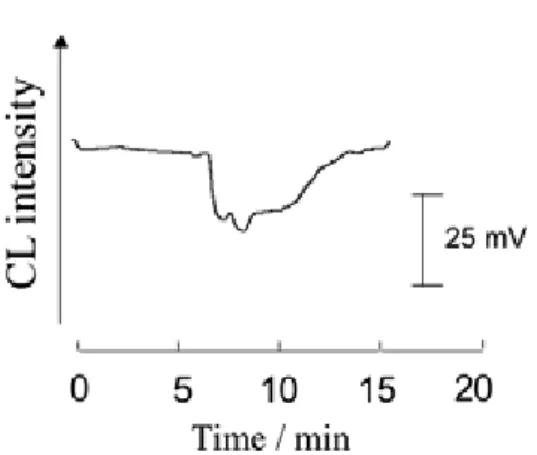
Fig. 4. Electropherogram of catechin (catechin mixture from green tea) in the indirect detection method using the Cu(II) catalyst. Conditions: Catalyst solution, 10 mM Cu(II) (as copper (II) sulfate) (10 mM carbonate buffer including 200 mM potassium tartrate; pH 10.8); the flow rate, 2 ml min-1; and sample, 1 mM catechin (catechin mixture from green tea).
Catechin, a polyphenol derivative that reacts with radical species, such as hydroxyl radicals and superoxide radical anions, is a well-known antioxidant. The main species of catechin in green tea are epicatechin, epigallocatechin, epicatechin gallate, and epigallocatechin gallate, the contents of which are as follows: epigallocatechin gallate > epigallocatechin >
epicatechin gallate > epicatechin. Catechin (catechin mixture from green tea) was also analyzed by the indirect detection method. Although the catechin mixture from green tea was not separated by the present system under the conditions used, the negative CL peak was observed on the electropherogram around 7–12 min, as shown in Fig. 4. The relative peak area was estimated to be ca. 417.
The relationship between the catechin concentration and the relative peak area due to the negative CL peak was examined. The molecular weight of epigallocatechin gallate (458.37) was used as that of the mixture. The negative CL peak responded over the range of 310-5–110-3 M catechin with a limit of detection of 0.03 mM (S/N=3) (correlation coefficient: 0.999).
3.5 Analysis of catechin in green tea
We examined the amounts of catechin in two
commercial green tea beverages (A and B) using the present indirect detection system. Samples of green tea diluted tenfold were analyzed using this system.
Negative CL peaks were observed on the electropherograms at elution times of ca. 7–12 min. The commercial green teas, A and B, were found to include catechin at concentrations of 360 and 200 mM in the tenfold-diluted sample (average values from 4–7 measurements), respectively. The data obtained with the present method were consistent with the manufacturers’
reported catechin concentrations for A and B (3.4 and 1.7 mM, respectively).
Acknowledgments
This work was supported by a Grant-in-Aid for Scientific Research (C) from the Ministry of Education, Culture, Sports, Science, and Technology, Japan. This study was also supported by the Academic Frontier Research Project on “New Frontier of Biomedical Engineering Research” of the Ministry of Education, Culture, Sports, Science, and Technology, Japan.
References
[1] A. M. Garcia-Campana, F. J. Lara, Anal. Bioanal.
Chem. 387 (2007) 165.
[2] K. Tsukagoshi, Sci. Engi. Rev. Doshisha Univ. 45 (2005) 165.
[3] X. Huang, J. Ren, TrAC 25 (2006) 155.
[4] M. Hashimoto, K. Tsukagoshi, R. Nakajima, and K.
Kondo, J. Chromatogr. A 832 (1999) 191.
[5] K. Tsukagoshi, K. Nakahama, R. Nakajima, Anal.
Chem. 76 (2004) 4410.
[6] T. Hara, K. Tsukagoshi, Anal. Sci. 6 (1990) 797.
[7] M. Macka, W.-C. Yang, P. Zakaria, A. Shitangkoon, E. F. Hilder, P. Andersson, P. Nesterenko, P. R. Haddad, J. Chromatogr. A 1039 (2004) 193.
[8] K. Tsukagoshi, K. Sawanoi, and R. Nakajima, J.
Chromatogr. A 1143 (2007) 288.
[9] K. Tsukagoshi, K. Sawanoi, R. Nakajima, Talanta 68 (2006) 1071.
[10] K. Tsukagoshi, T. Nakamura and R. Nakajima, Anal.
Development of Capillary Electrophoresis Equipped with a Novel PTFE Cell for Chemiluminescence Detection 191
Chem., 74 (2002) 4109.
[11] F. Haber, J. Weiss, Proc. Roy. Soc., 333 (1934) A147.
[12] O. S. Fedorova, S. E. Olkin, V. M. Berdnikov, Z.
Phy. Chem. 263 (1982) 529.
Misa YANAGIUCHI, Kosuke SUZUKI, Masahiko HASHIMOTO and Kazuhiko TSUKAGOSHI 192