*1 八重瀬会同仁病院腎センター腎臓内科,*2 同 泌尿器科,*3 南部医療センター腎臓内科 (平成 24 年 2 月 8 日受理)
MSSA
感染を契機にネフローゼ症候群と
急速進行性腎炎を呈した 1 例
桑
江
紀
子
*1謝
花
政
秀
*2宮
里
朝
矩
*2和
気
亨
*3A case of nephrotic syndrome and rapidly progressive glomerulonephritis associated with MSSA infection
Noriko KUWAE*1, Masahide JAHANA*2, Tomonori MIYASATO*2, and Touru WAKE*3 *1Department of Nephrology, *2Department of Urology, Doujin Hospital,
*3Department of Nephrology, Southern Medical Center, Okinawa, Japan
要 旨
症例は 67 歳,男性。化膿性肩関節炎,敗血症にて近医入院,メチシリン感受性ブドウ球菌(MSSA)感染症にて 抗生物質投与後,炎症反応,培養とも陰性化したが,肉眼的血尿,全身浮腫,6 kg の体重増加を認め,低アルブ ミン血症,尿蛋白 24 g/日,Cre 2.91 mg/dL とネフローゼ症候群および腎機能障害を呈した。精査目的で第 50 病 日に当院紹介入院。腎生検施行,迅速病理の光顕にて管内性および管外性半月体形成を認めた。炎症反応および 各種培養が陰性であることを確認,ステロイドミニパルス(500 mg)1 クール施行後ステロイド(PSL)40 mg 経口 投与開始,二重膜血漿交換(DFPP)6 回施行。Cre は一時 2.44 mg/dL まで軽快するも徐々に上昇,Cre 3.29 mg/dL となったため,再度ステロイドミニパルス施行,同日全血漿交換 PE(新鮮凍結血漿 FFP45 単位使用),この時点 で MPO-ANCA,PR3−ANCA は陰性,返却された病理結果は蛍光抗体にてメサンギウム領域に IgA,C3 の沈着 を,電顕にてメサンギウム領域に deposit を認め,メサンギウム増殖性腎炎,IgA 腎症を示唆する所見であった。 その後,徐々に腎機能が悪化したため,さらにステロイドミニパルス 3 クール目を施行,経口 PSL 30 mg にミゾ リビン(MZB)150 mg を追加。4 カ月後には症状,検査値とも軽快,尿蛋白は 3 g/日程度,Cre 2 mg/dL 台となっ た。MZB 血中濃度をモニタリングしつつ適正 MZB 投与量を決定,PSL 10 mg,MZB 100 mg 投与で 6 カ月後 Cre 1.4 mg/dL と改善を認めた。経過から MSSA 感染関連腎炎と考えられた。The patient was a 67−year-old male. He was admitted to a local hospital due to septic shoulder arthritis and sepsis. He was treated with antibiotics due to a methicillin-sensitive Staphylococcus aureus(MSSA) infection. The inflammatory reaction and culture returned to negative;however, he showed macroscopic hema-turia, anasarca and gained 6 kg. The patient had nephrotic syndrome and renal impairment including hypoalbu-minemia,(urine protein of 24 g/day and creatinine(Cre)of 2.91 mg/dL). On day 50 of illness, the patient was referred and admitted to this hospital for detailed examinations. A renal biopsy was performed and a rapid pathological diagnosis indicated endocapillary and extracapillary crescentic glomerulonephritis. The inflamma-tory reaction and culture were confirmed to be negative and the patient received one course of steroid mini-pulse (500 mg)therapy, followed by oral administration of 40 mg steroid(prednisolone:PSL)and double filtration
plasmapheresis(DFPP)was performed 6 times. Although there was transient remission to 2.44 mg/dL, the Cre gradually increased to 3.29 mg/dL. Therefore, steroid mini-pulse therapy was performed again and total plasma exchange(fresh frozen plasma:FFP, 45 units)was carried out on the same day. MPO-ANCA and PR3−ANCA were negative and the fluorescent antibody technique showed IgA and C3 deposition in the mesangial area. Elec-tron microscopy found deposits in the mesangial area, and thus the patient was diagnosed with
1995 年,小山らがメチシリン耐性ブドウ球菌(MRSA)感 染性腎炎症例を報告して以来,MRSA 感染性腎炎について の文献報告は多数みられるが,メチシリン感受性ブドウ球 菌(MSSA)感染関連腎炎については症例が少ない。今回, MSSA 感染症を契機に腎機能が増悪,ネフローゼ症候群, 急速進行性腎炎(RPGN)を呈し,血漿交換(DFPP+PE),ス テロイド,免疫抑制薬が有用であったメサンギウム増殖性 腎炎症例を経験した。 患 者:67 歳,男性 主 訴:四肢の浮腫,体重増加,肉眼的血尿 家族歴:特記事項なし 生活歴:タバコ 20 本/日 40 年間,酒 3 合/日 30 年間, 独身,生活保護,一人暮らし はじめに 症 例 既往歴:通院歴なく,詳細不明 現病歴:これまで健診を受けたことがなかった。自宅で 倒れているのを隣人に発見され,救急病院へ搬送された。 入院当時ショック状態であった。入院後,左肩の痛みを訴 え,CT にて膿瘍形成を伴う左鎖骨遠位端骨折,左肩前方 脱臼,左化膿性肩関節炎,咽後膿瘍を認めた。関節穿刺施 行,膿および血液培養から MSSA が検出された。入院当初, 抗生物質バンコマイシン(VCM)およびセフォタキシム (CTX)の投与を開始していたが,MSSA と判明後はセファ ゾリン(CEZ)およびゲンタマイシン(GM)へ変更,投与 16 日目で皮疹出現のため,クリンダマイシン(CLDM)に変更 し,19 日間投与。培養陰性にて第 39 病日投与終了。入院 時クレアチニン(Cre)は 2.29 mg/dL,尿蛋白 2+,尿潜血 3+であったが,輸液により第 6 病日,Cre は 0.7 mg/dL ま で低下した。第 20 病日頃より尿所見の増悪がみられ,第 39 病日には四肢の浮腫,体重増加および肉眼的血尿を認め た。TP 6.0 g/dL,Alb 1.3 g/dL,尿蛋白 24 g/日,BUN 28.6 mg/dL,Cre 2.91 mg/dL とネフローゼ症候群および腎機能 ative nephropathy. There was continued gradual deterioration of renal function. Therefore, a 3−course steroid mini-pulse therapy was performed again and 150 mg/day mizoribine(MZB)was added to 30 mg/day oral PSL. The symptoms and test values were resolved after 6 months. Urine protein was 1+, Cre was 1.4 mg/dL by PSL 10 mg/day and MZB 100 mg/day. The disease course suggested that patient had MSSA infection-associ-ated nephritis.
Jpn J Nephrol 2012;54:543−549. Key words:MSSA infection-associated nephritis, endocapillary and extracapillary crescentic formation,
mesan-gioproliferative nephritis, IgA and C3 deposition, DFPP, PE, MZB
Table 1. Laboratory findings during the disease course(1)
Fe 68μg/dL TIBC 184μg/dL VB12 333 pg/mL CRP 4.64 mg/dL ESR 108 mm/h Urinalysis pH 6.5 Prot 3+ Occult blood 3+ RBC deformity (+) Hyaline cast 1∼2/HPF Granular cast 1∼2/HPF RBC >100/HPF WBC 20∼25/HPF U-Prot/Cre 19.05 g/gCre Bence-Jones protein (−) Blood chemistry TP 6.1 g/dL Alb 1.9 g/dL A/G 0.5 AST 19 IU/L ALT 7 IU/L LDH 316 IU/L ALP 387 IU/L CK 54 IU/L T−Cho 215 mg/dL BUN 31.7 mg/dL Cre 3.67 mg/dL HbA1c 4.8 % Na 137 mEq/L K 2.8 mEq/L Cl 97 mEq/L UA 7.9 mEq/L Blood cell count
WBC 5,800/μL Neu 59.4 % Lym 34.5 % Mo 3.5 % Eo 2.6 % Ba 0.0 % RBC Hb 9.0 g/dL Ht 26.4 % MCV 90.4 fL MCH 30.8 pg MCHC 34.1 % Plt 21.1×10⁴/μL
障害を呈し,第 49 病日精査目的で当院へ転院。 入院時現症:身長 160 cm,体重 66 kg(本人申告で 6 kg 増加),意識清明,体温 36.2℃,血圧 144/70 mmHg,脈拍 90/分,不整,呼吸数 12/分。眼瞼結膜軽度貧血あり,眼球 黄染なし。肺音:両側背部に crackle 聴取,心雑音なし。左 腕脱力あり,挙上不可。四肢に皮疹および紫斑なし,前脛 骨部圧痕浮腫,アキレス腱反射正常で末 W神経障害なし。 転院時検査所見(Table 1):当科転院時,BUN 31.7 mg/ dL,Cre 3.67 mg/dL と高度の腎機能障害に加え,尿蛋白ク レアチニン比(Prot/Cre)19.05 g/gCre,TP 6.1 g/dL,Alb 1.9 g/dL,T-Cho 215 mg/dL とネフローゼの状態であった。尿 沈渣では多量の赤血球を認め,CRP 4.64 mg/dL,ESR 108 mm/h と炎症反応は亢進,高度貧血を認めた。免疫学的検 査(Table 2)では IgA 646 mg/dL,IgG 2,132 mg/dL と上昇,
Table 2. Laboratory findings during the disease course(2) Autoantibody
ANA <×40
MPO-ANCA <10 EU
PR3−ANCA <10 EU
anti GBM antibody <10 EU anti ds-DNA antibody <2.5 IU/mL anti RNP antibody <7.0 U/mL
Cryoglobulin (−) Others Ferritin 841 ng/mL Ferric acid 7.9 ng/mL Hyaluroic acid 145 ng/mL Ⅳ type collagen 4.4 ng/mL Hepaplastin 83 %
Direct, Indirect coombs (−) Haptoglobin 83 mg/dL Stool/Occult blood (−) Infection HBsAg (−) HBVAb (−) RPR (−) TPHA (−) Q-FERON (+) Strongyloidiasis(−) Serological study C3 103 mg/dL C4 41 mg/dL IgG 2,132 mg/dL IgA 646 mg/dL IgM 65 mg/dL
Fig. 1. The findings of abdominal ultrasound
肝炎ウイルスはすべて陰性,肝線維化マーカーも正常で あった。胸部 X 線像では心胸郭比の増大なく,胸部 CT で は両側胸水貯留,および心電図では心房細動を認めた。腹 部超音波では両腎臓の萎縮なく,むしろ腫大していた(Fig. 1)。心臓超音波では vegetation は認められなかった。 臨床経過(Fig. 2):転院数日で Cre 4.9 mg/dL と腎機能 が急速に悪化,尿沈渣上の赤血球増加から急速進行性腎炎 が疑われた。治療方針決定のため,腎生検による組織診断 が必要と判断,心房細動に対して前医より投与されていた ワルファリンを中止,第 55 病日,超音波ガイド下経皮的
Fig. 3. Light microscopic findings of the renal biopsy a.Endocapillary proliferation, crescent formation of the
glomeruli with tubular atrophy and interstitial fibrosis (PAS)
b.Fibrinoid tuft necrosis 9 o’clock, edematous mesan-gial change 8 o’clock and adhesion to Bowman’s capsule 1 o’clock are seen.(HE)
c.Enlargement of the mesangial area with double con-tour, endocapillary cell accumulation and small extracapillary cell proliferation are observed.(PAS)
b a c
b a
Fig. 4. Fluorescent antibody technique findings
a.Granular deposition of IgA in the mesangial area and segmentally on the capillary wall b.Granular deposition of C3 in the mesangial area and segmentally on the capillary wall
腎生検を施行した。迅速標本で,光顕で細胞性および線維 性半月体病変に管内増殖性病変の混在を認めた。この時点 で感染徴候なく,CRP および各種培養はすべて陰性である ことを確認,メチルプレドニゾロン 0.5 g/日のステロイド パルス療法を 3 日間施行,後療法として経口プレドニゾロ ン 40 mg/日を投与開始するとともに,腎生検病理がまだ明 らかでなく,ANCA 関連腎炎も疑われたため,二重膜血漿 交換(DFFP)を 6 回施行した。その間,臨床上および画像上, 結核を疑う所見はみられなかったが,コンティフェロンが 陽性であったため経口イソニコチン酸ヒドラジド(INH) 100 mg/日を併用投与した。Cre 値は 2.04 mg/ dL へ軽快,蛋白尿は 12 g/日から 3 g/日へ軽 減したが,第 80 病日より Cre が上昇(3.49 mg/dL)してきたため,2 回目のメチルプレド ニゾロンミニパルス療法を施行,同時に全血 漿交換(PE:血漿 45 単位)を 1 回施行した。 その後 Cre は 2 mg/dL 台と改善した。その後 返却された検査結果では,MPO-ANCA,PR 3-ANCA,クリオグロブリンなどは陰性で あった。 第 85 病日,病理結果が判明。光顕で糸球 体は 16 個含まれており,全節性硬化を 1 個 認めた。線維細胞性半月体が 1 個,また,約 半数の糸球体に癒着および線維性半月体を 7 個伴っていた。管内増殖性変化もみられ, 係蹄内腔では遊走細胞が散見された。尿細管 の萎縮は 20 %程度。尿細管炎および間質には 好酸球,リンパ球の浸潤も認めた。係蹄壁の フィブリノイド壊死を含む管内増殖性腎炎の 像が見られ,ボウマン *と係蹄壁の癒着など 急性病変,およびメサンギウム基質の増加と 係蹄壁の二重化などの慢性病変が認められた (Fig. 3)。蛍光抗体法では IgA と C3 を主体と した陽性像が見られた(Fig. 4)。電子顕微鏡で はメサンギウム領域に高密度 deposit および 足細胞の脱落,扁平化が著明であり,微絨毛 の形成および全周性のメサンギウム細胞の間 入が見られた(Fig. 5)。組織学的重症度分類で は GradeⅡ∼GradeⅢ,軽度の activitiy,中等度 の chronicity を示した。IgA 腎症を示唆する 所見であった。 第 88 病日,再度 Cre が 3 mg/dL 台へ上昇 し始めた。メチルプレドニゾロンのミニパル ス療法をさらに 1 クール施行後,経口プレドニゾロン 30 mg にミゾリビン 150 mg を朝 1 回投与で追加した。ミゾリ ビン血中濃度測定にてトラフ 3.78μg/mL,ピーク 6.24μg/ mL と双方とも高めであったためミゾリビンを 100 mg/日 へ減量,トラフ 2.44μg/mL,ピーク 3.90μg/mL となった。 トラフ値は若干まだ高めであったが,副作用は認められな かったため,これを維持量とした。その後 Cre は 2 mg/dL 台で推移。第 130 病日には Cre 2.14 mg/dL,尿 Prot/Cre 9.14 g/gCre,尿潜血 3+,尿沈渣赤血球 2∼3/HP で,肉眼 的血尿も消退した。経過中,尿路感染症に罹患したが,抗 a b
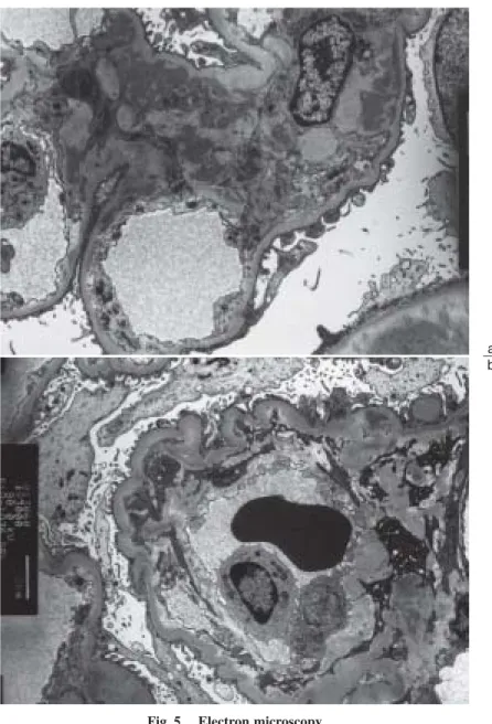
Fig. 5. Electron microscopy
a.Electron-dense deposits in the mesangial area and flattening of the footprocess of podocytes with villi formation
b.Circumferential mesangial interposition and mesangial bridge for-mation with mononuclear cell in the lumen
生物質投与にて回復した。起炎菌は肺炎桿菌であった。現 在,ステロイド 10 mg/日,ミゾリビン 100 mg/日投与で Cre は 1.4 mg/dL 前後を維持している(Fig. 2)。 本症例は健診歴なく,メサンギウム増殖性腎炎の既往の 有無は定かではないが,入院時より蛋白尿,潜血を認め, MSSA 感染後より肉眼的血尿,ネフローゼ症候群,RPGN を呈したことに関して,以前よりメサンギウム増殖性腎炎 が存在した可能性は否定できない。また腎病理組織では, 光顕 HE で係蹄壁のフィブリノイド壊死を含む管内増殖性 腎炎像,ボウマン *と係蹄壁の癒着などの急性病変,メサ ンギウム基質の増加と係蹄壁の二重化(メサンギウム間入) という慢性の病変が急性病変なしに,あるいは共存して認 められ,電顕ではメサンギウム領域の拡大に沈着物,単球, マクロファージの集積像に加え,全周性のメサンギウム間 入の完成像が見られた。これらの所見は急性の感染症およ び既往のメサンギウム増殖性腎炎,IgA 腎症などが同時に 存在することを示唆するものとも考えられる。 従来,メサンギウム増殖性腎炎の一型である IgA 腎症の 組織像には多様性が存在することが知られており,本例に みられたような管内増殖性糸球体腎炎は,IgA 腎症症例に 共通な初期像であるとする見解の報告もある1)が,一方, 管内増殖性糸球体腎炎を呈する症例は補体 C3 活性の点で 他の症例と異なり,溶連菌感染後糸球体腎炎と同様な系が IgA 腎症症例でも活性化,発動しているとする報告もあ る2∼4)。Satoskar ら5)も,MRSA あるいは MSSA 腎炎に伴っ て発症した腎炎が primary IgA nephropathy とよく似た病理 像を示すことを述べている。
MRSA 感染性糸球体腎炎では,T 細胞の著明な活性化,
考 察
特定の TCR-V β陽性細胞の増加,高サイトカイン血症が 認められる。MRSA の外毒素(Staphylococcal enterotoxin)が スーパー抗原として関与,病理組織所見では IgA 腎症関連 腎炎と考えられている6∼10)。 Handa ら11)は,MSSA 感染症後にネフローゼ症候群を呈 した症例を報告した。組織では管内増殖性腎炎像と著しい IgA 沈着がメサンギウム領域および係蹄壁に見られ,9 カ 月後に施行した腎生検では,臨床症状の改善とともにこれ ら所見は消失し,これにより,MRSA 感染後糸球体腎炎と 同様な機序で MSSA 感染性もしくは感染関連糸球体腎炎 が惹起されることが示唆された。 文献的にわれわれが検索しえた限りでは,MSSA 感染性 腎炎症例の治療に関して,全例(アトピー性皮膚炎で経口プ レドニゾロン 40 mg を投与していた Handa ら11)の症例お よ び Henoch-Schönlein purpura に 合 併 し た 詳 細 不 明 の Hirayama ら12)の症例を除いて)で,抗生物質の投与(Table 3)のみにとどまっているのに対して,本症例では CRP が陰 性となった後もネフローゼ,腎機能障害の急速な進展がみ られ,ステロイド,血漿交換,免疫抑制薬などが必要であっ た。 以上より,感染症が終焉,感染徴候は認められず,炎症 反応陰性,培養陰性となったにもかかわらず,病理で半月 体形成を認めるような急速進行性腎炎症例では,ステロイ ド,免疫抑制薬および DFPP,PE などの免疫抑制療法も有 用と思われた。 MSSA 感染を契機に,著明なネフローゼ症候群,急速進 行性糸球体腎炎を呈した症例を経験した。感染陰性化後の ステロイドパルス,血漿交換,経口ステロイドおよびミゾ 結 語
Table 3. Patients with methicillin-sensitive S. aures infection-associated nephritis
Outcome Treatment S-complement Clinical presentation Origin of infection Age Sex No. Recovered Antibiotics+PSL Low normal NS Infected dermatitis 57 F 111) Cre 1.9 Low NS Foot ulcer+cellulitis 59 M 213) Died on HD Low RPGN Foot ulcer+osteomyelitis 58 F 3 HD Low RPGN Foot ulcer 50 M 4 Cre 1.8 Antibiotics Low NS Endocarditis 56 F 5 HD Antibiotics Normal U/Prot>200 mg/dL Infected wound 75 M 6 Cre 2.0 Antibiotics Normal NS Infected wound 62 M 75) Cre 1.4 PSL+DFPP+PE+MZ Normal NS Osteomyelitis 67 M 8*
MSSA:methicillin-sensitive S. aureus, S-complement:serum complement, RPGN:rapidly progressive glomerulonephritis, NS:nephrotic syndrome, HD:hemodialysis, PSL:prednisolone, MZ:mizoribin, DFPP:double filtration plasma pheresis, PE:plasma pheresis, U/Prot:urine protein, *:our case
リビン投与は有用であった。 謝 辞 本症例執筆の際には,信州大学医学部特命教授(病理学)重松秀一先 生,および自治医科大学腎臓内科教授湯村和子先生にご助言をいただ きました。この場をお借りして深謝申し上げます。 利益相反自己申告:申告すべきものなし 文 献 1.竹林茂男,清保 博,久野 敏,上杉憲子,笹冨佳江.IgA 腎症早期病変とその治療および予後成績―急性期のステロ イドが奏効する.腎と透析 1999;46:55−59.
2.Nakagawa H, Suzuki S, Haneda M, Gejyo F, Kikkawa R. Sig-nificance of glomerular deposition of C3c and C3d in IgA nephropathy. Am J Nephrol 2000;20:122−128.
3.Masutani K, Mizumasa T, Iwanaga T, Shinozaki M, Yanagida T, Kashiwagi M, Fukuda K, Kanai H, Katafuchi R, Hirakata H. Superimposition of poststreptococcal acute glomerulonephri-tis on the course of IgA nephropathy:predominance of Th1 type immune response. Clin Nephrol 2002;58:224−230. 4.Okada K, Saitoh S, Sakaguchi Z, Zhang R, Kuhara T,
Yasu-tomo K, Kuroda Y. IgA nephropathy presenting clinicopa-thological features of acute post-streptococcal glomerulo-nephritis. Eur J Pediatr 1996;155:327−330.
5.Satoskar AA, Nadasday G, Plaza JA, Sedmak D, Shiaham G, Hebert L, Nadasday T. Staphylococcus infection-associated glomerulonephritis mimicking IgA nephropathy. Clin J Am Soc Nephrol 2006;1:1179−1186.
6.小林正貴,小山哲夫.MRSA 感染後腎炎.腎生検病理診断 標準化への指針.日本腎臓学会,2007:45.
7.Koyama A, Kobayashi M, Yamaguchi N, Yamagata K, Takano K, Nakajima M, Irie F, Goto M, Igarashi M, Iitsuka T, Aoki Y, Sakurai H, Sakurayama N, Fukao K. Glomerulonephritis asso-ciated with MRSA infection:a possible role of bacterial superantigen. Kidney Int 1995;47:207−216.
8.Koyama A, Sharmin S, Sakurai H, Shimizu Y, Hirayama K, Usui J, Nagata M, Yoh K, Yamagata K, Muro K, Kobayashi M, Ohtani K, Shimizu T, Shimizu T. Staphylococcus aureus cell envelop antigen is a new candidate for the induction of IgA nephropathy. Kidney Int 2004;66:121−132.
9.Yoh K, Kobayashi M, Yamaguchi N, Hirayama K, Ishizu T, Kikuchi S, Iwabuchi S, Muro K, Nagase S, Aoyagi K, Kondoh M, Takemura K, Yamagata K, Koyama A. Cytokine and T cell response in superantigen-related glomerulonephritis following methicillin-resistent Staphylococcus aureus infection. Nephrol Dial Transplant 2000;15:1170−1174.
10.Hoshino C, Satoh N, Sugawara S, Kuriyama C, Kikuchi A, Ohta M. Community-acquired Staphylococcus aureus pneumo-nia accompanied by rapidly progressive glomerulonephritis and hemophagocytic syndrome. Intern Med 2007;46:1047− 1053.
11.Handa T, Ono T, Watanabe H, Takeda T, Muso E, Kita T. Glomerulonephritis induced by methicillin-sensitive
Staphylo-coccus aureus infection. Clin Exp Nephrol 2003;7:247−249. 12.Hirayama K, Kobayashi M, Muro K, Yoh K, Yamagata K,
Koyama A. Specific T-cell receptor usage with cytokinemia in Henoch Schönlein purpura nephritis associated with
Staphylo-coccus aureus infection. J Intern Med 2001;249:289−295. 13.Nasr SH, Markowiz GS, Whelan JD, Albanese JJ, Rosen RM,
Fein DA, Kim SS, Dagati VD. IgA-Dominant acute poststaphy-lococcal glomerulonephritis complicated diabetic nephropathy. Human Pathol 2003;34:1235−1241.