53:1159
<シンポジウム(3)-3-1 >脳梗塞急性期治療の最前線
脳梗塞急性期の病態と治療のターゲット
田中耕太郎
1) 要旨: 脳は虚血に対し大変脆弱であり,脳血流量低下のレベルに応じて種々の細胞機能障害が出現する.虚血 開始後,虚血中心部から周辺部にかけて,虚血性脱分極,グルタミン酸や Ca2+による興奮性細胞傷害,フリーラ ジカルなどによる酸化的傷害,二次的微小循環障害,炎症反応,アポトーシスという一連の反応が,カスケード 的に進行する.また,虚血巣周辺部の外縁では,内在的保護機転も賦活化される.脳梗塞急性期には,虚血巣中 心部の周囲にペナンブラと称される領域が存在し,この領域の救済が脳梗塞急性期治療の重要なターゲットの一 つとなっている.脳梗塞急性期治療は,ペナンブラの有無を迅速に画像診断したうえで,脳血流の超早期回復(血 栓の溶解除去),二次的微小循環障害抑制(抗血栓療法),血液脳関門保護および側副血行の促進という血管系に 標的を当てた治療と,脳組織細胞傷害の抑制(脳保護療法)の二つの主軸からなる. (臨床神経 2013;53:1159-1162) Key words: 虚血性脳組織傷害,ペナンブラ,興奮性細胞傷害,酸化的傷害,二次的微小循環障害 急性脳虚血傷害の病態 脳血流低下と脳組織の反応 脳梗塞が他の代表的神経疾患,たとえばアルツハイマー病 やパーキンソン病などと根本的にことなるのは,その直接病 因が血管閉塞と明白であること,治療が時間との勝負である こと,タイムリーに血流を回復すれば神経症状は完全回復可 能である点である.脳梗塞は,脳血流量が脳の正常な機能維 持に必要なレベルより,一定時間以上にわたって低下するこ とで脳組織が急激に死滅する疾患である. 脳組織の構造と機能維持に必要なエネルギーは,通常,ブ ドウ糖の好気的代謝によって産生される ATP に全面的に依 存している.脳組織にはブドウ糖の貯蔵形態であるグリコゲ ンの貯蔵量はほとんど無く,脳血流によって果断無く運搬さ れてくるブドウ糖と酸素に脳組織は依存している1)2). 脳血流が急性に低下してくると,閾値的に脳組織は反応す る.ヒトの脳皮質では正常な脳血流量は約 70 ml/100 g/min であるが,50 ml/100 g/min 前後に低下すると,神経細胞での タンパク合成が抑制され始め,30 ml/100 g/min に低下すると ブドウ糖の嫌気性解糖が活性化され,脳組織の PCr や ATP 含有量が減少し,脳組織に乳酸が蓄積するようになり,細胞 質内の free Ca2+イオン濃度の上昇,神経終末からグルタミン 酸が放出されてくる.15 ml/100 g/min まで減少すると,神経 細胞の電気的活動は停止し(脳波の平坦化),6 ml/100 g/min 程度以下になると細胞膜イオンチャネルの活動が停止するた めに,神経細胞は脱分極して細胞外 K+濃度が上昇し,細胞 死を来す. 脳血流量 6 ~ 15 ml/100 g/min の間では,神経細胞は,活 動電位を発生する機能は停止しているが,細胞膜電位を維持 する細胞膜機能は保たれており,いわば休眠(冬眠)状態に あり,ペナンブラ(penumbra)と称される3).このペナンブラ は脳虚血急性期に虚血中心部の周囲に存在し,神経脱落症状 を呈してはいるが,適切な治療,とくに血流回復によって,症 状が回復可能な救済できる領域であり,急性期治療の主なター ゲットと考えられている.しかし,虚血発生後,比較的すみ やかに,ペナンブラ領域は梗塞化して神経後遺症を生ずるこ とが知られており,迅速なペナンブラ救済が必要である1)2). 臨床的なペンブラ 日常臨床でペナンブラを検出するために幾つかの方法が 利用されている4).代表的な方法として脳組織が不可逆 的損傷を受けている領域と脳血流量が有意に減少している領 域の差分が,臨床的なペナンブラに相当するとの考えから, diffusion-perfusion mismatchが 利 用 さ れ て い る.Diffusion-weighted imaging(DWI)が陽性である領域は原則として,す でに脳梗塞に陥っていると考えられる.一方,脳血流を測定 する方法としては脳血流 SPECT,perfusion CT や perfusion-weighted imaging(PWI)をもちいた perfusion MRI があるが, それぞれトレーサーとなる造影剤の静注が必要であり時間 的 に も 手 技 的 に も ハ ー ド ル が 高 い. し か し,DWI-PWImismatchを自動的に即座に計算可能なプログラムが開発さ
れているなど,種々の改良が進んでいる5).その他に
DWI-FLAIR mismatch(DWI 陽性病変の大きさと DWI-FLAIR 陽性病変 の大きさの差異),DWI-MRA mismatch(DWI 陽性病変の大き 1)富山大学附属病院神経内科〔〒 930-0194 富山県富山市杉谷 2630〕
臨床神経学 53 巻 11 号(2013:11) 53:1160 さと MRA での内頸動脈や中大脳動脈起始部の閉塞所見)な どの方法が使用されている.また脳血流低下の画像検査が比 較 的 時 間 が か か り 困 難 で あ る こ と か ら,clinical-diffusion mismatch(NIHSS などの神経症状スケールと DWI 陽性病変 の大きさの差異)も考案されているが精度はそれほど高くな い.
MRIの ASL(arterial spin labeling)法は造影剤を使用せず に,ある程度正確に脳血流低下を検出できるので,われわれ は脳梗塞急性期患者に使用している.Fig. 1 はその患者例で ある.発作性心房細動を有し,突然の意識障害を呈し重度な 心原性脳塞栓症を発症,その 75 分後の MRI では,ASL によ る脳血流低下領域が左中大脳動脈全域にわたっていたのに 対し,DWI の陽性領域は左前頭葉後部に限局的であった. FLAIRでは異常所見無く,MRA では左中大脳動脈 M1 に重 度な狭窄所見をみとめた.NIHSS は 24 点であり,DWI-ASL mismatch領域が広範にあることから,発症から 90 分後にエ ダラボンを投与すると共に,110 分後から t-PA の投与を開 始した.その後,急速に神経症状は改善し,発症から 24 時 間の時点で NIHSS は 14 点,DWI および FLAIR の陽性領域 は発症後 75 分の時点での DWI 陽性領域とほぼ同一であっ た.脳血流 SPECT では DWI および FLAIR の陽性領域で明 らかな低下があったが,それ以外の左中大脳動脈領域ではむ しろ過灌流(post-ischemic hyperperfusion)になっており,
MRAでは中大脳動脈の狭窄は完全に消失していた.以上よ
り,脳梗塞急性期治療のターゲットの検出方法として,
DWI-ASLは有用であると考えられるが,PWI に比し ASL は
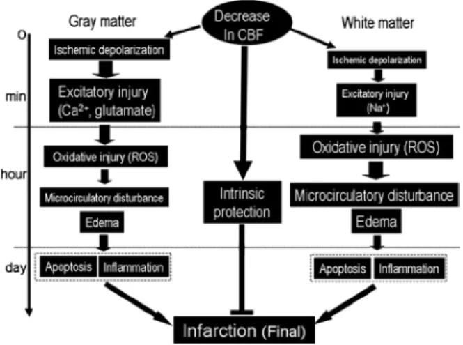
低灌流領域を過大評価する傾向も指摘され,今後の検討が必 要であろう6). 虚血性脳組織傷害のメカニズム(Fig. 2) 上記症例のように,超急性期に有効な血流回復がえられな かったばあいは,ペナンブラ領域では以下のメカニズムに よって不可逆性組織傷害に陥ってくる1)2).すなわち,虚血 発生直後から,虚血性脱分極,Ca イオンや Na イオンの細胞 Fig. 1 Diffusion-perfusion mismatch in a patient with acute cardioembolic infarction presenting NIHSS as 24.
MRI at 75 min after onset showed a limited positive area in DWI with extensive low flow area in ASL (arterial spin labeling) in the left cerebral hemisphere. FLAIR image did not show ant responsible lesion, whereas MRA revealed a marked decrease in blood flow in the territory of the left middle cerebral artery. Intravenous edaravone was given at 90 min after onset followed by intravenous t-PA at 110 min. At 24 hours after onset, NIHSS improved to 14, and DWI and FLAIR image showed a localized positive area, which was geometrically the same as the area detected by DWI at 75 min after onset. CBF (cerebral blood flow) SPECT revealed a marked recovery in blood flow with slight hyperperfusion in the left cerebral hemisphere except the DWI positive area. MRA showed a complete recovery of blood flow in the left middle cerebral artery.
脳梗塞急性期の病態と治療のターゲット 53:1161 内流入による興奮性細胞傷害,グルタミン酸放出,フリーラ ジカル反応,二次的微小循環障害,炎症反応,アポトーシス, DNA損傷,遺伝子発現変化という損傷メカニズムが,虚血 巣中心部から周囲組織に向かって急速に発現してくる.これ ら損傷メカニズムは灰白質と白質ではことなる点も多く,白 質ではグルタミン酸の関与はほとんどなく,フリーラジカル 反応が強いなど,部位的特異性も考慮する必要がある7). 脳梗塞急性期の治療のターゲット 脳梗塞の根本的治療ターゲットは単純明快である.できる だけ早く,脳血流量を脳の正常な機能維持に必要なレベルま で回復させることである.血栓溶解療法,血栓除去や血行再 建を目的とした血管内治療が第 1 の治療である.しかし,我 が国では,救急要請から病院到着まで平均 20 ~ 30 分かかっ ており,脳梗塞発症から治療開始までに虚血中心部ではすで に脳組織が不可逆性変化を示し始めている.虚血周辺部に存 在するペナンブラは早期に血流が回復しないばあいは,時間 と共に梗塞化し,神経障害は悪化固定化する. 血流再開や回復という根本的治療が成功しないと,脳実質 細胞の損傷のみならず,血管内皮・平滑筋の機能的・形態的 傷害も進行してくる.血管系の傷害は単に血栓の成長のみ ならず,出血性脳梗塞発生の基盤にもなる.脳実質細胞と 脳血管系を包括的に虚血性傷害から守るという立場から, Neurovascular unitの保護という戦略も必要である8).今後は 内因性保護因子の活性化など新しい治療法の開発も期待され る9). 以上より,脳梗塞急性期治療の基本戦略は,救済可能な領 域(ペナンブラ)の有無を迅速に画像診断した上で,脳血流 の超早期回復(血栓溶解除去),二次的微小循環障害抑制(抗 血栓療法)および側副血行の促進という血流にターゲットを 当てた治療と,Neurovascular unit 傷害の抑制(保護療法)の 二つからなる.具体的には,血栓溶解療法,フリーラジカル 除去療法,抗血小板療法・抗凝固療法,抗浮腫療法が虚血性 組織傷害機序に対応した治療となる. ※本論文に関連し,開示すべき COI 状態にある企業,組織,団体 はいずれも有りません. 文 献 1) 田中耕太郎.虚血性脳組織傷害の機序.田中耕太郎,高嶋 修太郎,編.必携脳卒中ハンドブック.東京:診断と治療社; 2008. p. 335-340. 2) 田中耕太郎.脳組織虚血性傷害メカニズムに基づく治療戦 略.田中耕太郎,編.脳卒中診療 Update 改訂第 2 版.大阪: 最新医学社;2010. p. 252-262.
3) Astrup J, Symon L, Branston NM, et al. Cortical evoked potential and extracellular K+ and H+ at critical levels of brain ischemia. Stroke 1977;8:51-57.
4) Janjua N. Use of neuroimaging to guide the treatment of patients beyond the 8-hour time window. Neurology 2012;79(13 Suppl 1):S95-S99.
5) Fisher M, Albers GW. Advanced imaging to extend the therapeutic time window of acute ischemic stroke. Ann Neurol 2013;73:4-9.
6) Zaharchuk G, El Mogy IS, Fischbein NJ, et al. Comparison of arterial spin labeling and bolus perfusion-weighted imaging for detecting mismatch in acute stroke. Stroke 2012;43:1843-1848. 7) 田中耕太郎.虚血性白質傷害の分子メカニズム.分子脳血
管病 2009;8:159-168.
8) Dirnagl U. Pathobiology of injury after stroke: the neurovascular unit and beyond. Ann N Y Acad Sci 2012;1268:21-25.
9) 田中耕太郎.内因性保護因子.Brain Medical 2010;22:83-92. Fig. 2 Time course of ischemic brain injury in the gray and the
white matters. ROS: reactive oxygen species.
臨床神経学 53 巻 11 号(2013:11) 53:1162
Abstract
Pathophysiology of brain injury and targets of treatment in acute ischemic stroke
Kortaro Tanaka, M.D., Ph.D.
1)1)Department of Neurology, Toyama University Hospital