CARBON NANOTUBES AS A THERAGNOSTIC TOOL
AGAINST BREAST CANCER(和訳:乳癌の診断・治療
のためのカーボンナノチューブの開発)
著者
PRASHANTI JEYAMOHAN
学位授与大学
東洋大学
取得学位
博士
学位の分野
生命科学
報告番号
32663甲第351号
学位授与年月日
2013-09-25
URL
http://id.nii.ac.jp/1060/00006459/
CARBON NANOTUBES AS A THERAGNOSTIC
TOOL AGAINST BREAST CANCER
Prashanti Jeyamohan
4R10101003
Doctor Course
Bio-Nano Science Fusion Course
Graduate School of Interdisciplinary New Science
Toyo University, Japan
Preface
The new field of nanotechnology, has opened up rapid advances in science and technology, and creates myriad new opportunities for advances in medical science and disease treatment in human health care. Since adequate cancer therapeutics remains elusive, it is required to develop new methods for efficient cancer diagnosis and therapy. Nanoparticle-mediated site targeted drug delivery offers an attractive option to enhance the therapeutic efficacy by delivering optimal drug concentrations to the targeted cancer cells, and with reduced toxicity to the healthy tissues. The integration of nano-materials into cancer therapeutics provides a way of smart drug delivery with high target specificity and greater therapeutic efficacy. Carbon nanotubes (CNTs), due to their unique intrinsic physiochemical properties are extensively explored as novel drug delivery systems for therapy and diagnosis.
In this thesis entitled “Carbon Nanotubes as a Theragnostic Tool Against
Breast Cancer” comprises of seven chapters and mainly focuses on the use of carbon
nanotubes as potential biomedical materials, which show great potential as a targeted and effective drug delivery system for cancer therapy, by grafting it with cell-specific receptors and intracellular targeting molecules.
Chapter 1 explains the scope of nanotechnology in developing target specific
carriers to achieve higher therapeutic efficacy, which will significantly contribute to the next generation of medical care and pharmaceuticals in the areas of disease diagnosis, disease prevention and most importantly cancer therapy. This review introduces specific physical and chemical properties of a series of nano-materials, such as nanoparticles, micelles, dendrimers, and with more emphasis on carbon nanotubes, which show great potential as effective drug delivery systems for cancer therapy. In addition to the ability of nanotubes to act as carriers for a wide range of
therapeutic molecules, they are also exploited for the use in photo-thermal destruction of cancer cells. Specifically, this article focuses on the current status, recent advancements, potentials and limitations of functionalized carbon nanotubes as targeted drug delivery carriers for efficient cancer nano-therapeutics.
Chapter 2 discusses the principles behind various instruments and techniques
used for characterization of nano-sized structures, and for analysis of biological samples used in our research work.
Chapter 3 explains about a targeted drug delivery system (DDS) based on
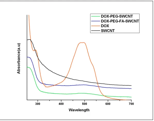
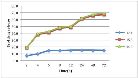
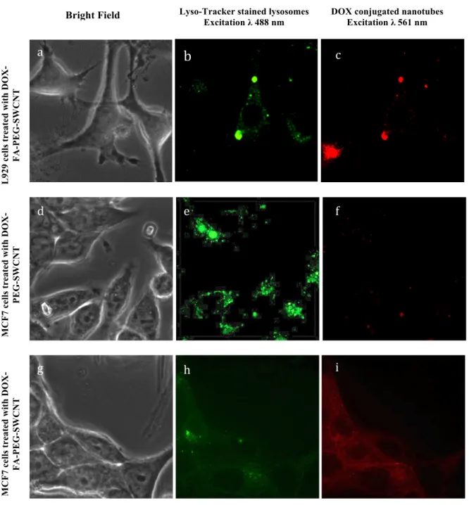
SWCNTs biofunctionalized with polyethylene glycol (PEG), conjugated with folic acid (FA) as targeting moiety and loaded with anticancer drug doxorubicin (DOX) for selective killing of tumor cells. The prepared nanotubes were characterized for their morphological features and their elemental composition. In this study, in vitro drug release analysis showed that the drug (DOX) binds at physiological pH (pH 7.4) and is released at a lower pH (lysosomal pH 4.0), which is the characteristic pH of the tumor environment. A sustained release of DOX from the SWCNTs was observed for a period of three days, which effectively caused the death of the breast cancer cells. The nanotubes were incubated with breast cancer cells and their biocompatibility and anti-proliferative effects were analyzed. We found that the targeted nanotubes were non-toxic to normal cells but toxic to tumor cells. Cellular uptake of the targeted nanotubes was studied in vitro using confocal microscopy. Time dependent drug effect was also studied on normal cells and tumor cells. Our results show significant internalization and retention of the nanotubes inside the tumor cells, inducing apoptosis. The results indicate that this study with folate conjugated and DOX loaded nanotubes can be a promising targeted delivery vehicle for cancer therapy.
Chapter 4 demonstrates that SWCNTs have a strong optical absorbance in the
near-infrared (NIR) region. An approach utilizing the photo-thermal effect of single-walled carbon nanotubes (SWCNTs), in combination with folate as a targeting moiety and DOX an anticancer drug doxorubicin, for selective and accelerated destruction of breast cancer cells is demonstrated. The results of our study show that, under laser irradiation at 800 nm, SWCNTs showed strong light-heat transfer characteristics. These optical properties of SWCNTs provide an opportunity for photo-thermal ablation in cancer treatment. Our observation also showed that the internalization and uptake of folate conjugated nanotubes into cancer cells was by a receptor mediated endocytosis mechanism. In our in vitro experiments, laser effectively caused destruction of cancer cells while sparing the normal cells. When the above laser effect was combined with DOX conjugated SWCNTs, an enhanced and accelerated killing of breast cancer cells was noted. Thus, this targeting nano drug delivery system (NDDS) could effectively kill and suppress the growth of breast cancer cells, showing much higher pharmaceutical efficiency compared to free DOX. This study suggests that Laser + drug + SWCNTs could prove to be a promising selective modality with high treatment efficacy and low side effects for future cancer therapy.
In Chapter 5 we present a dual drug delivery system consisting of PEG modified SWCNTs conjugated with FA and loaded with two anticancer drugs (DOX) and paclitaxel (PTX), forming DOX-PTX-FA-PEG-SWCNT conjugate. The combination of two or more therapeutic drugs is a feasible means to overcome the limitations of single chemotherapeutic drug in anti-tumor treatment, such as development of high toxicity, drug resistance, and limited regime of clinical uses. The prepared nanotubes were characterized for their morphological features and their elemental composition. Independent and combined drug release behaviors of the
drugs were studied. A time-dependent in vitro cytotoxicity studies were done for studying the cancer killing effect in comparison with free PTX and free DOX in breast cancer (MCF7) cells. A faster drug release was found to be associated with lower pH, which is advantageous for tumor targeted anticancer therapy. Confocal studies were carried out to demonstrate cellular uptake of the targeted nanotubes in tumor cells, while sparing the normal cells. This efficient suppression in tumor cell growth demonstrates a highly synergistic anti-proliferative activity of our developed system in breast cancer cells. The effect of this DDS in combination with laser was also analyzed, and enhanced destruction of breast cancer cells were observed. This nano-carrier might have important potential in clinical implications for co-delivery of multiple anti-tumor drugs with different properties.
Chapter 6 explains the application of another nano-carrier that could be
utilized as a drug delivery agent in cancer therapy. Here, an anticancer drug loaded poly-L-Lactic-co-glycolic acid (PLGA)-lecithin-PEG nanoparticles were synthesized and were functionalized with AS1411 anti-nucleolin aptamers for site-specific targeting against tumor cells, which over expresses nucleolin receptors. The results of this study confirmed that the aptamer-labeled PLGA-lecithin-PEG nanoparticles are potential carrier candidates for differential targeted drug delivery.
Chapter 7 concludes the thesis with the summary and future prospects of this
research work.
The need to develop target specific carriers to achieve higher therapeutic efficacy with minimal side effects to healthy tissues, is one of the most interesting and challenging endeavors faced in the pharmaceutical field.
Contents
Chapter 1Opportunities and challenges of single walled carbon nanotubes in cancer therapy
Abstract 1
Introduction 3
Types of nanotechnology based nano-carrier systems 4
Polymeric nanoparticles 5
Micelles 6
Dendrimers 8
Carbon nanotubes 8
Functionalization of carbon nanotubes for drug delivery and other
applications 10
CNTs as carriers for drugs, genes and proteins in cancer therapy 11
Drug delivery by CNTs 11
CNTs as carriers of immuno-active compounds, proteins and genetic
materials 15
CNT mediated photo-thermal therapy of cancer 18
CNT mediated photo-dynamic therapy 19
CNTs for other therapeutic applications 20
In vitro and in vivo toxicity of CNTs 20
Conclusion 22 References 24 Chapter 2 Instrumentation Abstract 35 Introduction 37 Microscopic techniques 37
Transmission electron microscope (TEM) 37
Scanning electron microscope (SEM) 39
Atomic force microscope (AFM) 41
Confocal laser scanning microscope (CLSM) 42
Phase contrast microscope 44
Spectroscopic techniques 45
UV-Vis spectroscopy 45
X-Ray photoelectron spectroscopy (XPS) 46
Other instrumentation techniques 48
Zetasizer 48
Particle size analyser 49
Conclusion 50
Chapter 3
Targeted drug delivery using PEG biofunctionalised folate conjugated SWCNTs against breast cancer cells
Abstract 51
Introduction 53
Purification of SWCNTs 55
Synthesis of PEGlyated SWCNTs 55
DOX loading onto the PEGlyated SWCNTs 56
Characterization of the modified nanotubes 56
Drug loading efficiency analysis 57 In vitro drug release studies 58
Cell culture studies 59
Biocompatibility studies of SWCNTs 59 In vitro cytotoxicity studies 60
Confocal microscopy 61
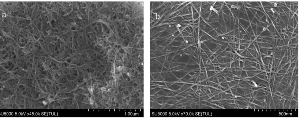
Results and Discussions 61 Nanotube characterization analysis 61
DOX loading onto the PEGlyated nanotubes 65 Drug loading efficiency studies 66 In vitro drug release studies 66
Biocompatibility studies 68 Confocal studies 71 In vitro cytotoxicity studies 72
Conclusion 75
References 77
Chapter 4 Accelerated killing of cancer cells using multifunctional SWCNTs based system for targeted drug delivery in combination with photo-thermal therapy Abstract 81 Introduction 83
Experimental details 86
Preparation and characterization of DOX loaded SWCNTs 87
Synthesis of fluorescent SWCNTs 87
Laser measurements 87
Cell culture 87
Selective internalization of SWCNTs into cancer cells 88
Cancer destruction using the NIR effect of SWCNTs 89
In vitro cytotoxicity assays of nanotubes under laser irradiation 89
Results and Discussions 90
Characterization of fluorescent SWCNTs 90
Temperature measurements during NIR irradiation 92
Selective internalization of SWCNTs into cancer cells 94
Cancer destruction using the NIR effect of SWCNTs 95
Conclusion 102
References 103
Chapter 5 Co-delivery of dual drugs using multifunctional carbon nanotubes for cancer therapy Abstract 107 Introduction 109 Experimental details 111 Purification of SWCNTs 111 Preparation of PEGylated SWCNTs 112
Formulation of drug loaded PEGylated SWCNTs 112
Particle characterization studies 113
Drug loading and in vitro release studies 114
Cell culture studies 115
Cellular imaging studies 115
In vitro cell viability studies 116
Cancer destruction using the NIR effect of SWCNTs 117
Cytotoxicity assays of nanotubes under laser irradiation in in vitro studies 117
Results and Discussions 117
Particle characterization and analysis 118
Drug loading and drug release studies 120
Cellular imaging studies 124
In vitro cell viability studies 124
Cancer destruction using the NIR effect of SWCNTs 126
Conclusion 131
References 133
Chapter 6 Nano-carriers for therapeutic applications Introduction 137
AS1411aptamer tagged PLGA-lecithin-PEG nanoparticles for tumor cell targeting and drug delivery 137
Chapter 7 Conclusion and Future Scope 139
Acknowledgement 143
Publications 147
Conferences 149
Chapter 1
Opportunities and challenges of single walled carbon nanotubes in
cancer therapy
Abstract
The scope of nanotechnology to develop target specific carriers to achieve higher therapeutic efficacy is gaining importance in the pharmaceutical and other industries. Specially, the emergence of nanohybrid materials is posed to edge over chemotherapy and radiation therapy as cancer therapeutics. This is primarily because nanohybrid materials engage controlled production parameters in the making of nanoparticles with specific size, shape and other essential properties. It is widely expressed that these materials will significantly contribute to the next generation of medical care technology and pharmaceuticals in areas of disease diagnosis, disease prevention and most importantly cancer therapy. This review firstly introduces the specific physical and chemical properties of a series of nanomaterials, such as nanoparticles, micelles, dendrimers and carbon nanotubes. It then places great emphasis on the carbon nanotubes, which show great potential as effective drug delivery systems for cancer therapy, as they can be grafted with cell-specific receptors and intracellular targeting molecules for the targeted delivery of therapeutics. In addition to the ability of nanotubes to act as carriers for a wide range of therapeutic molecules, they have also been exploited for use in photothermal destruction of cancer cells. Specifically, this article focuses on the current status, recent advancements, potentials and limitations of functionalized carbon nanotubes as targeted drug delivery carriers for efficient cancer treatment.
Introduction
Currently available technologies have made enormous advancement in cancer research, but an adequate therapy remains elusive.1 Cancer occurs at a molecular level when multiple subsets of genes undergo genetic alterations, either by activation of oncogenes or inactivation of tumor suppressor genes, allowing cancerous cells to grow and divide uncontrollably.2 These processes allow cancer cells to acquire
properties of malignant proliferations, tissue infiltrations through interactions with surrounding stromal cells, dysfunction of organs by the evasion of immune detection, self-sufficiency in growth signals and resistance to both anti-proliferative and apoptotic agents.3, 4 Also, in vivo delivery of the therapeutic agents has to overcome physiological barriers, including hepatic and renal clearance, enzymolysis and hydrolysis, as well as endosomal/lysosomal degradation.5, 6 In addition, the efficiency of anticancer drugs is limited by their unsatisfactory properties, such as poor solubility, narrow therapeutic window, and intensive cytotoxicity to normal tissues, which may be the causes of treatment failure in cancer.7 Therefore, it would be desirable to develop new methods to detect cancers at their early stage and also design novel therapeutic strategies capable of delivering chemical agents and other therapeutic materials specifically to tumor locations without affecting healthy tissues.8, 9, 10, 11 With the emerging field of nanotechnology, the integration of nanomaterial’s into cancer therapeutics is one of the rapidly growing fields in the development of nanoparticles, nanostructured surfaces and platforms for rapid and early diagnostics, smart drug delivery and for understanding the therapeutic efficacy.12, 13 Nanomaterials have sizes ranging from about one nanometer up to several hundred nanometers, Materials in this size range exhibit interesting physical
properties, presenting new directions for more effective diagnosis and therapy of cancer.14, 15
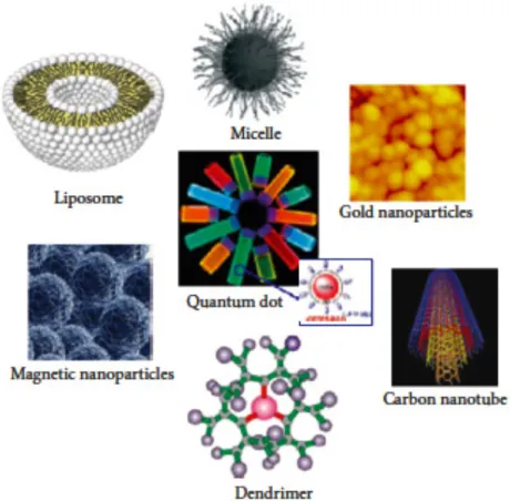
Types of nanotechnology based nanocarrier systems
Nanocarriers applied as drug delivery systems are synthesized from organic and inorganic materials such as polymeric nanoparticles, micelles, dendrimers, lipid nanoparticles, carbon nanotubes, quantum dots, and nanofibers16, 17 (Figure 1). They
show great potential in cancer therapy by enhancing the performance of medicine and reducing side effects to healthy tissues for improved therapeutic efficiency.
Polymeric nanoparticles
Polymeric nanoparticles are particles less than 1µm in diameter, synthesized from natural or synthetic polymers. Depending on the methods of preparation, matrix-type or reservoir-type structure, namely nanospheres or nanocapsules can be obtained.18 Natural polymers such as albumin, chitosan, heparin and synthetic polymers such as N-(2-hydroxypropyl)-methacrylamide copolymer (HPMA), polystyrene-maleic anhydride copolymer, polyethylene glycol (PEG), and poly-L-glutamic acid (PGA) have been material of choice for polymeric nanoparticles.19, 20, 21 They are considered as the promising carriers for drug delivery because they can improve the specificity of drug action by changing their tissue distribution and paharmacokinetics.22 They have also played important roles in delivering antitumor drugs in a targeted manner to the malignant tumor cells, thereby reducing the systemic toxicity and increasing their therapeutic efficacy. These nanoparticles due to the reticulo-endothelial system (RES) and the effect of enhanced permeation and retention (EPR) effect, can be used for passive delivery to the lymphatic system, brain, arterial walls, lungs, liver, spleen or for long-term systemic circulation.3, 22, 23 These nanoparticles are also amendable to
surface functionalization for active targeting to tumor tissues or cells and for stimulus-responsive controlled release of drug.24
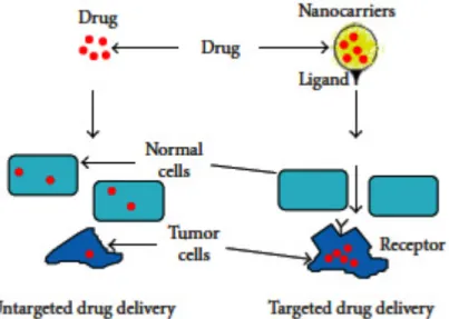
The receptor-mediated endocytosis (RME) reveals the selective recognition, high-affinity binding, and immediate internalization for the ligands at a cellular level.25 Various targeting moieties can be attached to the polymer backbone, which acts as a secondary uptake mechanism following EPR-based primary accumulation (Figure 2) .4, 26 Liang et al. developed paclitaxel-loaded nanoparticles with galactosamine conjugated on it for targeting liver cancer cells. The nanoparticles appeared most efficient in reducing the size of the tumor when injected into hepatoma-bearing nude
mice, through the specific interaction between galactosamine and asialoglycoprotein receptors.27 Thermosensitive magnetoliposomes (TMs) encapsulated with methotrexate (MTX) can achieve a good magnetic targeting effect and rapid drug release in response hyperthermia, which implies their great potential in cancer therapy.28
Micelles
Spontaneous assembly usually forms polymeric micelles into core-shell structures when its concentration is above critical micelle concentration (CMC). They have a number of unique features, including nanosize, easy manipulation of surface chemistry, core functionalities, as well as ease of fabrication, making them suitable carriers for encapsulation, and delivery of water insoluble agents.5 The hydrophobic
solid-like inner core region serves as a reservoir for hydrophobic drugs, whereas the hydrophilic shell region stabilizes the hydrophobic core and renders the polymers water-soluble.29 The hydrophilic shell helps the micelles in escaping the recognition of RES and prolongs the blood circulation of drugs. The small size (< 100 nm) allows the efficient accumulation of micelles in pathological tissues with permeabilized vasculature via the enhanced permeability and retention (EPR) effect.30 Mikhail presented a detailed review on the density and heterogeneity of the vasculature at tumor sites, interstitial fluid pressure, and found that transport of macromolecules in the tumor interstitium are responsible for the extravasation of micelles.31
Figure 2. Schematic of nanocarrier systems for site-targeted drug delivery. 25
Stimuli-responsive polymeric micelles are often designed for controlled release of drug into tumor tissue with external stimuli trigger, like temperature, pH, ultrasound and special enzymes.32, 33 Among these stimuli, pH and temperature are representative, because the external pH of cancerous tissue tends to be lower and the temperature is higher compared to the surrounding normal tissue, which are caused by abnormal metabolism of cancer tissues.5 Lower critical solution temperature (LCST) polymers, such as poly (N-isopropylacrylamide) (pNIPAAm) with a cloud point around 32°C or some other poly (N-alkylacrylamide) compounds, were investigated as components of temperature-responsive copolymer micelles.33 The micelles
exhibited rapid and temperature-responsive drug release in cancer cells, which was caused by the destruction of the hydrophobic-hydrophilic balance with the increase of temperature. Licciardi et al. synthesized novel folic acid- (FA-) functionalized diblock copolymer micelles for target delivery of antitumor drugs, and lowering the solution pH to 5 could trigger the rapid release of drug. This strategy combined the targeted delivery of therapeutics together with pH-controlled drug release, which provided a tumor-selective nanocarrier for the efficient delivery of anticancer drugs (Figure) .34
Dendrimers
Dendrimers are artificial macromolecules with tree-like structures in which the atoms are arranged in many branches and sub-branches radiating out from a central core.35, 36 They are synthesized from branched monomer units in a stepwise manner. Thus, able to control their molecular properties, such as size, shape, dimension, and polarity, which depend on the branched monomer units.36 They offer unique interfacial and
functional performance advantages due to their empty internal cavities and surface functional groups.37 So they have an enormous capacity for solubilization of hydrophobic drugs and can be modified or conjugated with various molecules. Based on these specific properties, the dendrimers have shown great development in anticancer drug delivery systems.36, 38 To actively target drugs to tumor tissues the
dendrimers are used for covalent attachment of special targeting moieties, such as sugar, folic acid, antibody, biotin, therapeutic drugs and epidermal growth factors.39,
34, 40, 36, 41 Choi and coworkers synthesized a fifth generation polyamidoamine (G5
PAMAM) dendrimers conjugated to fluorescein and folic acid, linked together by complementary DNA oligonucleotides to produce clustered molecules for targeting cancer cells that overexpress the high-affinity folate receptor. In vitro studies indicated that the DNA-linked dendrimer clusters could specifically bind to KB cells and could be used as imaging and therapeutics agents for cancer therapy.42
Carbon Nanotubes
Carbon nanotubes (CNT) are synthetic nanomaterials made from carbon and belong to the fullerene family. Carbon nanotubes are made up of thin sheets of benzene ring structured carbon rolled up into the shape of a seamless tubular structure. They can be classified into two general categories based on their structure: single-walled carbon nanotubes (SWCNTs) with a single cylindrical carbon wall having a diameter of 1-2
nm, and length ranging from 50 to several hundred nanometers. Multiwalled carbon nanotubes (MWCNTs) have multiple walled-cylinders nested within other cylinders with a diameter ranging from 5-100 nm.43 Figure 3 CNTs are generally produced by three major techniques: electric arc discharge, 44 laser ablation45 and thermal or plasma enhanced chemical vapor deposition (CVD) 46. These methods involve synthesis at high temperature and pressure in the presence of reaction catalysts, leading to fine structures of CNTs along with some impurities like graphite debris and catalytic particles. Various methods such as oxidation, acid treatment, chromatography, filtration and functionalization are involved in the purification.47 Compared to other nanomaterials, CNTs have unique physical and chemical properties such as high aspect ratio, ultrahigh surface area, light weight and distinct optical properties, such as, high absorption in the near-infrared (NIR) range and a strong Raman shift, which makes them advantageous for biomedical applications linked to detection and imaging.48 The combination of these characteristics makes CNT a unique nanomaterial with the potential for diverse applications, in areas of gene therapy, drug delivery, thermotherapy, imaging and anticancer treatments, as explained in the subsequent sections.25, 49
Figure 3. Structure of single-walled carbon nanotubes (SWCNTs) and multiwalled
carbon nanotubes (MWCNTs), as well as drug loaded carbon nanotubes (adapted from http://www-ibmc.u-strasbg.fr/ict/vectorisation/nanotubes eng.shtml and http://www-ibmc.u-strasbgfr/ict/vectorisation/vectorisation eng.shtml).
Functionalization of carbon nanotubes for drug delivery and other applications
CNTs are hydrophobic in nature and thus insoluble in water, which limits their application in biomedical and medicinal chemistry. Therefore, various functionalization methods like adsorption, electrostatic interaction and covalent bonding are being utilized with a number of compounds and polymers to render a hydrophilic character to CNTs so as to avoid aggregation and to facilitate their use in biomedical applications.1 Covalent modifications are carried out by reacting carbon atoms on the sidewall of carbon nanotubes to a therapeutic molecule. In biological applications, oxidation and grafting polymers on the sidewalls of carbon nanotubes are widely employed. During oxidation, pristine CNTs are refluxed in nitric acid, which results in oxygenated functional groups, mainly carboxyl acids.50, 51 Another
covalent modification of CNTs involves 1, 3-dipolar cycloaddition reactions.52 Using this method, azomethineylides are added on the graphite surface of CNTs, forming pyrrolidine-fused rings. These covalent modifications lead to the generation of various functional moieties on the ends and sidewall of CNTs and enable the conjugation of various fluorescent dyes, drugs, and peptides.53, 54 Other biocompatible
polymers like polyethylene glycol (PEG) can be grafted forming SWCNT-PEG graft copolymer that enhances the solubility of the nanotubes.55 Figure 4 Non-covalent functionalization of CNTs has also been explored with materials like surfactants, natural polymers, synthetic polyelectrolytes and amphiphilic block polymers or polymeric micelles. The hydrophobic moiety binds to the CNT sidewalls via π-π interactions, helping the CNTs to disperse in aqueous solutions. It was demonstrated that the non-covalent conjugation of pyrene with glycodendrimers via π-π stacking on the sidewall of CNTs, enhances their water solubility.56 Amphiphilic polymer phospholipid linked PEG (PL-PEG) was used to non-covalently bind to the sidewalls
of SWCNTs, giving rise to a number of biomedical applications.57
CNTs as carriers for drugs, genes and proteins in cancer therapy
Most of the research on CNTs has been focused on their potential for anticancer drug delivery. This might be attributed to the transporting capabilities of CNTs combined with appropriate surface modification and their unique physiochemical properties, which lead to the development of a new kind of nanomaterial for cancer therapy.11
Drug delivery by CNTs
Functionalized CNTs can cross the mammalian cell membrane by endocytosis or other mechanisms with the help of specific peptides or ligands on their surface to recognize cancer-specific receptors on the cell surface.58 Cancer cells overexpress folic acid (FA) receptors, and many research groups have designed CNT carriers with engineered surfaces to which FA derivatives can be attached. CNTs are used for targeting lymph node cancers, as they can be retained in the lymph nodes for longer periods of time compared to spherical nanocarriers.59-62 In a recent study, Yang et al.63 have loaded the anticancer molecule gemcitabine into magnetic MWCNTs and injected subcutaneously and, they reported high activity against lymph node metastasis in mice. In another study, the poorly water-soluble anticancer drug, camptothecin, has been loaded into polyvinyl alcohol-functionalized MWNTs and was reported to be potentially effective in the treatment of breast and skin cancers.64 In another approach, PAA-grafted MWCNTs were prepared and subcutaneously injected into the left rear footpad of rat.65 The biopsy result showed that left popliteal
lymph nodes were blacker than other regions, 24 h after the injection. Biopsy experiments did not identify the presence of PAA-g-MWCNTs in the major internal organs such as liver, kidney and lung, which suggests that the PAA-g-MWCNTs may
have the potential to be used as a vital staining dye and can be used for lymph node identification during surgery, even when they are very small.66
Dhar and coworkers developed the longboat delivery system, (Figure 5) in which cisplatin and FA conjugate was attached to a functionalized SWCNT via a number of amide bonds to form a “longboat” which is reported to be taken up by cancer cells via endocytosis, followed by the release of the drug and its subsequent interaction with the nuclear DNA. Another platinum anticancer drug, carboplatin, was incorporated into CNT and has been shown to inhibit the proliferation of urinary bladder cancer cells in vitro.67
Figure 4. Synthetic scheme of SWNT-PEG graft copolymers. The carboxylic acid
functionalized Single Wall Carbon Nanotubes (SWNT–COOH) react with oxalyl chloride to form the acylchloride intermediate, which is further reacted with PEG to form SWNT–PEG. 148
Figure 5. The “longboat” anticancer system in which one end of the
chemotherapeutic agent cisplatin is attached from to the FA derivative and the opposite end to a SWCNT via an amide link. 67
CNTs can carry therapeutic drugs more safely and effectively into the cells, which make them ideal candidates for drug delivery. Recently, novel SWCNT- based tumor-targeted drug delivery systems (DDS) have been developed. These delivery systems generally consist of three parts: functionalized SWCNTs, tumor targeting ligands and anticancer drugs. The complex is taken up efficiently and specifically by cancer cells with subsequent intracellular release of anticancer drugs, which suppressed cancer cell growth more effectively than untargeted controls containing the same drug.68 Hence, with this novel approach, serious and harmful side effects to healthy tissues can be avoided. Paclitaxel is a poorly water-soluble anticancer drug. In the commercialized paclitaxel product (Taxol), Cremophor EL is used to solubilize the drug. Unfortunately, Cremophor EL itself is toxic. Therefore, finding a suitable alternative is of high priority. Moreover, the circulation time of Taxol is very short. Coating the nanocarriers with hydrophilic polymers such as polyethylene glycol (PEG) has been established as a strategy to prolong the circulation of the nanocarrier in blood by making the carrier highly evasive to the uptake by the macrophages in blood.69, 70 PEGylation of paclitaxel increases the circulation time in the blood over Taxol.71 In the research performed by Liu Z. et al., 72 functionalized SWNTs conjugated with paclitaxel through branched PEG chains via a cleavable ester bond was synthesized. This resultant complex was more effective in suppressing tumor growth in vivo than Taxol or paclitaxel-PEG conjugate in a 4T1 breast cancer animal model. PEGylation of the nanotubes is able to prolong the circulation time and greatly enhance cellular uptake of the drug by the cancer cells. Similar findings of anti- cancer activity have been reported when paclitaxel was loaded into PEGylated SWCNTs or MWCNTs using HeLa and MCF-7 cancer cell lines.73, 74
Multidrug resistance is a significant obstacle to successful anticancer drug therapy since the P-glycoprotein transporter can interfere with the accumulation of anticancer drugs in the target cells, resulting in reduced effectiveness of therapy.75, 76 Recently, confocal microscopy has shown the accumulation of PEGylated MWCNTs in multidrug resistant hepatoma cell lines.77 Liu and coworkers reported that although PEGylation of SWCNTs can prolong blood circulation time of the associated anticancer drug, it may cause an accumulation of the nanotubes in the dermal tissues of mice, suggesting that the extend of PEG coating require optimization.78
Biotin-functionalized SWCNTs conjugated with the anticancer agent taxoid using a cleavable linker have also been designed. The drug is transported via endocytosis, released in the cell and interacts with microtubules as evaluated by flow cytometry. This resulted in the formation of a stable microtubule-taxoid complex, which finally caused apoptosis and cell death.74
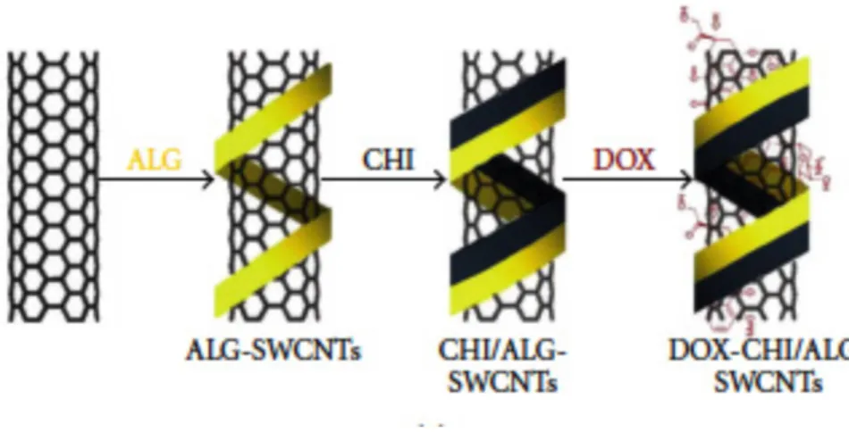
A targeted delivery system of FA-tethered SWCNTs-doxorubicin (DOX) has been designed. Bioadhesive polymers such as chitosan (CHI) and sodium alginate (ALG) were used to enhance the aqueous dispersibility of the nanotubes, while FA was used to improve the targeting properties of the nanotubes (Figure 6). Transmission electron microscopy (TEM) indicated that the drug was released at the low lysosomal pH of the HeLa tumor cells after being transported into the tumor but not at the normal physiological pH of 7.4.58 Recently, a novel approach by covalently attaching PAMAM dendrimers to FA-treated MWCNTs has been introduced and flow cytometry and confocal microscopy indicated possible targeting of cancer cells that overexpress FA receptors.79 Li and coworkers designed a novel system referred to as “dual-targeted drug nanocarrier” by conjugating MWCNTs with iron nanoparticles and folate moiety. This system was efficiently loaded with doxorubicin and
demonstrated superior delivery to HeLa cells when compared to free doxorubicin.80 A
list of CNT-based tumor-targeted drug delivery systems (DDS) is given in Table 1.
CNTs as carriers of immunoactive compounds, proteins, and genetic materials
Gene therapy aims to use genetic material to treat diseased cells by repairing the cause of the disease. Since genetic materials are not able to cross the biological membranes, the use of viral or nonviral vectors to carry the gene and internalize it into the cell is necessary. Nonviral vectors are less efficient than viral vectors81 and short lived, 82 but they are far safer.83, 84 Pantarotto and coworkers developed novel functionalized SWCNT-DNA complex and reported high DNA expression compared with naked DNA.85
Figure 7. Preparation of SWNTs-DOX after inclusion of bioadhesive
Table 1. CNT-based tumor- targeted drug delivery systems (DDS)
Functionalized SWCNTs have been used as suitable non-viral carriers of macromolecules and internalization of such macromolecules into living cells by CNTs has been reported to take place via energy-dependent endocytosis.86 Confocal microscopy and flow cytometry results have shown much greater fluorescent activity of protein and DNA when conjugated to SWNTs as compared to the naked macromolecules indicating that CNTs are promising vectors for gene and protein.87
Cai and coworkers introduced an approach to gene delivery named as “carbon nanotube spearing”.88 Plasmid DNA with a fluorescent protein were immobilized onto nickel-embedded CNTs and was “speared” into Bal 17 B lymphoma cells using a magnetic field, which produced high transfection in the target cells.88 In another study, HeLa cells were exposed to DNA attached SWCNTs at 37°C which resulted in gene internalization. However, at lower temperature (e.g., 4°C), no internalization took place, possibly because gene transport occurs by energy-dependent endocytosis.87
Gene silencing, a recent strategy in gene therapy, involves the use of small interfering RNA (siRNA) in the treatment of many diseases, including various cancers. In cancer, antitumor immunity might be inhibited by suppression of cytokine signaling 1 (SOCS1). siRNA can be conjugated to phospholipid-functionalized SWCNTs using a cleavable disulfide linker, resulting in efficient gene silencing and subsequent death
Reference CNTs Drug Tumor-targeted modules Tumor Chen et al. [74] SWNTs Taxoid Biotin and a spaces Leukemia Heister et al. [149] SWNTs Doxorubicin A monoclonal antibody Colon cancer Bhirde et al. [150] SWNTs Cisplatin Epidermal growth factor Squamous carcinoma Dhar et al. [67] SWNTs Cisplatin Folate
Nasopharyngeal epidermoid carcinoma etc.
Liu et al. [73] SWNTs Doxorubicin Rgd
Breast cancer, Glioblastoma Zhang et al. [58] SWNTs Doxorubicin Folate Cervical carcinoma Mc Devitt et al. [151] SWNTs Radionuclide Thiolated-antibody Burkitt lymphoma
of the targeted cell.89 In another study, amino-functionalized MWNTs-siRNA
complexes have shown successful suppression of tumor and prolonged survival in lung tumor of an animal model.90 Functionalized SWCNTs have been designed as carrier for siRNA for internalization into K562 cells and subsequent inhibition in the production of cyclinA(2) and treatment of chronic myelogenous leukemia. Many types of cancer (e.g., leukemia) can overexpress cyclinA(2), and suppression of this material using siRNA-CNTs can promote apoptosis in the targeted tumor.91 In another finding, functionalized SWNTs have been conjugated to telomerase reverse transcriptase (TERT) siRNA which successfully silenced the target gene, and tumor growth was inhibited in vitro using murine tumor cell lines and in vivo using a mouse model.92 Similarly in another study CNTs cationically functionalized with
polyethylene imines have been shown capable of complexing with siRNA and generating a silencing activity of up to 30% and cytotoxicity of up to 60%.93
Streptavidin is a protein that has anticancer activity.94 However, due to its very large molecular weight, it does not penetrate the cells. Kam et al.95 used a conjugate of streptavidin with SWCNT-biotin, which resulted in internalization of the protein into model cancer cells by adsorption-mediated endocytosis. Transmission electron and confocal microscopy have shown that MWCNTs can act as transporters of the recombinant ricin A chain protein, resulting in high death rates of cancer cells.96 Immunotherapy may be an alternative to gene therapy in cancer treatment. Antitumor immunotherapy using CNTs has gained interest among researchers. Tumor-specific monoclonal antibodies, radiometal ion chelates, and fluorescent probe have been conjugated to SWCNTs and successful targeting to tumor has been reported.97 The antitumor immune response was increased when MWCNTs were conjugated to tumor lysate protein as an antigen.98
Glioma is a brain tumor that is able to evade the host immune system.99, 100
Macrophages have a preferential affinity towards CNTs when compared to glioma cells.101 Van Handel and coworkers102 developed an immunotherapy approach using MWNTs against GL261 murine intracranial glioma cancer model, based on the fact that macrophages prefer to engulf CNTs compared with glioma cells. An increase in the influx of macrophages into the glioma cells with MWNTs was reported. Also, an increase in the levels of IL-10 expression was observed, suggesting that immunomodulation using CNTs is a possible strategy to treat cancer.
Reduction in the tumor volume and prolonged survival in animal models was reported when angiogenesis targeting antibodies E4G10 were attached to SWNTs via radiometal ion chelates.103 Oxidized MWCNTs has been reported to retard tumor
growth when injected subcutaneously to a hepatocarcinoma bearing animal to induce an immune response.104 This suggests that CNTs themselves could possibly be surface-engineered to have anticancer activity by inducing an immune response against tumor.
A major hurdle to effective anticancer therapy is the multidrug resistance caused by enhanced efflux of anticancer drugs by the overexpressed p-glycoprotein, resulting in poor anticancer effect.75, 76 Li et al. reported that SWCNTs functionalized with p-glycoprotein antibodies and loaded with the anticancer drug doxorubicin demonstrated higher cytotoxicity against K562R leukemia cells compared with free doxorubicin.105
CNT mediated photothermal therapy of cancer
CNTs are also welcomed in the field of thermal therapy and considered to be a non-invasive, harmless and highly efficient technique.106 CNTs are able to absorb light in
the near infrared (NIR) region, resulting in heating of the nanotubes. This unique property of CNTs has been exploited as a method to kill cancer cells via thermal effects.86, 107-119 According to Gannon et al.107 The incubation of the nanotubes functionalized using kentera (a polyphenylene ethynylene-based polymer) were studied using hepatic tumor cells followed by application of radiofrequency field, which demonstrated a concentration-dependent thermal destruction and complete necrosis of the tumor cells. By contrast, tumor cells that were injected with the Kentera alone (without CNTs) were viable after the application of the radiofrequency field. Both in vitro and in vivo tests have shown thermal destruction of cancer cells in the presence of SWNTs with exposure to radiofrequency. A formulation based on FA-PL-PEG functionalized SWCNTs was used for targeting cancer cells, which over-express folate receptors.109 Selective cancer cell destruction was achieved by continuous NIR radiation of SWCNTs. The NIR triggered cell death has been shown to remain nontoxic for neighboring normal cells. 86 This research presented a novel application of CNTs in cancer therapy. Another study utilizing the NIR characteristic of CNTs involved the destruction of breast cancer cells by non-covalently attaching monoclonal antibodies against membrane markers: insulin like growth factor 1 receptor (IGF1R) and human endothelial receptor 2 (HER2). The antibodies were attached using π-π interactions of pyrene rings onto the sidewalls of SWCNTs.120
CNTs mediated photodynamic therapy (PDT)
PDT could potentially be an ideal cancer treatment therapy capable of efficiently destroying tumors while at the same time sensitizing the immune system to seek out and destroy metastases.121 Singlet oxygen is one of the most important agents generated during PDT, which can react rapidly with cellular molecules and mediate cellular toxicity to cause cell damage, ultimately leading to cell death. However the
lifetime and diffusion distance of singlet oxygen are very limited. Zhu et al.122
synthesized a novel molecular complex photosensitizer, an ssDNA aptamer, and SWCNTs, which were able to control and regulate singlet oxygen generation, whereby more efficient, reliable and selective PDT could be guaranteed.
CNTs for other therapeutic applications
The use of CNTs in therapeutic applications other than cancer is also developing. Surface-engineered CNTs may be able to capture pathogenic bacteria in liquid medium.123-125 Thus, CNTs themselves might have antimicrobial activity since microorganisms may be adsorbed onto the surfaces of CNTs. It has been reported that the electronic properties of SWNTs may regulate the antibacterial activity in E. coli. The antibacterial effect was attributed to CNT induced oxidation of the intracellular antioxidant glutathione, resulting in increased oxidative stress on the bacterial cells and eventual death.126 Functionalized CNTs can attach covalently to amphotericin B and transport it into mammalian cells, thus demonstrating their ability to act as carriers for antimicrobial agents.127, 128
In vitro and in vivo toxicity of CNTs
Although there are exciting prospects for the application of CNTs in medicine, concerns over adverse and unanticipated effects on human health have also been raised. So far, many studies have been performed to evaluate the toxicological effects of CNTs both in vitro and in vivo.11 The CNT cytotoxicity was attributed to variability in the doses, physical form like length and type of CNTs, 129 effect of metal
catalyst impurities, 130 degrees of functionalization and dispersion of CNTs.131 Metal catalysts are the main source of cytotoxicity in CNTs.132, 133 Iron, the most common catalyst for growing CNTs, may boost the free radical reactions in the living cells.134
Becker et al.135 proved that CNTs shorter than 189 +/- 17 nm have greater
cytotoxicity. Furthermore, CNTs are required to be hydrophilic as drug carriers. Therefore, the surface chemistry plays an important role in improving the biocompatibility of CNTs. Few reports have demonstrated significant reduction in the cytotoxicity of CNTs due to the high degree of functionalization on CNT side walls.130, 136, 137 Functionalized SWCNTs (f-SWCNTs) are much less toxic than
surfactant-stabilized SWCNTs.138 In a typical experiment, immunoregulatory cells (macrophages, B and T lymphocytes) were incubated in two types of amino group f-SWCNTs, one being highly soluble and another forming stable suspension in aqueous solution. The activities of the immunoregulatory cells are not influenced by the highly soluble CNTs, whereas proinflammatory cytokines are secreted by macrophages in the CNT suspension.139 Another group studied the influence of SWCNTs with different degrees of agglomeration on primary cultures derived from chicken embryonic spinal cord (SPC) or dorsal foot ganglia (DRG). 140 Significant decrease in DNA content was more pronounced when cells were exposed to highly agglomerated SWCNTs as compared with better-dispersed SWNT bundles.
Bottini et al.141 compared the toxicity of pristine and oxidized MWCNTs on human T cells and found that the MWCNTs were more toxic and induced massive loss of cell viability through programmed cell death.
Pulskamp et al.133 did not observe any acute toxicity on cell viability upon incubation with all CNT products. However, an increase in reactive oxygen species was observed due to the presence of metal particles as well as metalloids in commercial nanotubes, while treatment with a highly ultra-purified form of CNTs had no biological adverse effects.
intravenously exposed mice. No acute toxicity was reported by Zeni et al.143 on
administration of SWCNTs. Similarly, other reports show low or no cytotoxic effects due to exposure to CNTs. Huezko and Lange reported null risk of skin irritation and allergy on dermatological trials of CNTs.
Functionalized SWCNTs have been used as drug carriers with paclitaxel.143, 144 Paclitaxel, being insoluble in water, is currently clinically administered as a highly toxic formulation with Cremophor EL. However, using SWNTs as a carrier allowed for a prolonged circulation of the drug and a 10-fold higher uptake by tumor cells.144 The high uptake of SWNT complexes by the reticuloendothelial system may endanger liver and spleen as they may suffer toxic effects from SWCNT deposition and accumulation. One of the main problems faced with intravenous administration is the rapid clearance through blood, and another is the toxic effect to other organs and tissues.145 Another study tested the toxicity of intravenously administered functionalized SWCNTs in mice and demonstrated no evidence of toxicity. PEG-SWCNTs were observed to be gradually removed from the body following an intravenous administration in mouse models with no evidence of toxicity during the excretory process.146
Zhuang et al. used PEGylated SWNTs conjugated to RGD peptide that specifically binds to integrin αvβ3, expressed on the surface of numerous tumor cells. This
formulation was intravenously injected into U87MG tumor bearing mice. The RGD – bound SWCNTs showed a higher tumor uptake as compared to SWCNTs alone.147
Conclusion
Nano delivery systems hold great potential to overcome many of the present obstacles of drug delivery, of which carbon nanotubes have been proposed and actively
explored as multipurpose innovative carriers for drug delivery and diagnostic applications. They have many unique physical, mechanical and electronic properties, and these distinct and exceptional properties have made it possible to exploit CNTs as drug delivery systems for biomedical applications. CNTs are promising carriers of both small drug molecules as well as macromolecules such as genes and proteins. Functionalization of CNTs with an organic moiety has made possible their use in diagnostics for imaging as well as for targeting purposes, especially in cancer therapy. Nanotube drug delivery holds future promise for high treatment efficacy combined with minimal side effects for cancer therapy with low drug doses.
Even though CNTs are playing a larger and most promising role in the field of nanomedicine, more research is required to guarantee safety in drug delivery. Functionalized CNTs have been considered biocompatible and safe for drug and biomolecular delivery applications, as they are soluble in physiological media and nontoxic. They have shown no accumulation in the tissues and can be readily excreted through the renal route. Toxicity studies are critical to establish the full in vivo potential of CNTs for drug delivery before their actual application and marketing and in order to understand the toxic impact of the systemic delivery of carbon nanotubes in greater detail, more extensive toxicity, safety and efficacy studies on animal models and in humans over a longer time frame need to be performed. The effects of CNTs aggregation, size, length, functionalization, metal impurities and polymers safety require more thorough research. Functionalization of SWCNTs and its effects on aggregation and consequent genotoxicity also needs to be evaluated. Overall, the use of CNTs for delivery of drugs and biomolecules is a significant development in the field of therapeutic nanomedicine.
References
1. S Prakash, M Malhotra, W Shao, et al. Advanced Drug Delivery Reviews. 63 (2011) 1340-1351.
2. FH Sarkar, S Banerjee, YW Li. Toxicology and Applied Pharmacology. 224 (2007) 3, 326-336.
3. JD Byrne, T Betancourt, L Brannon-Peppas. Advanced Drug Delivery Reviews. 60 (2008) 15, 1615-1626.
4. AK Iyer, G Khaled, J Fang, H Maeda. Drug Discovery Today. 11 (2006) 17-18, 812-818.
5. N Wiradharma, Y Zhang, S Venkataraman, Jl Hedrick, YY Yang. Nano Today. 4 (2009) 4, 302-317.
6. L Jabr-Milane, LV Vlerken, H Devalapally, et al. Nano Today. 130 (2008) 2, 121-128.
7. M Pulkkinen, J Pikkarainen, T Wirth, et al. European Journal of Pharmaceutics and Biopharmaceutics. 70 (2008) 1, 66-74.
8. Y Fukumori, H Ichikawa. Advanced Powder Technology. 17 (2006) 1-28.
9. JK Vasir, V Labhasetwar. Advanced Drug Delivery Reviews. 59 (2007) 718-728. 10. AH Faraji, et al. Medicinal Chemistry. 17 (2009) 2950-2962.
11. S Ji, C Liu, B Zhang, et al. Biochimica et Biophysica Acta. 1806 (2010) 29-35. 12. KK Jain. Clinica Chimica Acta. 358 (2005) 1-2, 37-54.
13. W Qiao, B Wang, Y Wang, et al. Journal of Nanomaterials. (2010) 796303. 14. TC Yih, C Wei. Nanomedicine: Nanotechnology, Biology, and Medicine. (2005) 191-192.
15. I Brigger, C Dubernet, P Couvreur. Advanced Drug Delivery Reviews. 54 (2002) 631-651.
16. W Cai, AR Hsu, ZB Li, X Chen. Nanoscale Research Letters. 2 (2007) 6, 265-281.
17. W Cai, X Chen. Small. 3 (2007) 11, 1840-1854.
18. AK Bajpai, SK Shukla, S Bhanu, S Kankane. Progress in Polymer Science. 33(2008) 11, 1088-1118.
19. C Li. Advanced Drug Delivery Reviews. (2002) 54, 695-713.
20. P Sabbatini, C Aghajanian, D Dizon, et al. Journal of Clinical Oncology. (2004) 22, 4523-31.
21. WJ Gradishar, S Tjulandin, N Davidson, et al. Journal of Clinical Oncology. (2005) 23, 7794-803.
22. C Fonseca, S Simoes, R Gaspar. Journal of Controlled Release. 83 (2002) 2, 273-286.
23. ML Hans, AM Lowman. Current Opinion in Solid State and Materials Science. 6 (2002) 4, 319-327.
24. N Sanvicens, MP Marco. Trends in Biotechnology. 26 (2008) 8, 425-433.
25. T Tanaka, S Shiramoto, M Miyashita, Y Fujishima, Y Kaneo. International Journal of Pharmaceutics. 277 (2004) 1-2, 39–61, 2004.
26. S Sandhiya, SA Dkhar, A Surendiran. Fundamental and Clinical Pharmacology. 23 (2009) 3, 263–269.
27. Y Fukumori, H Ichikawa. Advanced Powder Technology. 17 (2006) 1, 1–28. 28. M Johannsen, U Gneveckow, B Thiesen, et al. European Urology. 52 (2007) 6, 1653–1662.
29. ML Adams, A Lavasanifar, GS Kwon. Journal of Pharmaceutical Sciences. (2003) 92:1343-55.
30. LB Li, YB Tan. Journal of Colloid and Interface Science. 317 (2008) 1, 326–331. 31. AS Mikhail, C Allen. Journal of Controlled Release. 138 (2009) 3, 214–223. 32. GA Husseini, NY Rapoport, DA Christensen, et al. Colloids and Surfaces B. 24 (2002) 3-4, 253–264.
33. N Rapoport. Progress in Polymer Science. 32, 8-9, 962–990.
34. M Licciardi, G Giammona, J Du, et al. Polymer. 47 (2006) 9, 2946–2955.
35. KJ Morrow Jr, R Bawa, C Wei. Medical Clinics of North America. 91 (2007) 5, 805–843.
36. WJ Yang, YY Cheng, TW Xu, et al. European Journal of Medicinal Chemistry. 44 (2009) 2, 862–868.
37. S Svenson, DA Tomalia. Advanced Drug Delivery Reviews. 57 (2005) 15, 2106– 2129.
38. ER Gillies, JMJ Frechet. Drug Discovery Today. 10 (2005) 1, 35–43.
39. D Bhadra, AK Yadav, S Bhadra, NK Jain. International Journal of Pharmaceutics. 295 (2005) 1-2, 221–233.
40. AK Patri, A Myc, J Beals, et al. Bioconjugate Chemistry. 15 (2004) 6, 1174– 1181.
41. M Hussain, M Shchepinov, M Sohail, et al. Journal of Controlled Release. 99 (2004) 1, 139–155.
42. Y Choi, T Thomas, A Kotlyar, et al. Chemistry and Biology. 12 (2005) 1, 35–43. 43. F Liang, B Chen. Current Medicinal Chemistry. 17 (2010) 10-24.
44. DS Bethune, CH Klang, MS de Vries, et al. Nature. 363 (1993) 605-607. 45. A Thess, R Lee, P Nikolaev, et al. Science. 273 (1996) 483-487.
46. AM Cassel, JA Raymakers, J Kong, H Dai. Journal of Physical Chemistry B 103 (1999) 6484-6492.
47. T Park, S Banerjee, TH Bennya, SS Wong. Journal of Material Chemistry. 16 (2006) 141-154.
48. A Bianco, K Kostarelos, CD Partidos, M Prato. Chemical Communications. (2005) 571-577.
49. L Lacerda, A Bianco, M Prato, K Kostarelos. Advanced Drug Delivery Reviews. 58 (2006) 14, 1460–1470.
50. D Bonifazi, C Nacci, R Marega, et al. Nano Letters. 6 (2006) 1408-1414.
51. B Zhao, H Hu, A Yu, D Perea, et al. Journal of American Chemical Society. 127 (2005) 8197-8203.
52. EB Malarkey, RC Reyes, B Zhao, RC. Haddon, et al. Nano Letters. 8 (2008) 3538-3542.
53. EB Malarkey, RC Reyes, B Zhao, RC Haddon, et al. Nano letters. 9 (2009) 264-268.
54. CL Lay, HQ Liu, HR Tan, Y Liu. Nanotechnology. 21 (2010) 065101. 55. N Tagmatarchis, M Prato. Journal of Material Chemistry. 14 (2004) 437-439. 56. P Wu, X Chen, N Hu, et al. Angewandte Chemie International Edition. 47 (2008) 5022-5025.
57. Z Liu, K Chen, C Davis, et al. Cancer Research. 68 (2008) 6652-6660. 58. XK Zhang, LJ Meng, QH Lu, et al. Biomaterials. 30 (2009) 30, 6041-6047. 59. ST Reddy, A Rehor, HG Schmoekel, et al. Journal of Controlled Release. 112 (2006) 1, 26–34.
60. F Yang, DL Fu, J Long, and QX Ni. Medical Hypotheses. 70 (2008) 4, 765–767. 61. F Yang, J Hu, D Yang et al. Nanomedicine. 4 (2009) 3, 317–330.
62. Y Liu, KY Ng, KO Lillehei. Cancer Control. 10 (2003) 2, 138–147.
63. F Yang, C Jin, D Yang et al. European Journal of Cancer 47 (2011) 12, 1873– 1882.
64. NG Sahoo, H Bao, Y Pan et al. Chemical Communications 47 (2011) 18, 5235– 5237.
65. JJ Li, F Yang, GQ Guo, et al. Polymer International. 59 (2010) 169–174.
66. M Pramanik, KH Song, M Swierczewska, et al. Physics in Medicine and Biology. 54 (2009) 3291–3301.
67. S Dhar, Z Liu, J Thomale H Dai, SJ Lippard. Journal of the American Chemical Society. 130 (2008) 34, 11467–11476.
68. AA Bhirde, V Patel, J Gavard, et al. ASC Nano. 3 (2009) 307–316.
69. AL Klibanov, K Maruyama, VP Torchilin, L Huang. FEBS Letters. 268 (1990) 1, 235–237.
70. TM Allen, T Mehra, C Hansen, YC Chin. Cancer Research. 52 (1992) 9, 2431– 2439.
71. C Li, D Yu, T Inoue et al. Anti-Cancer Drugs. 7 (1996) 6, 642–648. 72. Z Liu, K Chen, C Davis, et al. Cancer Research. 68 (2008) 6652–6660. 73. Z Liu, XM Sun, NN Ratchford, et al. ACS Nano. 1 (2007) 50–56.
74. J Chen, S Chen, X Zhao, et al. Journal of the American Chemical Society. 130 (2008) 49, 16778–16785.
75. JY, W Chan, ACY Chu, et al. Life Sciences. 67 (2000) 17, 2117– 2124.
76. SS Suri, H Fenniri, B Singh. Journal of Occupational Medicine and Toxicology. 2 (2007) 1, 16.
77. J Cheng, MJ Meziani, YP Sun, and SH Cheng. Toxicology and Applied Pharmacology. 250 (2011) 2, 184–193.
78. X Liu, H Tao, K Yang, S Zhang, ST Lee, and Z Liu. Biomaterials. 32 (2011) 1, 144–151.
79. X Shi, HW Su, M Shen et al. Biomacromolecules. 10 (2009) 7, 1744–1750. 80. R Li, R Wu, L Zhao et al. Carbon. 49 (2011) 5, 1797–1805.
81. E Fortunati, A Bout, MA Zanta, D Valerio, et al. Biochimica et Biophysica Acta. 1306 (1996) 1, 55–62.
82. T Rochat, MA Morris. Journal of Aerosol Medicine. 15 (2002) 2, 229–235. 83. FD Ledley. Current Opinion in Biotechnology. 5 (1994) 6, 626–636. 84. C Coutelle R Williamson. Journal of Aerosol Medicine. 9 (1996) 1, 79–88. 85. D Pantarotto, R Singh, D McCarthy et al. Angewandte Chemie International Edition. 43 (2004) 39, 5242– 5246.
86. NWS Kam, M. O’Connell, JA Wisdom, and H Dai. Proceedings of the National Academy of Sciences of the United States of America. 102 (2005) 33, 11600–11605. 87. NWS Kam, Z Liu, and H Dai. Angewandte Chemie International Edition. 45 (2006) 4, 577–581.
88. D Cai, JM Mataraza, ZH Qin et al. Nature Methods. 2 (2005) 6, 449–454.
89. NWS Kam, Z Liu and H Dai. Journal of the American Chemical Society. 127 (2005) 36, 12492–12493.
90. E Podesta, KT Al-Jamal, MA Herrero et al. Small. 5 (2009) 10, 1176–1185. 91. X Wang, J Ren, and X Qu. Chem Med Chem. 3 (2008) 6, 940–945.
4939.
93. AK Varkouhi, S Foillard, T Lammers et al. International Journal of Pharmaceutics. 416 (2011) 2, 419–425.
94. SL Hussey and BR Peterson. Journal of the American Chemical Society. 124 (2002) 22, 6265–6273.
95. NWS Kam, TC Jessop, PA Wender, H Dai. Journal of the American Chemical Society 126 (2004) 22, 6850– 6851.
96. X Weng, M Wang, et al. Molecular BioSystems. 5 (2009) 10, 1224– 1231.
97. MR McDevitt, D Chattopadhyay, BJ Kappel et al. Journal of Nuclear Medicine. 48 (2007) 7, 1180–1189.
98. J Meng, J Duan, H Kong et al. Small. 4 (2008) 9, 1364–1370.
99. Y Liu, KY Ng, and KO Lillehei. Cancer Control. 10 (2003) 2, 138–147. 100. IF Parney, C Hao, and KC Petruk. Neurosurgery. 46 (2000) 4, 778– 792.
101. B Kateb, M Van Handel, L Zhang, MJ Bronikowski, H Manohara, and B Badie. NeuroImage. 37 (2007) 1, 9–17.
102. M VanHandel, D Alizadeh, L Zhang et al. Journal of Neuroimmunology. 208 (2009) 1-2, 3–9.
103. A Ruggiero, CH Villa, JP Holland et al. International Journal of Nanomedicine. 5 (2010) 1, 783–802.
104. J Meng, M Yang, F Jia et al. Nanotechnology. 21 (2010) 14, 145104.
105. R Li, R Wu, L Zhao, M Wu, L Yang, and H Zou. ACS Nano. 4 (2010) 3, 1399– 1408.
106. ME Brennan, JN Coleman, A Drury. Optics Letters. 28 (2003) 4, 266–268. 107. CJ Gannon, P Cherukuri, BI Yakobson et al. Cancer 110 (2007) 12, 2654–2665. 108. SV Torti, F Byrne, O Whelan et al. International Journal of Nanomedicine. 2
(2007) 4, 707–714.
109. F Zhou, D Xing, Z Ou, B Wu, DE Resasco, and W. R. Chen. Journal of Biomedical Optics. 14 (2009) 2, 021009.
110. NH Levi Polyachenko, EJ Merkel, BT Jones, et al. Molecular Pharmaceutics. 6 (2009) 4, 1092–1099.
111. CH Wang, YJ Huang, CW Chang, WM Hsu, CA Peng. Nanotechnology. 20 (2009) 31, 315101.
112. A Burke, X Ding, R Singh et al. Proceedings of the National Academy of Sciences of the United States of America. 106 (2009) 31, 12897–12902.
113. A Burlaka, S Lukin, S Prylutska et al. Experimental Oncology. 32 (2010) 1, 48– 50.
114. S Ghosh, S Dutta, E Gomes et al. ACS Nano. 3 (2009) 9, 2667–2673. 115. Y Xiao, X Gao, O Taratula et al. BMC Cancer. 9 (2009) 1471, 351. 116. HK Moon, SH Lee, and HC Choi. ACS Nano. 3 (2009) 11, 3707–3713.
117. N Huang, H Wang, J Zhao, H Lui, M Korbelik and H Zeng. Lasers in Surgery and Medicine. 42 (2010) 9, 638–648.
118. JD Jackson, Classical Electrodynamics, John Wiley & Sons, New York. 3 (1999).
119. J Xu, M Xiao, R Czerw, and DL Carroll. Chemical Physics Letters. 389 (2004) 4-6, 247–250.
120. N Shao, S Lu, E Wickstrom, B Panchapakesan. Nanotechnology. 18 (2007) 315101.
Photobiology. B: Biol. 96 (2009) 1–8.
122. Z Zhu, ZW Tang, JA Phillips. Journal of American Chemical Society. 130 (2008) 10856–10857.
123. L Gu, T Elkin, X Jiang et al. Chemical Communications. 7 (2005) 874–876. 124. ASB Esteyez, S Kang, M Elimelech. Small. 4 (2008) 4, 481–484.
125. LR Arias and L Yang. Langmuir. 25 (2009) 5, 3003–3012.
126. CD Vecitis, KR Zodrow, S Kang, M Elimelech, et al. ACS Nano. 4 (2010) 9, 5471–5479.
127. W Wu, S Wieckowski, G Pastorin et al. Angewandte Chemie International Edition. 44 (2005) 39, 6358–6362.
128. M Benincasa, S Pacor, W Wu, et al. ACS Nano. 5 (2011) 1, 199–208. 129. S Lanone, J Boczkowski. Current Molecular Medicine. (2006) 6, 651-666. 130. CM Sayes, F Liang, JL Hudsona. Toxicology Letters. 161 (2006) 135–142. 131. P Wick, P Manser, LK Imbach. Toxicology Letters. 168 (2007) 121–131.
132. VE Kagan, YY Tyurina, VA Tyurin, et al. Toxicology Letters. (2006), 165(1): 88-100.
133. K Pulskamp, S Diabata, HF Krug. Toxicology Letters. (2007) 168(1), 58-74. 134. FQ Schafer, SY Qian, GR Buettner. Molecular Cell Biology. (2000) 46(3): 657-62.
135. ML Becker, JA Fagan, ND Gallant, et al. Advance Material. (2007) 19(7): 939-45.
137. X Chen, UC Tam, JL Czlapinski, et al. Journal of American Chemical Society. (2006) 128(19): 6292-3.
138. H Dumortier, S Lacotte, G Pastorin, Marega et al. Nano Letters. (2006) 6(7): 1522-8.
139. LW Zhang, L Zeng, A Barron, et al. International Journal of Toxicology. (2007) 26(2): 103-13.
140. L Belyanskaya et al. NeuroToxicology. (2009) 30:702-711. 141. M Bottini et al. Toxicology Letters. (2006) 160, 121-126. 142. S Yang et al. Toxicology Letters. (2008) 181, 182-189 143. O Zeni et al. Sensor. (2008) 8, 488-499.
144. W Wu, R Li, X Bian, et al. ACS Nano. 3 (2009) 2740–2750.
145. ML Schipper, N Nakayama-Ratchford, CR Davis, et al. Nature Nanotechnology. 3 (2008) 216–221.
146. L Zhuang, C Davis, W Cai, et al. Proceedings of the National Academy of Sciences. 105 (2008) 1410–1415.
147. Z Liu, W Cai, L He, et al. Nature Nanotechnology. 2 (2007) 47–52.
148. CMJ Hu, S Kaushal, HST Cao, et al. Molecular Pharmacology. 7 (2010) 914– 920.
149. E Heister, V Neves, T Carmen, et al. Carbon. 47 (2009) 2152–2160 150. AA Bhirde, V Patel, J Gavard, et al. ACS Nano. 3 (2009) 307–316.
151. MR McDevitt, D Chattopadhyay, BJ Kappel1, et al. Journal of Nuclear Medicine. 48 (2007) 1180–1189.
Chapter 2
Instrumentation
Abstract
An understanding of the structure, surface morphology and elemental composition of the nanoparticles is essential to know the specific properties of the nano-materials under study. This chapter describes the principles of different instruments, which were employed for various characterizations of nanoparticles (NPs) and also analytical instruments used in biological studies. Various microscopic techniques, spectroscopic methods and other characterization techniques are being discussed in this chapter. In addition, the application of nanoparticles in vitro is also visualized with the help of various microscopic techniques. The techniques used for the characterization studies and nanoparticle applications are explained in this chapter.
Introduction
Proper characterization of nanomaterials is highly essential for their accurate analysis. Microscopic techniques and spectroscopic techniques are the common characterization methods for the characterization of various nanoparticles (NPs). Microscopic techniques including both optical and electron microscopes were used in our research. For studying the internal structure and surface morphology of nanoscale materials electron microscopic techniques are inevitable. NPs were characterized for their morphology using Transmission Electron Microscope (TEM), Scanning Electron Microscope (SEM) and Atomic Force Microscope (AFM). The elemental composition studies of the nano samples were carried out using X-ray Photoelectron Spectroscopy (XPS or ESCA), and UV-visible spectrophotometer (UV-Vis spectroscopy) was used to study the electronic excitations. Zetasizer has been used for measuring the zeta potential of the samples. We are discussing in detail about the general working principle and sample preparation of various electron microscopic methods, optical microscopic methods, spectroscopic techniques and other characterization techniques employed to analyze the NPs.
Microscopic techniques
Transmission Electron Microscope (TEM)
Transmission Electron Microscope (TEM) is a scientific microscopy technique, which utilizes a beam of electrons instead of light to analyze the ultra-thin specimens at a significantly higher resolution. This enables the examination of samples even a small single column of atoms can be