Structures and Bioactivities of Triterpene Glycosides from Three Plants (Bitter Gourd, Passion Flower, and Shea)
[三種の植物(ニガウリ,パッションフラワー,シア)由来トリテルペン配糖体 の構造と生物活性]
January 2015
Materials and Applied Chemistry Major Graduate School of Science and Technology
Nihon University
Jie Zhang
Thesis Title: Structures and Bioactivities of Triterpene Glycosides from Three Plants (Bitter Gourd, Passion Flower, and Shea)
Author Name: Jie Zhang
Degree: Doctor of Philosophy (Engineering)
Examining Committee: Dr. Takashi Sawaguchi Professor
School of Science and Technology Nihon Univesity
Dr. Yasunori Kushi Professor
School of Science and Technology Nihon Univesity
Dr. Atsuyoshi Nishina Professor
School of Science and Technology Nihon Univesity
I
Contents
Chapter 1: Introduction
1.1 General Introduction ... 1
1.2 Triterpene Glycosides ... 3
1.3 Momordica charantia (Bitter Gourd) ... 7
1.4 Passiflora edulis (Passion Flower) ... 7
1.5 Vitellaria paradoxa (Shea) ... 8
Chapter 2: Experimental
2.1 General Experimental Procedure ... 92.1.1 Chromatography ... 9
2.1.2 Determination of Physical Constants and Spectroscopy ... 11
2.2 Plant Materials ... 12
2.3 Chemical Reagents ... 13
2.4 Extraction and Isolation ... 14
2.4.1 Momordica charantia (Bitter Gourd) Leaves ... 14
2.4.2 Passiflora edulis (Passion Flower) Leaves ... 17
2.4.3 Vitellaria paradoxa (Shea) Kernels ... 20
2.5 Cell Lines and Culture Conditions ... 24
2.6 Bioassay ... 24
2.6.1 Assay of Melanin Content ... 24
2.6.2 Mechanism of Melanogenesis Inhibition ... 25
2.6.3 DPPH Free Radical-Scavenging Activity ... 26
2.6.4 TPA-Induced Inflammation Ear Edema in Mice ... 28
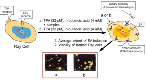
2.6.5 TPA-Induced EBV-EA Activation... 30
2.6.6 Two-Stage Carcinogenesis on Mouse-Skin ... 31
II
2.6.7 Assay of Cytotoxicity ... 32
2.6.8 Apotosis Detection ... 33
Chapter 3: Structure Elucidation and Identification
3.1 Introduction ... 343.2 Constituents of Momordica charantia Leaves ... 40
3.2.1 Spectral Data of Known Compounds from Momordica charantia Leaves . 40 3.2.2 Structure Elucidation of New Compounds from Momordica charantia Leaves... 45
3.3 Constituents of Passiflora edulis Leaves ... 54
3.3.1 Spectral Data of Known Compounds from Passiflora edulis Leaves ... 54
3.3.2 Structure Elucidation of New Compounds from Passiflora edulis Leaves... 59
3.4 Constituents of Vitellaria paradoxa Kernels ... 65
3.4.1 Spectral Data of Known Compounds from Vitellaria paradoxa Kernels .... 65
3.4.2 Structure Elucidation of New Compounds from Vitellaria paradoxa Kernels ... 75
3.5 Chemical Modification ... 82
3.5.1 Acid Hydrolysis of Glycosides and Sugar Identification ... 82
3.5.2 Preparation of MTPA Ester Derivatives (Mosher’s Method) ... 83
Chapter 4: Bioactivity Evalution
4.1 Introduction ... 854.2 Anti-Melanogenesis Activities ... 89
4.2.1 Melanogenesis Inhibitory Activities of Extracts ... 89
4.2.2 Melanogenesis Inhibitory Activities of Compounds ... 91
4.2.3 Western Blot Analysis of Melanogenesis-Related Proteins ... 96
4.3 Anti-Oxidant Activities ... 97
III
4.3.1 DPPH Free Radical-Scavenging Activities of Extracts ... 97
4.3.2 DPPH Free Radical-Scavenging Activities of Compounds ... 98
4.4 Anti-Inflammatory Activities ... 100
4.4.1 Anti-Inflammatory Activities of Extracts in Mice ... 100
4.4.2 Anti-Inflammatory Activities of Compounds in Mice ... 100
4.5 Anti-Tumor Promoting Activities ... 102
4.5.1 Inhibitory Effects on EBV-EA induction in Raji Cell Lines ... 102
4.5.2 In Vivo Two-Stage Carcinogenesis ... 106
4.6 Cytotoxicities ... 108
4.6.1 Cytotoxic Activities against Human Cancer Cell Lines... 108
4.6.2 Apoptosis-Inducing Activities... 112
Chapter 5: Conclusion
... 114References
... 117Acknowledgments
... 138Appendix
1. List of Compounds in This Dissertation ... 1392. List of Abbreviation ... 147
3. List of Publications ... 152
4. List of Presentations ... 156
Supplementary Data
1. NMR Spectral Data of New Compounds
2. MS and MS-MS Spectral Data of New Compounds
- 1 -
Chapter 1 Introduction
1.1 General Introduction
Natural products are a precious gift from nature to health and beauty of human. And, they are characterized as secondary metabolites with small-molecule structure that originate from plants, microorganisms, and animals, tend to present more structurally diverse “biologically friendly” molecular qualities than pure synthetic compounds at random, and are an important origin of new and original lead structures for the synthetic combinatorial chemistry aspects of antimelanogenesis cosmetic and antitumor agents [1–3]. Despite certain technical limitations inherent in the investigation of the small-molecule natural constituents of organisms using modern drug and cosmetic discovery platforms, improvements in automated high-throughput bioactivity screening techniques and technologies applied in the processes of constituent analysis, purification, and structural elucidation have significantly speeded up the natural product bioassay-guided fractionation procedure [4, 5]. The application of biotechnological techniques has allowed selected natural product metabolites to be produced in a relatively controlled manner, and to be less limited by sourcing conditions caused by environmental, seasonal, and geographical effects [6]. It has been concluded recently that natural products from all types or organisms offer an
“unlimited” resource for future drug discovery [7].
The purpose of this study is to develop new lead compounds for skin whitening, antioxidant, and antitumor agents based on natural triterpene glycosides and other polar compounds isolated from the methanol (MeOH) extracts of bitter gourd (Momordica charantia; Cucurbitaceae), passion flower (Passiflora edulis;
Passifloraceae), and defatted shea kernels (Vitellaria paradoxa; Sapotaceae).
- 2 -
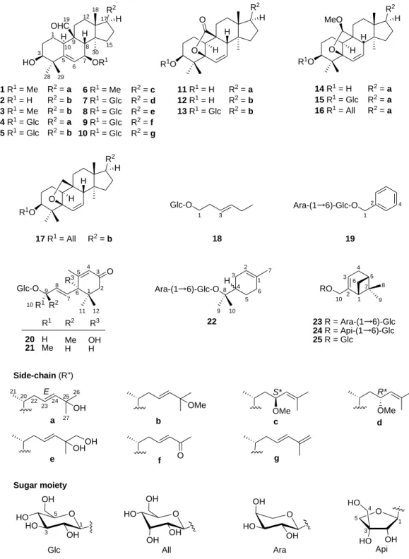
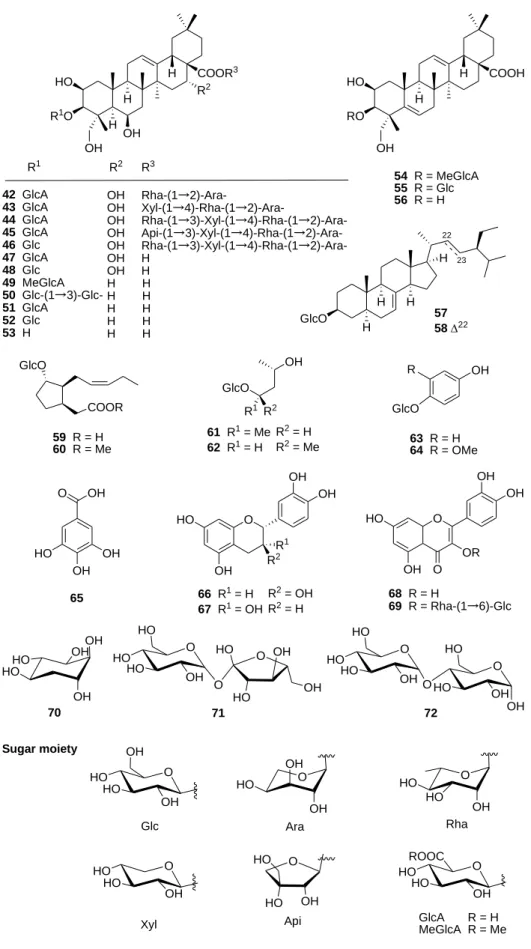
Seventeen cucurbitane-type triterpene acids and their glycosides, 1–17, eight cycloartane-type triterpene glycosides, 30–37, and fifteen oleanane-type triterpene acids and their glycosides, 42–56, two steroid glycoisdes, 57 and 58, eleven phenolic compounds and flavonoids, 26–29 and 63–69, sixteen other glycosidic compounds, 18–25, 38–41, and 59–62, and four sugars, 70–73, were isolated. Among these, sixteen compounds, 1, 6–9, 12, 22, 24, 27, 32, 33, 42, 43, 49, 50, and 54, were new naturally occurring compounds.
Fifty-three compounds, 18–70, against melanogenesis in B16 melanoma cells induced by -melanocyte-stimulating hormone (-MSH), forty-six compounds, 20 and 26–70, against generation of 1,1-diphenyl-2-picrylhydrazyl (DPPH) free radicals, and eighteen compounds, 42–46, 49–53, 55, 56, 59, 60, 63, 65, 68, and 69, against inflammation induced by 12-O-teradecanoylphorbol-13-acetate (TPA) in mice were evaluated for their biological activities. From the viewpoints of cancer chemopreventive and anticancer properties, sixty-three compounds, 1–17, 20, and 26–
70, against the TPA-induced Epstein-Barr virus early antigen (EBV-EA) activation in Raji cells, and forty-six compounds, 1–17 and 42–70, against proliferation of HL60, A549, AZ521, and SK-BR-3 human cancer cell lines also were evaluated.
Eighteen compounds, 19–21, 23–27, 47, 48, 52, 54, 55, 59, 61, 62, 67, and 69, exhibited potent inhibitory activities against melanogenesis (42.0–71.5% melanin content) with no or very low toxicity to the cells (84.9–107.3% cell viability) at a concentration of 100 M, and, among which, compounds 24, 27, 54, and 59 were further analyzed for their antimelanogenesis mechanisms by Western-blotting. Five phenolic compounds and flavonoids, 65–69, exhibited strong radical-scavening activities (IC50 5.8–12.9 M) which were more potent than reference compound, tocopherol (IC50 27.1 M). Furthermore, twelve triterpenes, 42–46, 49–53, 55, and 56, exhibited potent inhibitory activities against TPA-induced inflammation (1g/ear) with ID50 values in the range of 0.02–0.38 mol/ear. In addition, most of the triterpenoids and flavonoids, i.e., 1–3, 6–8, 11, 12, 14–17, 30–32, 34, 35, 47–49, 51–
- 3 -
56, and 66–68, exhibited potent inhibitory effects on EBV-EA induction with IC50
values in the range of 242–387 molar ratio/32 pmol TPA, and four compounds, 1, 11, 58a, and 59, on skin-tumor promotion in an in vivo two-stage mouse-skin carcinogenesis test based on 7,12-dimethylbenz[a]anthracene (DMBA) as initiator and with TPA as promoter. Furthermore, compounds 2 and 6 against HL60 cell line, compounds 6, 7, 15, 17, 44, and 45 against A549 cell line, compound 2 against SK-BR-3 cell line, exhibited potent cytotoxic activities (IC50 1.7–19.7 M).
Compound 44 was further evaluated for its apoptosis inducing activity in A549 cell line.
A literature review, which has been done on the topics of triterpene glycosides and three plant materials used in this study, viz., bitter gourd (Momordica charantia), passion flower (Passiflora edulis), and shea (Vitellaria paradoxa) kernels, was described below (Sections 1.3–1.5 and Section 2.2).
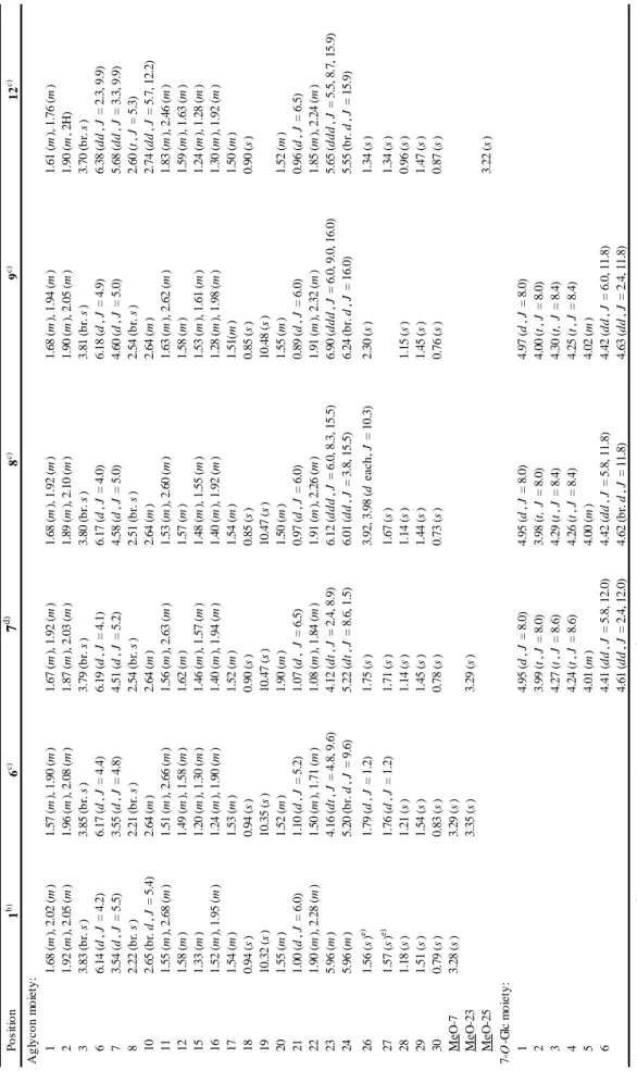
1.2 Triterpene Glycosides
Triterpene glycosides are triterpenoids belonging to the group of saponin compounds, which are high-molecular-weight complicated glycosides, containing a sugar group attached to either a triterpene. The aglycon of triterpene glycoside is a type of terpene containing thirty carbon atoms, assembling from six isoprene unit.
Triterpene glycosides are an important bioactive class of natural prouducts, and the biosynthesis of triterpene glycoside was described below.
The aglycon of triterpene glycoside, built-up from C5 units, isopentenyl diphosphate (IPP), which is supplied from the cytosolic mevalonic acid (MVA) pathway (Figure 1-1) [8]. Sesquiterpene (C15; 3 C5 units) and triterpene (C30; 6 C5 units) are biosynthesized via the MVA pathway, whereas monoterpene (C10; 2 C5 units), diterpene (C20; 4 C5 units), and tetraterpene (C40; 8 C5 units) are biosynthesized via the methylerythritol phosphate (MEP) pathway. The first diversifying step in triterpene
- 4 -
biosynthesis is the cyclization of 2,3-oxidosqualene catalyzed by oxidosqualene cyclase (OSC) [9].
S O CoA
Acetyl-CoA S
O O
CoA
Acetoacetyl-CoA S
O CoA
HO
COOH
3-Hydroxy-3methyl- glutaryl-CoA (HMG-CoA)
Acetyl-CoA
2 NADPH 2 NADP
HO OHO
COOH 3, 5-dihydroxy-3-methyl- pentanoic acid (MVA)
H2O5P2 O HO
COOH Mevalonyl-PP
H2O5P2 O
Isopentenyl-PP (IPP; C5)
2 ATP 2 ADP ATP ADP
CO2
H2O5P2 O
Dimethylallyl-PP (DMAPP; C5) Geranyl-PP
(GerPP; C10) H2O5P2 H2O5P2
Farnersyl-PP (FPP, C15)
IPP IPP
NADPH NADP
O
O2
Squalene 2,3-Oxidosqualene
Figure 1-1. Mevalonic acid (MVA) pathway.
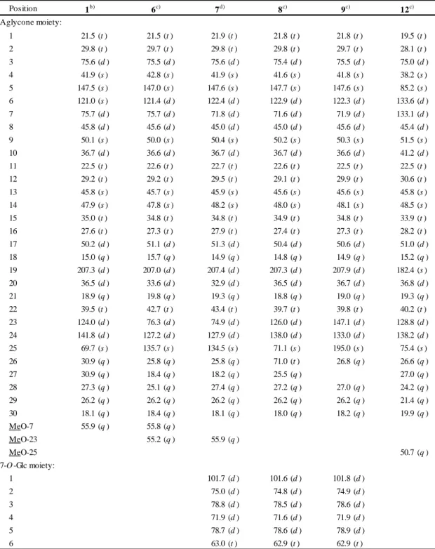
Generally, plants and animals have only one OSC, lanosterol synthase (LAS), for sterol biosynthesis. However, higher plants have several OSCs not only for sterol
- 5 -
biosynthesis, such as cycloartenol synthase (CAS) and LAS [10], but also for triterpene biosynthesis. The molecular diversity of OSCs enables more than 100 skeletal variations of triterpene in plants [11]. Such as ginsenosides which were main pharmacologically active compounds in the ginseng [12], major ginsenosides have a dammarane skeleton constructed by an OSC, dammarenediol-II synthase (PNA). In addition, 2,3-dihydro-2,5-dihydroxy-6-methyl-4H-pyran-4-one (DDMP) saponins and their derivatives have beneficial effects on human health, but some saponins are unfavorable because of their astringent taste [13]. To reduce the astringent taste of soybean, transgenic soybean plants with suppressing β-amyrin synthase (AS).After an OSC constructs the basic triterpene skeleton, the skeleton is modified to a hydrophobic aglycon called sapogenin. The first modification is oxidation catalyzed by cytochrome P450 monooxygenase (P450), and this step enables further modifications such as O-glycosylation. P450 is highly diverse and catalyzes several kinds of chemical reactions committed to the secondary metabolism [14].
Glycosylation is essential for saponin biosynthesis. Glycosylation increases the water solubility and changes the biological activity of triterpene. Uridine diphosphate (UDP)-dependent glycosyltransferases (UGT) recognize a wide range of natural products as acceptor molecules. P450 species and UGTs belong to multigene families and are the key factors for explosive diversification of other natural products in plants (Figure 1-2) [15].
Triterpene glycosides, refered to the attachment of various sugar moiety to the triterpene unit. These sugar moieties can be cleaved off in the gut by bacteria, as well as the aglycon of triterpene glycoside to be absorbed into the blood stream or to insert into cell membranes [16]. Typically, triterpene glycosides have detergent properties, readily form foams in water, have a bitter taste, and toxic to fish. Most of plants that contain triterpene glycosides were used as soapbars, such as soapnut (Sapindus mukurossi; Sapindaceae), soapbark (Quillaja saponaria; Rosaceae), soapwort (S a p o n a r i a o f f i c i n a l i s; C a r y o p h y l l a c e a e ) , s o a p b e r r y (S a p i n d u s s a p o n a r i a; S a p i n d a -
- 6 -
O
HO
HO HO
HO OH
HO
HO OH OH HO
H HO
H COOH OH
HO
H COOH HOOC
HO
H COOH HOOC
HO HO
H
COOH OHO
CHO OH
OH
O
HO
H OH
OH HO
H OH
OH
OH O
H OH
O
OH
HO
O OH O
HOO
OH OH HO O HOHO
OHHO OHO H
O O
OH O
O OH OHO HO OH
OH
HOO
OH OH O
HOO
OH OH HO OHO HO O
OH OAcO AcO OAc
OHO HO O
HOOC O
HO HO O
OH OHO H
O OH
OH O
H OH
O OHO HO O
HOOC O
HO HO O
OH O
HO HOOH
O
O OH O
CHO OH
OH
O O
O HO OOHO H
O OH
OH
OHO HO OH
OHNHCH3
OH O O O
H
COOH O OHO HO O
HOOC OHO H
O OH
HOOC
O
H C HOOC
O
OH HO OOHO HO OH
OHOHO H
O OH
OH
O O
HO
H HOOC
C OHO O OH
O
O O OHO HO OH
OH
OHO H
O OOHO H
O OH
OH
OHOOCO OH
O
H HOOC
HO OHO HO OH
OHC OHO H
O OH
OH
O O
(20S)-Protopanaxatriol 2,3-Oxidosqualene
Dammarenediol-II(20S)-Protopanaxadiol
Ginsenoside Rg-1 Ginsenoside Rb-1 Soyasaponin Aa (group A saponin)
Soyasapogenol ASoyasapogenol B
-amyrin Medicagenic acid Gypsogenic acid Hederagenin Glycyrrhetinic acidSoyasoponin bg (DDMP saponin) Vaccaroside BGlycyrrhizinAvenacin A-1
PNA AS
OSC P450 UGT
Mevalonic acid pathway Figure 1-2. Triterpene glycoside biosynthetic pathway. 2,3-oxidosqualene catalyzed by OSC (blue arrows), a triterpene undergoes various modifications including P450-catalyzed oxidation (red arrows) and UGT-catalyzed glycosylation (green arrows).
- 7 -
ceae), and soaproot (Chlorogalum pomeridianum; Asparagaceae) [17]. Because of their various beneficial properties for health and beauty of humans, the triterpene glycosides are used in wide-ranging applications in addition to medicinally [15].
1.3 Momordica charantia (Bitter Gourd)
The plant Momordica charantia L. (bitter gourd; Cucurbitaceae), commonly called
“nigauri” or “goya” in Japan, is cultivated throughout the world for use as a vegetable as well as medicine. M. charantia has been used in traditional medicines in developing countries, mostly for healing diabetes, and as a carminative and in the treatment of colic [18–20]. Previous investigations have shown that crude extracts of the fruits of M.
charantia possess antidiabetic activity [21, 22], and many cucurbitane-type triterpenoids have been isolated from the fruits [23–29], seeds [30, 31], leaves and vines [32–35], roots [36, 37], and stems [38–41] of M. charantia.
1.4 Passiflora edulis (Passion Flower)
Passiflora edulis (passion flower; Passifloraceae), known also as the passion vines, is a genus of about 500 species of herbaceous vines or trees distributed mainly in tropical America, with a smaller number of species occurring in Southeast Asia, India, Malaysia, and Australia [42]. Plants of Passiflora species are very popular, not only because of their fruits (passion fruits), but also because the tea of their leaves has been largely used in American and European countries, in popular medicine, as a sedative, diuretic, tonic, and also in the treatment of hypertension and skin diseases [42]. The chemical constituents of leaf extract of P. edulis have been extensively studied, showing the predominance of alkaloids [43, 44], cyanogenic glycosides [45], saponins [46–49], and polyphenols [50–53]. As part of an ongoing study in this laboratory on the plant metabolites possessing melanogenesis-inhibitory activities [54–64], the
- 8 -
constituents of the extract of P. edulis leaves have been investigated in this study.
1.5 Vitellaria paradoxa (Shea)
The Vitellaria paradoxa C.F. Gaertner [shea tree; synonyms Butyrospermum paradoxum (C.F. Gaertn.) Hepper, Butyrospermum parkii (G. Don) Kotschy;
belonging to family Sapotaceae] is indigenous to the savanna belt extending across sub-Saharan Africa north of the equator, ranging from Senegal in the west to Ethiopia and Uganda in the east [65–67]. The fruit of the tree is edible and nutritious, while the most valued product of shea is shea butter, the edible fat extracted from the seed kernel, consisting of an olein fraction and a stearin fraction along with non- saponifiable (non-lipid) compounds. Fractionated shea stearin is used primarily as a cocoa butter substitute or extender in chocolate manufacture [68].These applications are due to properties imparted by the structures of its component triacylglycerols. In addition, shea butter is increasingly popular as component of skin care products and cosmetic product formulations, in part due to the unusually high level of non- saponifiable lipid (NSL) constituents in the fat [69]. In order to characterize and quantify the constituents of shea butter among widely dispersed V. paradoxa populations, the contents and compositions of triterpene alcohol fractions from the NSL, and fatty acid, triacylglycerol, and triterpene ester compositions of the kernel lipids (n-hexane extracts) from 36 shea kernel samples from seven sub-Saharan countries has recently been determined [70, 71]. In addition, it has been demonstrated that cinnamyl and acetyl triterpene esters isolated from the kernel fat could be valuable as anti-inflammatory agents and chemopreventive agents in chemical carcinogenesis [72]. From a perspective, I have been interested in the evaluation of pharmacological and cosmeceutical potentials of the constituents of defatted shea kernel, since there seems to be little industrial utilization of defatted shea kernel (residue), other than as fuel.
- 9 -
Chapter 2 Experimental
2.1 General Experimental Procedure
2.1.1 Chromatography
(1) Analytical thin-layer chromatography (TLC): Silica gel 60 F254 aluminum sheets (SiO2, 20 cm × 20 cm; Merck & Co., Inc., Darmstadt, Germany), and Silica gel 60 RP-18 F254S (ODS, 20 cm × 20 cm; Merck & Co., Inc.) were used for TLC.
(2) Open column chromatography (CC): Diaion HP-20 (Mitsubishi Chemical Co., Tokyo, Japan), silica gel (SiO2, 230–400 mesh; Merck & Co., Inc.), octadecyl silica gel (ODS, 100–200 mesh; Fuji Silysia Chemical Ltd., Aichi, Japan), and Sephadex LH-20 (Amersham Biosciences AB, Uppsala, Sweden) were used for CC.
(3) Reversed-phase (RP) preparative high-performance liquid chromatography (HPLC): HPLC was carried out under the following conditions: (i) on ODS columns (25 cm 10 mm i.d.) at 25 ºC: on a TSK ODS-120A column (Toso Co., Ltd., Tokyo, Japan) with MeOH/H2O/AcOH [90:10:0.1 (HPLC system M.c.I) or 80:20:0.1 (HPLC system M.c.II)], or on a Pegasil ODS-II 5 m column (Senshu Scientific Co., Ltd., Tokyo, Japan) with MeOH/H2O/AcOH [85:15:0.1 (HPLC system M.c.III), 80:20:0.1 (HPLC system M.c.IV) or 50:50:0.1 (HPLC system M.c.VI)], or MeCN/H2O/AcOH [70:30:0.1 (HPLC system M.c.V)], or on a Capcell pak AQ 5 m column (Shiseido Co., Ltd., Tokyo, Japan) with MeCN/H2O/AcOH [10:90:0.1 (HPLC system M.c.VII) or 45:55:0.1 (HPLC system M.c.VIII)], at flow rate of 2.0 ml min-1 of mobile phase, or on a Capcell pak AQ 5 m column with MeCN/H2O/AcOH [35:65:0.1 (HPLC system
- 10 -
M.c.IX)], at flow rate of 3.0 ml min-1 of mobile phase.
(ii) on a Pegasil ODS-II 5 m column with MeCN/H2O [9:1 (HPLC system P.e.I)], or on a Capcell pak AQ 5 m column with MeCN/H2O [1:1 (HPLC system P.e.II) or 7:13 [HPLC system P.e.III)], or with MeCN/H2O/AcOH [25:75:0.1 (HPLC system P.e.IV), 22:78:0.1 (HPLC system P.e.V) or 10:90:0.1 (HPLC system P.e.VI)], or with MeCN/H2O/HCOOH [57:43:0.1 (HPLC system P.e.VII) or 17:83:0.1 (HPLC system P.e.VIII)] as mobile phase with a flow rate of 2.0 ml min-1.
(iii) on a Pegasil ODS SP100 column (Senshu Scientific Co., Ltd.) with MeCN/H2O/AcOH [30:70:0.2 (HPLC system V.p.I), 28:72:0.2 (HPLC system V.p.II), or 100:0:0.2 (HPLC system V.p.III)], or with MeOH/H2O/AcOH [78:22:0.2 (HPLC system V.p.IV), or on a Capcell Pak C18 column (Shiseido Co., Ltd.) with MeCN/H2O/AcOH [28:72:0.2 (HPLC system V.p.V)] or with MeOH/H2O/AcOH [48:52:0.2 (HPLC system V.p.VI), 20:80:0.2 (HPLC system V.p.VII), or 2:98:0.2 (HPLC system V.p.VIII)], at flow rate of 2.0 ml min-1 of mobile phase.
(4) Evaporative light-scattering detector (ELSD) HPLC: Consisted of a SSC-3461 gradient pump (Senshu Seientific Co., Ltd.), and a Sedex Model 55 ELSD system (regulation temperature: 40 °C; air pressure: 2.7 bar, Sedere, France); and a reversed-phase column, Senshu Pak NH2-1251-N (25 cm × 4.6 mm i.d.; column temperature: 30 °C, Shiseido Co., Ltd.), with MeCN/H2O [mobile phase A: MeCN 100%; mobile phase B: H2O; drift tube temperature: 85 °C; elution was performed as follows: solvent A/solvent B (4:1, 0 min) → solvent A/solvent B (4:1, 40 min)]
(HPLC system V.p.IX), flow ratio: 1.0 ml min-1.
(5) Gas-liquid chromatography (GLC): Shimadzu GC-2014 instrument on a DB-17 fused silica glass capillary column (30 m × 0.32 mm i.d.; column temperature:
200°C; injection and detector temperature: 270°C; He flow rate: 0.4 ml min-1; split ratio: 1:75, Agilent Technologies, Inc., Santa Clara, CA, USA).
- 11 -
2.1.2 Determination of Physical Constants and Spectroscopy
(1) General: Crystallizations were performed in MeOH, and melting points were determined on a Yanagimoto micro melting point apparatus and uncorrected. Optical rotations were measured on a JASCO P-2200 polarimeter in EtOH at 25 °C. UV spectra, on a JASCO V-630Bio spectrophotometer, and IR spectra, using a JASCO FTIR-300 E spectro- meter, were recorded in EtOH and KBr disks, respectively.
(2) Nuclear magnetic resonance (NMR) spectroscopy: Acquired with a JEOL ECX-400 (1H, 400 MHz; 13C, 100 MHz), with a JEOL ECX-500 (1H, 500 MHz; 13C, 125 MHz), or with a JEOL ECX-600 (1H, 600 MHz; 13C, 150 MHz) spectrometer in CD3OD, C3D6O, C5D5N, or DMSO-d6. Chemical shift () values are given in ppm with tetramethylsilane (TMS; = 0 ppm) as internal standard, and coupling constants (J) in Hz.
(3) High-resolution (HR)-electrospray ionization mass spectrometry (ESIMS) and atmospheric-pressure chemical ionization mass spectrometry (APCIMS):
Recorded on an Agilent 1100 LC/MSD TOF (time-of-flight) system [polarity mode:
positive or negative; nebulizer pressure: 35 psi; drying gas (N2) flow: 12 l min-1; drying gas temperature: 325 °C; capillary voltage: 3000 V; fragmentor voltage: 225 V]; Electrospray ionization with tandem mass spectrometry (ESIMSMS) were recorded on an Agilent 6530 LC/QTOF (quadrupole time-of-flight) system [polarity modes: positive or negative; nebulizer pressure: 50 psi; drying gas (N2) flow: 10 l min-1; drying gas temperature: 350 °C; fragmentor voltage: 150 V; mass range: 100–
1200; acquisition rate: 1.5 spectra sec-1; HPLC instrument: Agilent 1200 series;
column : ZORBAX Eclipse Plus C18 (100 × 2.1 mm i.d., 1.8 μm); mobile phase A: 5 mM CH3COONH4 (with 0.1% CH3COOH); mobile phase B (%): MeCN with gradient (5→50→90→90, 0→10→10.1→15 min); flow rate: 0.3 ml min-1; column
- 12 - temperature: 40 °C; injection volume: 5 l].
2.2 Plant Materials
(1) Momordica charantia: The plants of M. charantia (bitter gourd, Figure 2-1) were cultivated at Kyann, Itoman-shi (Okinawa, Japan) and collected on 14th March, 2006. The plant material was authenticated by Mr. Kei-ichiro Inafuku, and voucher specimen has been deposited in the Laboratory for Biological and Natural Resource, College of Science and Technology, Nihon University.
Figure 2-1. Momordica charantia (Family: Cucurbitaceae).
(2) Passiflora edulis: The plants of P. edulis (passion flower, Figure 2-2) were cultivated on a farm at Tamil Nadu state in India and harvested in April, 2007. A voucher specimen (Registry No. SH0709-SB2183) of the plant has been deposited in the Research Laboratory of Ichimaru Pharcos Co., Ltd. (Motosu-shi, Gifu, Japan).
Authentication was done by Mr. Norihiro Banno (Ichimaru Pharcos Co., Ltd.).
Figure 2-2. Passiflora edulis (Family: Passifloraceae).
- 13 -
(3) Vitellaria paradoxa: The kernels of V. paradoxa (shea, Figure 2-3) were collected and identified by Mr. Eliot T. Masters on behalf of the World Agroforestry Centre (ICRAF), an international research institute constituted under the Consultative Group for International Agricultural Research (GGIAR), in parallel to project CFC/FIGOOF/23 ‘Improving Product Quality and Market Access for Shea Butter originating from sub-Saharan Africa’ (ProKarité). Near the geographic center of a regional sampling mission undertaken from Senegal to South Sudan during the 2006 shea season (May through July), the specific sample described in this study was collected as fresh fruit gathered from fallow ground beneath the crown of a healthy mature tree at a site (longitude E 7°27ʹ9″, latitude N 9°40ʹ53″, elevation 365 m) in central Nigeria [70].
Figure 2-3. Vitellaria paradoxa (Family: Sapotaceae).
2.3 Chemicals and Reagents
Chemicals and reagents were purchased as follows: (–)-2-Methoxy-2-phenyl-2- (trifluoromethyl) acetic acid (MTPA) and (+)-MTPA chlorides and N,N-dimethyl- 1,3-propanediamine from Tokyo Chemical Industry Co., Ltd. (Tokyo, Japan); TPA from ChemSyn Laboratories (Lenexa, KS, USA); the EBV cell culture reagents and butanoic acid from Nacalai Tesque, Inc. (Kyoto, Japan); fetal bovine serum (FBS), RPMI-1640 medium, antibiotics (100 units ml-1 penicillin and 100 g ml-1
- 14 -
streptomycin), and non-essential amino acid (NEAA) from Invitrogen Co. (Carlsbad, CA, USA); D-arabinose, D-xylose, DMBA, Dulbecco’s modified Eagle’s medium (DMEM), Eagle’s minimal essential medium (MEM), 4-hydroxy-phenyl -D-glucopy- ranoside (arbutin), -tocopherol, -melanocyte stimulating hormone (-MSH),
D-glucuronic acid, indomethacin, and 3-(4,5-dimethylthiazol-2-yl)-2,5-diphenyl-2H- tetrazolium bromide (MTT) from Sigma-Aldrich Japan Co. (Tokyo, Japan). Formic acid, L-glucose, L-arabinose, 5-fluorouracil, cisplatin, and -carotene from Wako pure Chemical Industries, Ltd. (Osaka, Japan); D-glucose and L-cysteine methyl ester hydrochloride from Kanto Chemical Co., Inc. (Tokyo, Japan); and L-rhamnose from Nacalal Tesque, Inc. (Tokyo, Japan). All other chemicals and reagents were of analytical grade.
2.4 Extraction and Isolation
2.4.1 Momordica charantia (Bitter Gourd) Leaves
Chopped and air-dried leaf materials of M. charantia (4.8 kg) was extracted with MeOH (1 kg 4 l-1, reflux, 3 h, 3) to give a crude extract (399 g). The extract was suspended in H2O (40 g l-1) and then extracted with ethyl acetate (AcOEt) (3 1 l).
The AcOEt fraction was further partitioned between n-hexane/MeOH/H2O (19:19:1), which yielded n-hexane- (48 g) and MeOH/H2O- (75 g) soluble fractions. On the other hand, the H2O layer was further extracted with n-BuOH, which yielded n-BuOH- (40 g) and H2O- (170 g) soluble fractions (Scheme 1).
(1) MeOH/H2O-Souble fraction: A portion (47 g) of the MeOH/H2O-soluble fraction was chromatographed on a SiO2 (977 g) column with a stepwise gradient of n-hexane/AcOEt (1:0→1:4) and AcOEt/MeOH (1:0→1:4) as eluent, which yielded nine fractions, Frs. M1–9, listed in increasing order of polarity. Fr. M2 (9.5 g, eluted
- 15 -
with n-hexane/AcOEt 1:1) was chromatographed on a Diaion HP-20 (150 g, MeOH/H2O gradient 7:3→9:1) column to yield purified Fr. M2 (5.6 g). This fraction was chromatographed on a SiO2 (250 g, n-hexane/AcOEt gradient 19:1→0:1) column to yield five fractions, Frs. M2-1–M2-5. Fr. M2-2 (3.17 g, eluted with n-hexane/
AcOEt 1:1) was further chromatographed on a SiO2 (150 g, n-hexane/AcOEt gradient 7:3→3:7) column to yield eight fractions, Frs. M2-2a–M2-2h. Chromatography on an ODS (30 g, MeOH/H2O gradient 7:3→8:2) column of Fr. M2-2d (1292 mg) yielded purified Fr. M2-2d (241 mg). Preparative HPLC (system M.c.I) of this fraction yielded 6 (2.4 mg; tR 16.0 min), and 3 (5.6 mg; tR 17.3 min), 12 (7.9 mg; tR 18.0 min), and 14 (1.6 mg; tR 28.4 min). Preparative HPLC (system M.c.III) of Fr. M2-2e (254 mg) gave 11 (28.4 mg; tR 10.2 min) and 1 (22.1 mg; tR 12.4 min). Fr. M2-3 (720 mg, eluted with n-hexane/EtOAc 3:7) was subjected to ODS CC (30 g, MeOH/H2O gradient 4:1→9:1) column to yield purified Fr. M2-3 (97.0 mg), which upon preparative HPLC (system M.c.I) yielded 2 (5.1 mg; tR 13.2 min). Fr. M5 (12.8 g), eluted with n-hexane/EtOAc (0:1), was subjected to ODS CC (250 g, MeOH/H2O gradient 3:7→1:0) column to yield eleven fractions, Frs. M5-1–M5-11. Isolation of the following eleven compounds was performed by preparative HPLC: compound 4 (139.8 mg; tR 16.1 min) from Fr.
M5-3 (0.49 g; HPLC system M.c.II); compounds 5 (46.4 mg; tR 26.7 min) and 7 (15.6 mg; tR 40.7 min) from Fr. M5-5 (0.50 g; HPLC system M.c.II); compounds 10 (7.8 mg; tR 19.0 min), 16 (3.4 mg; tR 30.8 min), 15 (14.2 mg; tR 34.3 min), and 13 (3.8 mg;
tR 40.7 min) from Fr. M5-7 (0.36 g; HPLC system M.c.II); compounds 8 (4.6 mg; tR 4.8 min), 9 (4.2 mg; tR 8.0 min), 4 (4.4 mg; tR 12.0 min), and 10 (6.8 mg; tR 52.0 min) from Fr. M5-8 (0.21 g; HPLC system M.c.IV); and compound 17 (13.2 mg; tR 3.7 min) from Fr. M5-9 (0.11 g; HPLC system M.c.V).
- 16 -
Scheme 1.Extraction and isolation procedures of 1–25 from the MeOH extract ofM. Charantia Leaves.
- 17 -
(2) n-BuOH-Soluble fraction: A portion (39 g) of n-BuOH-soluble fraction was chromatographed on a Diaion HP-20 (1226 g; with step gradient MeOH/H2O 1:9→1:0) column, which yielded three fractions, Frs. B1–3. Fr. B2 (6.9 g, eluted with H2O/MeOH 7:3 and 1:1) was further chromatographed on a SiO2 (160 g, n-hexane/AcOEt gradient 1:0→0:1, and AcOEt/MeOH gradient 1:0→0:1) column to yield fifteen fractions, Frs. B2-1–B2-15. Preparative HPLC (system M.c.VI) of Fr.
B2-3 (101 mg, eluted with n-hexane/AcOEt 3:7) yielded compound 18 (3.0 mg, tR
14.8 min). Fr. B2-6 (1.6 g, eluted with n-hexane/AcOEt 7:3) was chromatographed on an ODS (48 g, H2O/MeOH gradient 3:7→0:1) column to yield eight fractions, Frs.
B2-6a–B2-6h. Preparative HPLC (system M.c.VII) of Fr. B2-6c (193 mg, eluted with MeOH/H2O 3:7) yielded compounds 19 (4.4 mg, tR 27.2 min) and 20 (27.5 mg, tR 33.6 min). Fr. B3 (5.5 g, eluted with H2O/MeOH 3:7 and 0:1), was chromatographed on an ODS (144 g) column to yield nine fractions, Frs. B3-1–B3-9. Preparative HPLC (system M.c.VIII) of Fr. B3-2 (94 mg, eluted with MeOH/H2O 6:4) yielded compound 21 (2.1 mg, tR 32.0 min). Fr. B3-3 (1326 mg) was chromatographed on an ODS (48 g;
MeOH/H2O gradient 3:7→0:1) column to yield thirteen fractions, Frs. B3-3a–B3-3m.
Preparative HPLC (system M.c.IX) of Fr. B3-3c (97 mg, eluted with MeOH/H2O 1:1) yielded compounds 22 (3.1 mg, tR 25.6 min), 23 (3.2 mg, tR 27.6 min), 24 (6.4 mg, tR
3 0 . 4 m i n ) , a n d 2 5 ( 7 . 4 m g , t R 4 1 . 6 m i n ) .
2.4.2 Passiflora edulis (Passion Flower) Leaves
The air-dried and sliced leaves of P. edulis (2.2 kg) were extracted with MeOH (reflux, 3 h, 3) to yield a MeOH extract (399 g). This was suspended in H2O and partitioned with AcOEt. The AcOEt layer was further partitioned between n-hexane/H2O/MeOH (19:19:1), which yielded n-hexane- (65 g) and MeOH/H2O- (71 g) soluble fractions. On the other hand, the H2O layer was further extracted with
- 18 -
n-BuOH to yield n-BuOH- (151 g) and H2O- (58 g) soluble fractions (Scheme 2).
(1) MeOH/H2O-Soluble fraction: A portion (32 g) of MeOH/H2O-soluble fraction (71 g) was subjected to SiO2 CC (700 g; with a step gradient of AcOEt/MeOH 1:0→0:1), which yielded fifteen fractions, Frs. M1–M15. Fr. M9 (10.1 g, eluted with AcOEt/MeOH 1:1) was further subjected to SiO2 CC (500 g; AcOEt/MeOH gradient 1:0→0:1) to yield eight fractions, Frs. M9a–M9h. Fr. M9d (1.2 g, eluted with AcOEt/MeOH 4:1 and 3:2) was further chromatographed on SiO2 (60 g;
AcOEt/MeOH gradient 1:0→0:1) column to give seven fractions, Frs. M9d-1–M9d-7.
Application of HPLC (system P.e.I) to Fr. M9d-2 (156 mg, eluted with AcOEt/MeOH 19:1), yielded compounds 32 (5.3 mg; tR 30.0 min) and 33 (6.5 mg; tR 40.0 min). Fr.
M9d-4 (399 mg, eluted with AcOEt/MeOH 7:3→1:1), upon preparative HPLC (system P.e.VII), gave compounds 34 (10.5 mg; tR 58.0 min), 35 (16.0 mg; tR 62.4 min), 30 (4.0 mg; tR 70.0 min), and 31 (3.0 mg; tR 75.6 min).
(2) n-BuOH-Soluble fraction: A portion of the n-BuOH-soluble fraction (22 g) was subjected to Diaion HP-20 CC (1 kg). A step gradient elution was conducted with MeOH/H2O (0:1→1:0) to give ten fractions, Frs. B1–B10. A portion (3.6 g) of Fr. B7 (4.3 g, eluated with MeOH/H2O 1:1) was further subjected to SiO2 CC (210 g;
CHCl3/MeOH gradient 4:1→0:1) to yield nine fractions, Frs. B7a–B7i. Fr. B7a (144 mg, eluted with CHCl3/MeOH 4:1→7:3), was subjected to HPLC (system P.e.VI) which yielded compound 41 (26.7 mg; tR 46.0 min). HPLC (system P.e.IV) of Fr. B7b (103 mg, eluted with CHCl3/MeOH 7:3) yielded compounds 20 (2.4 mg; tR 14.0 min) and 38 (11.2 mg; tR 18.0 min). Fr. B7e (1.55 g, eluted with CHCl3/MeOH 3:2) was constituted with one compound 39 (cf.: tR 14.5 min on HPLC system P.e.IV). SiO2 CC (160 g; CHCl3/MeOH gradient 1:0→1:1) of Fr. B8 (3.1 g) yielded eight fractions, Frs.
- 19 -
Scheme 2.Extraction and isolation procedures of 20, and 26–41 from the MeOH extract ofP. edulis Leaves.
- 20 -
B8a–B8h. Fr. B8c (498 mg, eluted with CHCl3/MeOH 4:1) was further separated by ODS CC (16 g; MeOH/H2O gradient 0:1→1:0) which gave 10 fractions, Frs. B8c-1–
B8c-10. HPLC (system P.e.VII) of Fr. B8c-5 (91 mg) yielded compound 40 (4.1 mg;
tR 34.0 min). Fr. B8d (1.47 g, eluted with CHCl3/MeOH 9:1) was passed through ODS CC (35 g; MeOH/H2O gradient 1:19→1:0) to afford 10 fractions, Frs. B8d-1–B8d-10.
HPLC (system P.e.VII) of Fr. B8d-6 (210 mg) yielded compounds 29 (21.7 mg; tR
32.0 min) and 28 (49.0 mg; tR 64.0 min), while HPLC (system P.e.V) of Fr. B8d-8 (421 mg) afforded compound 26 (7.1 mg; tR 58.0 min). Fr. B9 (4.4 g, eluted with MeOH/H2O 7:3 and 9:1) was chromatographed on a SiO2 (265 g; CHCl3/MeOH gradient 4:1→0:1) column to give twelve fractions, Frs. B9a–B9l, among which Fr.
B9c (1.86 g, eluted with CHCl3/MeOH 3:2) was further chromatographed on SiO2
(110 g; CHCl3/MeOH gradient 4:1→0:1) to give nine fractions, Frs. B9c-1–B9c-9.
HPLC (system P.e.II) of Fr. B9c-2 (138 mg) yielded compound 27 (5.7 mg; tR 16.7 min), whereas HPLC (system P.e.III) of Fr. B9c-3 (1.07 g) afforded compounds 37 (59.5 mg; tR 16.0 min) and 36 (95.4 mg; tR 50.0 min).
2.4.3 Vitellaria paradoxa (Shea) Kernels
Whole kernels were oven-dried at 60 °C over 72 h and decorticated. Dried kernels were crushed into powder first. The pulverized samples were weighed (3705 g), and extracted with n-hexane (reflux, 3 h, 3×) which gave an extract (1737 g). The defatted residue was then extracted with MeOH (reflux, 3 h, 3×) to yield a MeOH extract (450 g) which was suspended in H2O, and partitioned successively with AcOEt and n-BuOH to yielded AcOEt- (69.0 g), n-BuOH- (134.0 g), and H2O- (191.0 g) soluble fractions sequentially (Scheme 3).
- 21 -
(1) AcOEt-Soluble fraction: A portion of the AcOEt-soluble fraction (60 g) was subjected to SiO2 CC (800 g). Step gradient elution was conducted with n-hexane/
AcOEt (1:0→0:1) and AcOEt-MeOH (1:0→0:1) to give fourteen fractions, Frs. A1– A14. A portion of Fr. A9 (200 mg, eluated with AcOEt) was crystallized from MeOH to yield crystalline material (45 mg) which was acetylated in acetic anhydride/pyridine.
HPLC (system V.p.III) of the resulting acetate yielded compounds 58Ac (the tetraacetate derivative of 58; 1.7 mg, tR 17.0 min) and 57Ac (the tetraacetate derivative of 57; 2.0 mg, tR 24.0 min). A portion of the Fr. A10 (403 mg, eluated with AcOEt) was subjected to HPLC (system V.p.VI) giving compounds 68 (7.2 mg, tR 33.0 min) and 69 (6.6 mg, tR 36.0 min). A portion of the Fr. A11 (650 mg, eluated with EtOAc/MeOH 19:1) was passed through a SiO2 CC (20 g; n-hexane/EtOAc 7:3→0:1) to give a purified fraction (100 mg), from which was obtained compound 65 (61.9 mg) by crystallization from MeOH.
(2) n-BuOH-Soluble fraction: A portion (130 g) of the n-BuOH-soluble fraction (134 g) was subjected to Diaion HP-20 CC (1 kg; step-gradient elution with MeOH/H2O 0:10→10:0) to give nine fractions, Frs. B1–B9. A portion (5.0 g) of the Fr.
B2 (29.6 g, eluted with H2O) was crystallized from MeOH to yield purified Fr. B2, from which was obtained compounds 70 (3.9 g, tR 7.8 min) and 71 (20.0 mg, tR 11.2 min) by HPLC (system V.p.IX), respectively. Fr. B3 (4.6 g, eluted with H2O) was passed through an ODS CC (120.0 g; MeOH/H2O 0:1→7:3) to afford eight fractions, Frs. B3-1–B3-8. Crystallization of the Fr. B3-2 (1.7 g, eluted with H2O) from MeOH yielded compound 63 (137.6 mg). Fr. B3-7 (140.0 mg, eluted with MeOH/H2O 17:3) was subjected to SiO2 CC (10 g; CHCl3/MeOH 19:1→0:1) to afford a purified fraction (100 mg) from which were isolated compounds 66 (1.3 mg, tR 11.0 min) and 67 (3.9 mg, tR 12.0 min) by HPLC (system V.p.V). Fr. B4 (4.8 g, eluted with MeOH/H2O 1:9)
- 22 -
Scheme 3.Extraction and isolation procedures of 42–56, 57Ac, 58Ac, and 59–73 from the MeOH extract of defatted Shea (V. paradoxa) Kernels.
- 23 -
was applied to a SiO2 CC (150 g; CHCl3/MeOH 1:0→7:3) to yield nine fractions, Frs.
B4-1–B4-9. Fr. B4-5 (424 mg, eluated with CHCl3/MeOH 9:1), upon ODS CC (MeOH/H2O 0:1→3:17), yielded a fraction (36.0 mg) from which were obtained compound 64 (3.0 mg, tR 15.0 min) and a mixture (5.0 mg, tR 17.0 min) by HPLC (system V.p.VII). Further HPLC (system V.p.VIII) of the mixture yielded compounds 62 (0.8 mg, tR 57.0 min) and 61 (1.4 mg, tR 58.5 min). A protion (26.0 g) of Fr. B7 (27.6 g, eluated with MeOH/H2O 7:3) was subjected to ODS CC (700 g; MeOH/H2O 0:1→1:0) to give nine fractions, Fr. B7-1–Fr. B7-9. Further SiO2 CC (100 g;
CHCl3/MeOH 1:0→13:7) of Fr. B7-5 (3.4 g, eluated with MeOH/H2O 3:2) yielded nine fractions, Frs. B7-5a–B7-5i. Fr. B7-5f (371 mg, eluted with CHCl3/MeOH 4:1) was purified by SiO2 CC (12.0 g; CHCl3/MeOH 10:0→9:1) to afford compounds 59 (50.0 mg) and 60 (9.6 mg). HPLC (system V.p.I) of Fr. B7-5h (430.8 mg, eluted with CHCl3/MeOH 7:3) yielded compounds 42 (12.5 mg, tR 122.0 min), 43 (14.9 mg, tR
130.0 min), and 44 (17.1 mg, tR 140.0 min). Fraction B8 (20.9 g, eluted with MeOH/H2O 9:1) was subjected to an ODS CC (200 g; MeOH/H2O 0:1→1:0) to afford six fractions, Frs. B8-1–B8-6. SiO2 CC (CHCl3/MeOH 1:0→0:1) of a portion (1.4 g) of Fr. B8-4 (8.8 g, eluted with MeOH/H2O 7:3) gave eight fractions, Frs. B8-4a–B8-4h.
HPLC (system V.p.IV) of the Fr. B8-4b (50.0 mg, eluted with CHCl3/MeOH 9:1) yielded compounds 53 (18.6 mg, tR 41.0 min), 54 (4.0 mg, tR 43.9 min), and 55 (9.3 mg, tR 42.6 min). Fr. B8-4f (155 mg, eluted with CHCl3/MeOH 7:3) upon repeated on a SiO2 CC (CHCl3/MeOH 1:0→1:1), eventually afforded compounds 47 (1.2 mg), 48 (1.0 mg), 49 (25.0 mg), 50 (35.5 mg), 51 (12.3 mg), and 52 (24.7 mg). An AcOEt-soluble portion (26 mg) of the fraction B8-5 (766 mg) was passed through a SiO2 CC (n-hexane/AcOEt 1:0→3:2) which afforded compound 56 (4.0 mg).
(3) H2O-Soluble fraction: A portion of the H2O-soluble fraction (90.0 g) was
- 24 -
subjected to Sephadex LH-20 CC (150 g; MeOH/H2O 0:1→1:1) which yielded seven fractions, Frs. H1–H7. Fr. H2 (2.1 g) and Fr. H3 (53.9 g), both from the eluates of H2O, were crystallized from MeOH yielding 72 (1.6 g) and 73 (1.4 g), respectively. Fr. H4 (20.4 g, eluated with H2O) was further subjected to ODS CC (196 g; MeOH/H2O 0:10→5:5) to yield eight fractions, Frs. H4-1–H4-8. Fr. H4-7 (723.8 mg) was further subjected to SiO2 CC (25 g; CHCl3/MeOH 1:0→13:7) to afford a purified fraction (105 mg) from which were isolated compounds 46 (16.2 mg, tR 36.0 min) and 45 (20.0 mg, tR 48.0 min) by HPLC (system V.p.II).
2.5 Cell Lines and Culture Conditions
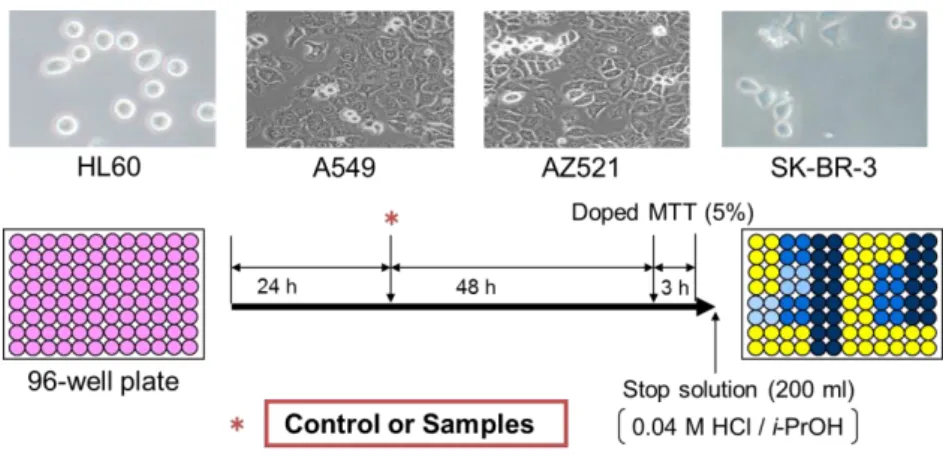
B16 4A5 (mouse melanoma) cell line and four human cancer cell lines, HL60 (human leukemia), AZ521 (duodenum), A549 (lung), and SK-BR-3 (breast), were obtained from Riken Cell Bank (Ibaraki, Japan). HL60 and SK-BR-3 cell lines were grown in RPMI 1640 medium, while B16, A549, and AZ521 cell lines were grown in DMEM and in 90% DMEM + 10% MEM + 0.1 mM NEAA, respectively. The medium was supplemented with 10% FBS and antibiotics. Cells were incubated at 37C in a 5% CO2 humidified incubator. The cell lines were cultured as described in [56, 80, 81].
2.6 Bioassay
2.6.1 Assay of Melanin Content
Melanogenesis-inhibitory assay in -MSH-stimulated B16 melanoma cells was performed as described in Figure 2-4 [56]. The B16 cells, plated at 5 × 10³ cells well-1
- 25 -
in a 24-well plate, were preincubated for 24 h. The samples dissolved in DMSO at the final concentration of 10–100 µM, and α-MSH (100 nM) were added to the medium and cultured for 96 h. The medium was removed and the cells were dissolved in 200 µl of 2 M NaOH in 10% DMSO. The amount of melanin was determined spectrophotometrically by a Sunrise-Basic microplate reader at the wavelength of 405 nm. The experiments were performed in triplicate. Arbutin used as reference compound.
Figure 2-4. Outline of melanin content assay.
2.6.2 Mechanism of Melanogenesis Inhibition
Mechanism of melanogenesis (Figure 2-5) inhibition was analyzed based on Western blot analysis, which was performed according to the method reported in [60]
with a slight modification. Briefly, B16 melanoma cells (1×10⁵ cells) were exposed to the test, sample (30 and 100 m), supplemented with -MSH (0.1 m) for 48 h. Cells were collected and lysed. Lysates of total protein were separated by 15% sodium dodecyl sulfate (SDS)-polyacrylamide gels and transferred to polyvinylidene
- 26 -
difluoride (PVDF) membranes. After blocking, the membranes were incubated with anti-microphthalmia-associated transcription factor (anti-MITF), anti-tyrosinase, anti-tyrosinase-related protein-1 (anti-TRP-1), anti-TRP-2, and anti--actin primary antibodies overnight. The percentages of Western blot analysis were calculated according to the following equation: Inhibition (%) = 100 – (Asample / Acontrol) × 100.
The blots were then detected with enhanced chemiluminescence (ECL) plus Western blotting detection system (GE Healthcare, Chalfont St Giles, UK).
Figure 2-5. Mechanisms of melanin production.
2.6.3 DPPH Free Radical-Scavenging Activity
The free radical-scavenging activity assay using DPPH, a stable free radical, has been widely used to monitor the free radical-scavenging abilities (the ability of a compound to donate an electron) or hydrogen donating activities of various compounds since it is a simple, rapid, and sensitive method [179, 180]. DPPH, a radical generating substance, has a deep violet color due to its unpaired electron. Free radical-scavenging ability can be followed by the loss of the absorbance at 515 nm as the pale yellow non-radical form is produced. After DPPH solution has been reacted
- 27 -
with the samples, the absorbance of the resulting solutions is measured and compared with the absorbance of DPPH in the absence of sample solution. Lower absorbance represents higher activity. The reaction of the DPPH radical in the presence of the antioxidant compound during the DPPH assay is shown in Figure 2-6.
N
.
N
NO2 NO2
O2N NH
N
NO2
NO2
O2N
DPPH Radical (Purple)
+ RH + R
.
Non-Radical form (Yellow)
Figure 2-6. Reaction of DPPH free radical in the presence of antioxidant.
The DPPH free radical-scavenging activity of extracts and three fractions was determined by the method described previously with a slight modification [82]. Briefly, 50 µl of the five serial concentration extracts [0.001–10 mg ml-1 dissolved in MeOH and 20% v/v DMSO (1:1)] and 50 µl of ethanol (EtOH) solution of DPPH were put into each well of a 96-well microplate. The reaction mixture was allowed to stand for 30 min at 27 ± 2 °C, and the absorbance was measured at 515 nm by a well reader against a blank [MeOH mixed with 20% v/v DMSO (1:1)]. -Tocopherol (0.001–10 mg ml-1) was used as a positive control. The experiments were done in triplicate. The IC50 value which was the concentration of the sample that scavenged 50% of the DPPH radical was determined. The percentages of DPPH radical scavenging activity were calculated according to the following equation:
% Scavenging = 𝐴𝑏𝑠 𝑐𝑜𝑛𝑡𝑟𝑜𝑙−𝐴𝑏𝑠 𝑠𝑎𝑚𝑝𝑙𝑒
𝐴𝑏𝑠 𝑐𝑜𝑛𝑡𝑟𝑜𝑙 × 100
- 28 -
where Abscontrol was the absorbance of the control and Abssample was the absorbance of the sample.
In addition, free radical-scavenging activity of isolated compounds was determined by DPPH with a slight modification of the method previously described [83]. An amount of 10 µl of the samples in a DMSO, 200 µl of EtOH, 190 µl of 0.1 M acetate buffer (pH 5.5), and 100 µl of 500 µM DPPH in EtOH were mixed in a test tube. For the negative control, DMSO was used instead of the sample solution. The reaction mixtures were mixed at 30 °C for 30 min. The absorbance at 517 nm of the mixture was measured by a microplate reader. Each sample was measured in triplicate. IC50 values were determined by the method of probit-graphic interpolation of six concentration levels. -Tocopherol was used as a positive control. The free radical scavenging activity was calculated according to the equation as described above in the determination of % scavenging.
2.6.4 TPA-Induced Inflammation Ear Edema in Mice
Six-week-old specific pathogen-free female ICR mice were obtained from Japan SLC (Shizuoka, Japan). The animals were housed, five per polycarbonate cage, in an air-conditioned specific pathogen-free room at 24 ± 2°C. Food and water were available ad libitum.
TPA (1 g, 1.7 nmol) dissolved in acetone (20 l) was applied to the right ear of female ICR mice by means of a micropipette. A volume of 10 l was delivered to both the inner and outer surfaces of the ear. The test samples were dissolved in CHCl3/MeOH/H2O (1:2:1), MeOH/C5H5N (9:1), or CHCl3/MeOH (1:1) and were applied topically (20 l) about 30 min before TPA treatment. Control treatments consisted of the carrier only (CHCl3/MeOH). For ear thickness determinations, a
- 29 -
pocket thickness gauge with a range of 0–9 mm, graduated at 0.01 mm intervals and modified so that the contact surface area was increased to reduce the tension, was applied to the tip of the ear. The ear thickness was measured before treatment (a), and 6 h after TPA treatment (b = TPA alone; b′= TPA plus sample) (Figure 2-7). The following values were then calculated:
Edema A is induced by TPA alone (b – a).
Edema B is induced by TPA plus sample (b′ – a).
Inhibitory ratio (%) = [(Edema A – Edema B)/Edema A] × 100
Each value was the mean of individual determinations from five mice. The 50%
inhibitory dose (ID50) values and their 95% confidence intervals (CI 95%) [84] were determined by nonlinear regression using the GraphPad program 5.0 (Intuitive Software for Science, San Diego, CA, U.S.A.).
Figure 2-7. Outline of TPA-induced inflammation assay.