Acta Med. Nagasaki 43: 50-54
Pedunculated Hepatoma ; A Clinical Study of 7 Cases
Takatoshi SHIMOYAMA1) , Hiroyuki YAMAGUCHI2), Hiroshi ISHIKAWA2), Teruhisa SHIMIZU2), Thoru NAKAGOE2), Hiroyoshi AYABE2), Tadahiro YOKOYAMA1), Hideki YAMASHITA1) , Takatomo YAMAYOSHI1) , Shintaro YOSHIDA3) 1) Department of Surgery, Higashisaga National Hospital
2) First Department of Surgery, Nagasaki University School of Medicine 3) Department of Radiology, Higashisaga National Hospital
Pedunculated hepatoma (hepatocellular carcinoma ; HCC) showing an extrahepatic growth is very rare. In order to clarify the clinical and pathological features and problems presented by this tumor, seven patients in our hospitals were reviewed. All patients were men and the average age was 64 years. Tumors originated from the lateral segment in 3 patients, anterior segment in 2, and medial segment and quadrate lobe in one patient each, and ranged in size from 7 cm to 21 cm with pedicles (2-7 cm) corresponding to the liver tissue. Underlying liver disease was found in 6 patients ; cirrhosis and chronic hepatitis in 3 patients each. Initial symptoms were abdominal pain and palpable abdominal mass. Imaging modalities including US, CT and angio- graphy could not adequately demonstrate definite findings of this tumor arising from the liver. Serum AFP was normal or slightly elevated in 3 patients. Pathologically, pedunculated tumors showed expansive growth with septum formation and complete capsule, with a portal vein thrombus and/or frequent satellite tumors. The postoperative outcomes of the 5 patients were excellent in only one patient. The other 4 patients died of hepatic recurrence of the tumor within 3 years. This study suggests that early detection of small pedunculated HCC and an adequate resection margin are the most important factors for the prognosis of patient.
Key words : pedunculated hepatoma, accessory liver, hepatocellular carcinoma
Introduction
Although hepatocellular carcinoma (HCC) is not a rare disease in Japan, extrahepatic growth of HCC is ex- tremely uncommon. Edmondson and Steiner, in their histopathologic study of primary liver cancers, described macroscopically an extrahepatic growth of HCC as pedunculated hepatoma1 . The morphological and etiolo- gical events of the lesion have been frequently discribed,
but no clinical features relevant to the etiology of pedunculated HCC have been identified"). This paper reviews our experience of 7 patients with pedunculated HCC with particular reference to clinical features and pathological factors affecting patient survival').
Patients and Materials
Of 165 hepatocellular carcinoma patients treated in our hospitals since 1972, seven patients (4.2%) with peduncu- lated hepatoma were retrospectively reviewed. All patients were histologically confirmed as HCC in 6 resected speci- mens and one autopsy. Clinical and pathological findings were evaluated on the basis of the General Rules for the Clinical and Pathological Study of Primary Liver Cancer').
Address Correspondence :
Dr. Takatoshi Shimoyama, Department of Surgery, Higashisaga National Hospital, 7324 Nakabarucho, Miyakigun, Saga, 849-0101 Japan
Results
Clinical Features
All patients were men ranging from 53 to 83 years in age, with an average of 64 years (Table 1). Primary sites of the tumor were the lateral segment in 3 patients, anterior segment in 2, and medial segment and quadrate lobe in one, each. Initial symptom and physical findings were usually nonspecific. All patients had a palpable mass on the upper abdomen. Five patients presented with abdominal pain and fever. Two patients (cases 4 and 6) were admitted with sudden onset of abdominal pain due to spontaneous rupture of HCC. One patient (case 7) had rupture of a metastatic nodule in the liver. The other patient (case 6) presented with a subcapsular rupture of a pedunculated tumor originating from the median segment of the liver.
Ultrasonography (US) and computed tomography (CT) were performed in 5 and angiography in 6 patients (Table 2), but definite findings of the tumor arising from the liver could not be adequately demonstrated in these images (Fig. 1). In one patient presenting with a mobilable tumor
Table 1. Clinical Features of Pedunculated Hepatoma
No. 1 2 3 4 5 6 7
Age •Sex 63•M 53•M 83•M 57•M 62•M 56•M 71•M
Tumor
Location A(S5) A(SS) L(S3) Q(So M(S4) L(S3) L(S3)
Size(cm) 14 x 16 21 x 11 7 x 6 10 x 10 7.8 x 6 7.5 x 7 21 x 18
Pedicle (cm) 7.0 5.0 3.0 7.0 3.2 3.5 2.0
Treatment HRo HR2(L,M) HRo TAE HRo HRo HR2(L,M)
Prognosis 3Y1M died 3Y1M died 3Y1M died 3Y1M died 3Y1M died 3Y1M died 3Y1M died Table 2. Clinical Examination on Admission
No. 1 2 3 4 5 6 7
Age •Sex 63•M 53•M 83•M 57•M 62•M 56•M 71•M
Angiogram + + NE + + + +
CT-scan NE NE + +(multiple) + + +
Ultrasonography NE NE + + + + +
AFP (ng/ml) 18 - - 4.3 23 48.8 559.6
HBs-Ag NE - - + - - -
HBs-Ab NE - - - - - -
NE: not examined, (+) : positive findings.
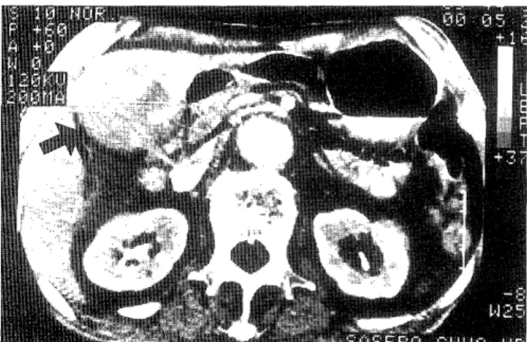
(case 3) CT scan showed a large tumor occupying the right upper abdominal cavity (Fig. 2). Operation revealed a pedunculated HCC arising from the left hepatic lobe and shifting to the left hypochondrium (Fig. 3).
The serum AFP and HBsAg values of patients are tabulated in Table 2. In 6 out of 7 patients, the serum AFP value was normal or slightly below 100 ng/ml. Only one patient (case 7) had abnormal levels of 559.6 ng/ml. One patient was positive for serum HBsAg.
Fig. 2. CT-scan of case 3 showing a large tumor in the reight upper abdominal cavity.
Fig. 1. Hepatic arteriography of case 2. The parenchymal blush of the branch of hepatic artery and middle colic artery are seen.
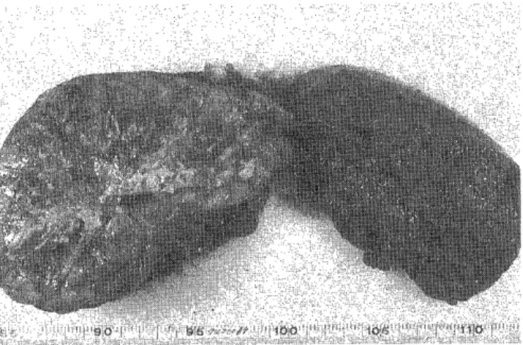
Fig. 3. Resected specimen of case 3 showing 7 x 6 cm encap- sulated massive tumor with 3 cm pedicle.
Table 3. Pathological Findings of Tumor
No. 1 2 3 4 5 6 7
Age •Sex 63•M 53•M 83•M 57•M 62•M 56•M 71•M
fc + + + + + + +
fc-inf + + + + + + + + + +
vp - + + + + - -
Edmondson's II III III I II II II III • N II• III
grade
Underlying PC LC none PC CH PC LC
liver disease
fc: : fromation of capsule, fc-inf : capsular invasiojn of cancer cells.
(-) : no invasion, (+) : intracapsular invasion, (+ +) : cancer invasion extending through capsule.
CH : chronic hepatitis, PC : precirrhosis, LC : liver cirrhosis.
cm to 21 cm with a tumor pedicle (2-7 cm) corresponding to the liver. Grossly, the tumors were firmly encapsulated by fibrous connective tissues, and were separated from the liver by this capsule in the pedicle of the tumor. The cut surface of the tumors also showed several septum toward the periphery from the core (Fig. 4). The invasion of HCC extending through the capsule was noted in all cases.
Microscopically, the presence of tumor thrombus in the portal vein (vpi) was detected in 4 cases (57%).
Fig. 4. Cut surface of the tumor (case 2) showing several septum toward the periphery from the centrum.
Operation and Prognosis
One patient with spontaneous rupture of HCC (case 7) was managed by lipiodolization with 20 mg of mitomycin C. The patient died of hepatic failure associated with DIC
7 days after TAE. Autopsy revealed a huge pedunculated HCC arising from left lobe and a rupture of metastatic tumor in anterior segment. Tumors in 6 out of 7 patients were surgically removed. Five patients underwent partial hepatic resection around the pedicle of the tumor (Fig. 5).
The operations were considered to be radical in all patients judged by intraoperative findings and US and/or CT. The patient who underwent right lobectomy (Case 2) subse- quently developed hepatic failure and died 6 days after surgery. The postoperative outcomes of the 5 patients were excellent in only one patient. The other 4 patients died of hepatic recurrence of the tumor within 3 years.
Fig. 5. Operative finding of case 1 showing a large tumor originating from right lobe of the liver.
Pathologic Features
Pathologic findings of the tumors removed at operation or autopsy are summarized in Table 3. Six out of 7 patients (86%) had underlying liver disease ; chronic hepatitis in one (14%), precirrhosis in 3 (43%) and cirrhosis in 2 (29%). There was one patient with no underlying liver disease. The maximum diameter of tumors ranged from 7
Discussion
Goldberg and Wallenstein first reported in 1934 an extrahepatic growth of HCC arising from the lower surface of the left lobe, with a pedicle of about 1.5 cm in diameter", and Edmondson and Steiner described this type of HCC as pedunculated hepatoma in 1954'). Since these descriptions, several reports have discussed the clinical and pathological features of pedunculated HCC. However, no relevant morphological or etiological events of tumor progression were identified. Goldberg et al. suggested that such an extrahepatically growing HCC might arise from congenitally displaced lobules in Glisson' capsule" .
Miyoshi et al. also suggested that a pedunculated HCC could arise from an accessory lobes). Accessory lobes are often found on the right main lobe, and are frequently attached by pedicles containing blood vessels and bile ducts. They are also often found on the inferior surface of the right lobe, and anatomically, they are found every- where on the hepatic lobes"). On the other hand, in pa- tients with HCC associated with cirrhosis, it is possible that the protruding site of liver cirrhosis may become an extrahepatically growing HCC3) A pedunculated HCC may also arise from ectopic liver tissue. Ectopic liver tissue has been found at various sites, including the triangular ligament of the liver, spleen, umbilical fossa, omentum, retroperitoneium near the celiac axis, and in the gallblad- der and its vicinitya9).
The present series in the pedunculated tumor which exhibited a mobile pedicle and no underlying liver tissues (case 4) appeared to arise from an accessory lobe. In the other 6 patients, pathologic findings of the tumors seem to be characteristic. The tumors had a long pedicle and showed expansive growth with formation of a septum toward the periphery from the tumor centrum and com- plete capsule formation. This may be morphological evidence that these tumors may also arise from an acces- sory lobe rather than developing from the surface of cirrhotic liver. Tumor-associated symptoms in patients with HCC are uncommon, even in advanced stages. In our series, unusual presentations, such as a palpable mass, intractable pain, fever and periodic follow-up examination for underlying liver disease led to tumor detection. Two patients represented spontaneous rupture of HCC. Several factors contributing to rupture of HCC have been pointed out') : pathologically, 1) encapsulated tumor, 2) capsule invasion of cancer cells, 3) portal tumor thrombus, 4 ) tumor size, and physiologically, 1) neovascularization, 2 ) increased intrahepatic shunting, 3) obstruction of bra- nches of the hepatic vein draining the tumor bearing area, 4) portal hypertension due to preexisting cirrhosis. In the current series, we recognized the presence of these patho- logical features in all ceses. The incidence of spontaneous rupture of HCC in Asia or Africa were 4-14.5%10.11), but spontaneous rupture of pedunculated HCC was uncomm- ona13). The physiological factors seems to play important roles in the pathogenesis of the spontaneous rupture.
Abdominal pain was also found in one patient presenting with a mobilable tumor. In patient with pedunculated HCC, abdominal symptoms might be caused by mobiliza- tion or torsion of tumor in the same genesis for sympto- matic accessory lobe of the liver,").
Serum AFP has been frequently used to screen for HCC to differentiate it from other abdominal tumors. In the Japan series, the positive rate of AFP for pedunculated HCC was 66% showing almost equal incidence of all HCC with 68% in 19971,14). However, serum AFP measurement was not the first diagnostic clue for pedunculated HCC in
any of our patients, and most of the patients (6 out of 7) showed normal or slightly elevated levels that remained below 100 ng/ml. Thus , initial and laboratory abnormali- ties are generally minimal with this tumor, so that the role of imaging procedures for diagnosis of the tumor is important.
According to our Japanese series, hepatic angiography, computed tomography, and ultrasonography, which were performed in approximately 90% of the HCC patients, demonstrated positive findings in 90% patients tested").
Angiography may provide useful information on the vascular supply the tumors with hypervascular encapsula- tion. However, it was difficult to differentiate peduncu- lated HCC from tumors of other organs which often infiltrated the liver with neovascularization from hepatic vessels",").
For operation of pedunculated HCC, the resectability rate was relatively high at 65% in this Japanese series".
Most tumors were removed by minute procedures such as wedge resection or subsegmentectomy. However, the prognosis was still poor despite curative resection. This was attributed to the advanced stage of the lesions due to delayed diagnosis. In previous reports, we have identified several important pathologic features of HCC affecting the prognosis'). These include the size of the tumor, the presence of tumor thrombus in portal vein, capsule inva- sion, intrahepatic metastasis, underlying liver diseases, and an adequate resection margin. In the current series, we confirmed this fact with a review of the literature.
In conclusion, early detection of small pedunculated HCC and an adequate hepatic resection for cirrhotic liver are most important for good prognosis of the patients.
References
1) Edmondson HA and Steiner PE : Primary carcinoma of the liver. A study of 100 cases among 48,900 necropsies. Cancer 7: 462-530, 1954 2) Goldberg SJ and Wallenstein H : Primary massive liver cell carcinoma.
Rev Gastroenterol 1: 305-313, 1934
3) Arakawa M, Kage M, Isomura T et al : Pathomorphological studies on hepatocellular carcinoma (HCC) - Seven cases of HCC with an
extrahepatic tumor growth, so-called "Pedunculated hepatoma". Acta
Hepatol Jpn 23: 942-948, 1982
4) The Liver Cancer Study Group of Japan : The general rules for the clinical and pathological study of primary liver cancer. Jpn J Surg 19:
98-129, 1989
5) Miyoshi M, Iwasa N, Fujii H et al : Acta hepatol Jpn 18: 765-772, 1977 6) Omanies S and Jablonsky I : Pedunculated accessory hepatic lobe. Arch
Surg 105: 792-794, 1972
7) Frase C : Accessory lobes of the liver. Ann Surg 035: 127-129, 1952 8) Horiuchi N, Kitamura T, Tateishi R and Wada A: Hepatoma
originated in the retroperitoneal space. Oncology 27: 235-243, 1973 9) Angquist KA, Boquist L, Domellof L : Ectopic liver lobule with portal
cirrhosis. Acta Chir Scand 141: 238-241, 1975
10) Chearanai 0, Plengvanit U, Asavanich C et al : Spontaneous rupture of primary hepatoma: report of 63 cases with particular reference to the
pathogenesis and rationale treatment by hepatic artery ligation.
Cancer 51: 1532-1536, 1983
11) Berman C : Primary carcinoma of the liver: a study in incidence, clinical manifestation, pathology and aetiology. London, H.K. Lews and Co.
Ltd P27,1951
12) Llorente J et al : Symptomatic accessory lobe of the liver associated with absence of the left lobe. Arch Surg 102: 221-223, 1971
13) Hashimoto M, Degawa H, Sakamoto M et al : A case of extrahepatic
growing Hepatocellular carcinoma. Syokakigeka (Japan) 12: 1473- 1477,1989
14) The Liver Cancer Study Group of Japan : Survey and follow-up study of primary liver cancer in Japan -report 11-. Acta hepatol Jpn 36:
208-218, 1995
15) The Liver Cancer Study Group of Japan : Primary liver cancer in Japan. Clinicopathological features and results of surgical treatment.
Ann Surg 211: 277-287, 1990
16) Kimura H, Inoue T, Konishi K et al: Hepatocellular carcinoma presenting as extrahepatic mass on computed tomography. J
Gastroenterol 32: 260-263, 1997
17) Kaneko T, Terabe K, Ito K et al : A case of extrahepatically growing hepatocellular carcinoma fed by the right middle adrenal artery. Jpn J
Gastroenterol Surg 25: 132-135, 1992
18) Shimoyama T, Fukuda Y, Takahira R et al : Clinicopathological study of Hepatocellular carcinoma in relation to tumor size. Acta Med
Nagasaki 33: 228-234, 1988