Acta Med. Nagasaki 46 : 77-79
Case Report
Large Benign Polyp of the Extrahepatic Bile Duct
(Mimicking Bile Duct Cancer)
Takayuki ASAKAWA 1), Masazumi TERADA, Hidenobu OSHIBUCHI, Hiroyoshi ABIRU, Takeshi MATSUO 2), Takashi KANEMATSU 3)
1) Department of Surgery, Oshibuti Hospital
2) Department of Pathology, Kyushu Microbiological Laboratory
3) Second Department of Surgery, Nagasaki University School of Medicine
An 85-year-old woman was admitted because of
epigastric pain. The endoscopic examination showed the presence of duodenal ulcer. Abdominal ultrasonography and computed tomography revealed a dilatation of the common bile duct, but no apparent stones were seen in the biliary tract including the gall bladder. In addition, endoscopic retrograde cholangiography showed a filling de- fect, 20x30 mm in size, in the extrahepatic bile duct. The laboratory data and serum tumor makers were within nor- mal limits. Carcinoma of the extrahepatic bile duct was tentatively made. At operation, an exploration using cholangioscopy and ultrasonography failed to reveal any malignancy. She underwent a local excision of the biliary tumor. Histological examination of the resected specimen showed a benign papillary adenoma. This case is herein re- ported because of rarity of the disease, which was inciden- tally found.
ACTA MEDICA NAGASAKIENSIA 46 : 77-79, 2001
Key Words: biliary tumor, papillary adenoma, local excision
ing from the extrahepatic bile duct.
Case report
An 85-year-old woman complained of epigastric pain. Physical examination revealed tenderness in the epigastric region. She had been operated on for a clo- sure of the duodenum due to a perforated ulcer on 1993.
The laboratory data, including the carcinoembryonic antigen and carbohydrate antigen 19-9 levels in the serum, were all within normal limits. The endoscopic examination revealed a duodenal ulcer. Ultrasonography showed dilatation of the extrahepatic bile duct.
Computed tomography revealed dilatation and a mass region in the extrahepatic bile duct (Fig. 1).
Endoscopic retrograde cholangiography disclosed a 20 x 30 mm filling defect in the common bile duct (Fig. 2).
At operation which was performed on August 21,1996, the common hepatic duct was found to be di- lated. A mass was palpated in the anterior wall of the
Introduction
The occurrence of benign polyps in the biliary tract are comparatively rare"'). Most of the early cancers of the bile duct reported have been polyps 3 ' 9 1. In addi- tion, several reports have suggested a relationship be- tween adenoma and cancer in the biliary tract 5.6 ). It is therefore difficult to determine whether biliary polyps are benign or malignant at preoperative examinations.
We herein report a case of papillary adenoma aris-
Address Correspondence: Takayuki Asakawa, M.D.
Department of Surgery, Oshibuti Hospital, 37-1 Satomen Mikuriya, Matsuura 859-4752, Japan
TEL: +81-956-75-0311 FAX: +81-956-75-1100
Figure 1. Computed Tomography showed the dilatation of
the extrahepatic bile duct.
Figure 2. Endoscopic retrograde cholangiogram showed a fill-
ing defect measuring 20 x30 mm in diameter the
extrahepatic bile duct.
bile duct at the junction of the cystic and common he- patic duct. There was no evidence of involvement of the perineural lymph node or direct invasion of the tumor into adjacent vascular system. No abnormality was seen in the liver or pancreas on intraoperative ultrasonography. The common hepatic bile duct was opened, and a 20 x 30 mm papillary polyp was re- moved by a wedge resection. Stones or other tumor in the gall-bladder or biliary tract was not seen on the intraoperative ultrasonography and choledochoscopy.
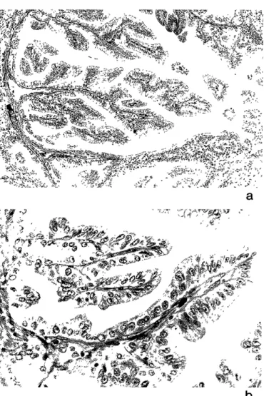
The repair of the biliary duct was done by inter- rupted sutures. Histopathologic diagnosis of surgical specimen was papillary adenoma with no evidence of malignancy (Fig. 3a,b).
The postoperative course has been uneventful and she has been doing well 54 months after surgery.
Discussion
Benign polyp of the common bile duct are rare.
Clinical findings of benign tumors in the bile duct are jaundice, pain and fever caused by luminal obstruc- tion and cholangitis. Our patient had epigastralgia but this was probably due to duodenal ulcer.
The biliary tumor is diagnosed preoperatively by a combination of ultrasonography, computed tomography and cholangiography. Endoscopic retrograde cholangio- graphy or percutaneous transhepatic cholangiography
Figure 3. a, b Microscopic features of the resected bile duct tumor (Hematoxylin and Eosion).a. low magnification view of the lesion (x 10). A papillary proliferation of the bile duct glands extending to the lumen without invasive growth.
b. high magnification view (x100). The epithelium is com- posed of tall and columnar cells which accompanied the po- larity of nucleus and thin stroma. No features of malignancy were observed.
are useful in diagnosing tumors of the bile duct based on the location, shape and spread of the tumor along the biliary tract. However it is difficult to determine whether a polyp is malignant or benign. Because most of the early bile duct carcinoma reported are polypoid and are similar to benign polyps in shape', 8) . Moreover, several reports have suggested the exis- tence of some relationship between adenoma and car- cinoma in gall bladders' and extrahepatic bile duct".
Kozuka et al'). reported that the benign adenomas ranged in size from 3 to 12 mm in diameter, while adenomas with malignant change ranged from 12 to 35 mm in diameter. Also biliary cancer has character- istic of involvement of perineural lymph space or di- rect invasion of the tumor into adjacent vascular
system by ultrasonography, computed tomography.
Otherwise, the preoperative and intraoperative find- ings were no invasion of the organ and lymph node metastasis around the biliary duct in spite of an inten- sive exploration. In this case, the tumor was also easy to resect because the tumor was on the anterior wall of the biliary duct and bile duct was dilated. This case has been done complete resection of the biliary tumor in pathological findings.
A local excision is a treatment of choice for solitary benign tumors of the biliary tree, with a tumor free margin to minimize the possibility of recurrence""', and intraoperative choledochoscopy and ultrasonography should also be performed to confirm a complete clear- ance of the biliary duct. This case has potential of other region of extrahepatic bile duct, gall bladder and intrahepatic bile duct because case of diffuse biliary papillomatosis has been reported"'. Postoperative ob-
servations using ultrasonography and computed
tomography must also be regularly performed.
References
1) Dowdy GS, Olin WG, Shelton EL, Waldron GW. Benign tumors of the extrahepatic bile ducts: Report of three cases and review of
the literature. Arch Surg 85: 503-513, 1962
2) Bruhans R, Myers RT. Benign neoplasms of the extrahepatic biliary ducts. Am Surg 37: 161-166, 1971
3) Yamaguchi K, Enjoji M, Nakayama F. Cancer of extrahepatic bile duct: A clinicopatholgic study of immunohistochemistry for CEA,
CA19-9, and p21. World J Surg 12: 11-17, 1988
4) Tsunoda T, Eto T, Koga M, et al. Early carcinoma of the extrahepatic bile duct. Jpn J Surg 19: 691-698, 1989
5) Kozuka S, Tsubone M, Yasui A, Hachisuka K. Relation of ade- noma to carcinoma in gall bladder. Cancer 50: 2226-2234, 1982 6) Kozuka S, Tsubone M, Hachisuka K. Evolution of carcinoma in
the extrahepatic bile ducts. Cancer 54: 65-72, 1984
7) Ouchi K, Matsuno S, Sato T. Long-term survival in carcinoma of the biliary tract. Analysis of prognostic factors in 146 resections.
Arch Surg 124: 248-252, 1989
8) Todoroki T, Okamura T, Fukao K, et al. Gross appearance of car- cinoma of the main hepatic duct and its prognosis. Surg Gynecol
Obstet 150: 33-40, 1980
9) Marsh JL, Dahms B, Longmire WP. Cystadenorna and cystadenocarcinoma of the biliary system. Arch Surg 109: 41-43,
1974
10) Short WF, Nedwich A, Levy HA, Howard JM. Biliary cystadenoma;
report of a case and review of the literature: Arch Surg 102: 78- 80, 1971
11) Neumann RD, LiVolsi VA, Rosenthal Ns, Burrell M, Ball TJ.
Adenocarcinoma in biliary papillomatosis. Gastroenterology 70:
778-782, 1976