Table 1. IVM/IVF of porcine oocytes.
Four replicated trials were carried out. Percentages are expressed as mean ± SEM.
aOocytes at the metaphase-II stage or penetrated oocytes with one first and one second polar body. bPenetrated oocytes with one first and one second polar body.
cMPN; male pronucleus(ei).
dWith both single MPN and female pronuclei. Total No. of
oocytes examined
No. (%) of matured oocytesa
No.(%) of penetrated oocytesb
Penetration MPN
c
formation Monospermy d
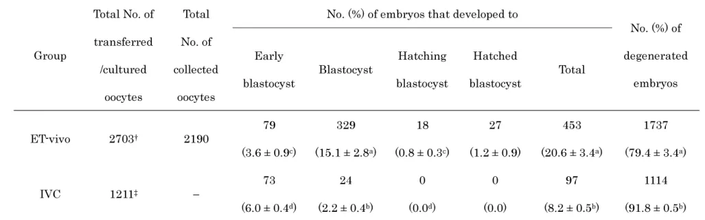
Table 2. Development of IVM/IVF oocytes to the blastocyst stage on Day 5 after transfer to recipients or culture in vitro.
Percentages are expressed as mean ± SEM of total number of examined oocytes.
Total of 2703 IVM/IVF oocytes were transferred to 10 recipients on Day 0 (in vivo culture) and embryos were recovered from 10 recipients on Day 5.
Total 1211 IVM/IVF oocytes were subsequently cultured in vitro without transfer until Day5 of IVF. Values in the same column with different superscripts differ significantly (a,bP < 0.01, c,dP < 0.05).
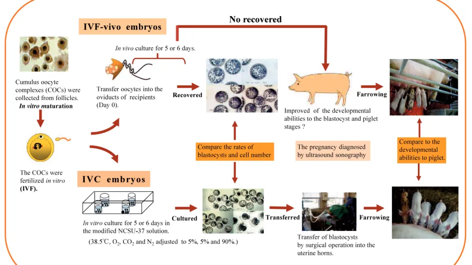
Figure 3. In vitro-matured and fertilized oocytes were transferred to recipients and recovered (ET-vivo), or subjected to continuous in vitro culture (IVC). Blastocysts after collection and culture were then evaluated.
A) ET-vivo blastocysts recovered on Day 5 (Day 0 = in vitro fertilization). An inner cell mass in some blastocysts was clearly confirmed (arrows).
B) ET-vivo hatched blastocysts on Day 5 (arrows).
C) ET-vivo blastocysts on Day 5. These had adherent external glass-like material (arrows). D) IVC blastocysts on Day 6 had degenerated cells (arrows). Scale bar = 100 µm.
A
B
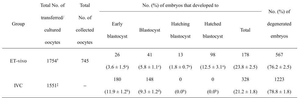
Table 3. Development of IVM/IVF oocytes to the blastocyst stage on Day 6 after transfer to recipients or culture in vitro. Group Total No. of transferred/ cultured oocytes Total No. of collected oocytes
No. (%) of embryos that developed to
No. (%) of degenerated embryos Early blastocyst Blastocyst Hatching blastocyst Hatched blastocyst Total ET-vivo 1754 745 26 (3.6 ± 1.5a) 41 (5.8 ± 1.1c) 13 (1.8 ± 0.7a) 98 (12.5 ± 3.1a) 178 (23.8 ± 2.5) 567 (76.2 ± 2.5) IVC 1551 180 (11.9 ± 1.2b) 148 (9.3 ± 1.2d) 0 (0.0b) 0 (0.0b) 328 (21.2 ± 1.8) 1223 (78.8 ± 1.8)
Percentages are expressed as mean ±SEM of the total number of examined oocytes.
Total of 1754 IVM/IVF oocytes were transferred to 8 recipients on Day 0 (in vivo culture) and embryos were recovered from 7 recipients on Day 6.
Total 1551 IVM/IVF oocytes were subsequently cultured in vitro without transfer until Day 6 of IVF. Values in the same column with different superscripts differ significantly (a,b P < 0.01, c,dP < 0.05).
Table 4. Embryo quality on Day 5 after transfer to recipients or culture in vitro.
Group Mean number of cells per blastocyst (No. of oocytes examined)
Early blastocyst Total
ET-vivo 44.6 ± 17.7a (25) 89.2 ± 37.3a (43) 72.8 ± 38.2a (68) IVC 21.0 ± 7.8b (64) 25.0 ± 8.8b (23) 22.1 ± 8.1b (87) Cell numbers are expressed as mean ± SEM.
IVM/IVF oocytes were transferred to 10 recipients on Day 0 and the blastocysts were recovered from 10 recipients on Day 5.
IVM/IVF oocytes were cultured in vitro until Day 5 of IVF. Replication was performed 8 times. a,bValues in the same column with different superscripts differ significantly (P < 0.01).
Table 5. Embryo quality on Day 6 after transfer to recipients or culture in vitro.
Group Mean number of cells per blastocyst (No. of oocytes examined)
Early Blastocyst Total
ET-vivo 25.5 ± 10.7 (18) 94.4 ± 47.1a (61) 78.7 ± 50.8a (79) IVC 29.4 ± 10.3 (44) 49.9 ± 16.0b (44) 39.7 ± 16.9b (89) Cell numbers are expressed as mean ± SEM.
IVM/IVF oocytes were transferred to 8 recipients on Day 0 and the blastocysts were recovered from 7 recipients on Day 6.
IVM/IVF oocytes were cultured in vitro until Day 6 of IVF. Replication were performed 10 times. a,bValues in the same column with different superscripts differ significantly (P < 0.01).
Figure 4. In vitro-matured and fertilized oocytes were transferred to recipients and recovered (ET-vivo), or subjected to continuous in vitro culture (IVC). Blastocysts after collection and culture were fixed, stained and evaluated.
A) ET-vivo blastocysts on Day 6 (Day 0 = in vitro fertilization). B) ET-vivo hatching blastocysts.
C) ET-vivo hatched blastocysts on Day 6. D) IVC blastocysts on Day 6. Scale bar = 100 µm.
A
B
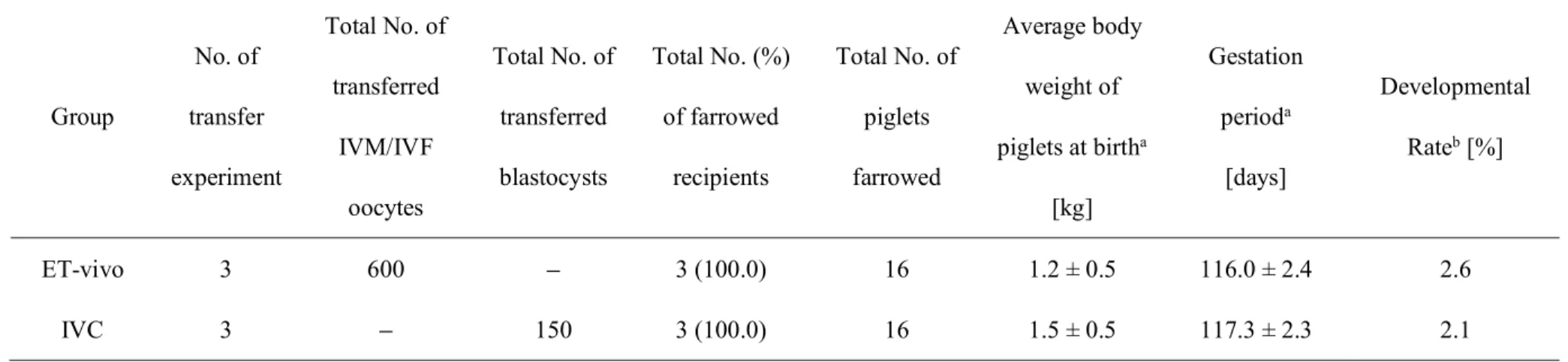
Table 6. Production of piglets derived from IVM/IVF oocytes (transferred on Day 0) and blastocysts cultured in vitro for 6 days. Group No. of transfer experiment Total No. of transferred IVM/IVF oocytes Total No. of transferred blastocysts Total No. (%) of farrowed recipients Total No. of piglets farrowed Average body weight of piglets at birtha [kg] Gestation perioda [days] Developmental Rateb [%] ET-vivo 3 600 3 (100.0) 16 1.2 ± 0.5 116.0 ± 2.4 2.6 IVC 3 150 3 (100.0) 16 1.5 ± 0.5 117.3 ± 2.3 2.1
Three recipients farrowed 5, 5 and 6 piglets in each group. aValues are expressed as mean ± SEM.
bDevelopmental rates were calculated from the total number of piglets per the total number of the 600 IVM/IVF oocysts transferred (200 per recipient) and 150 blastocysts (50 per recipient) that were cultured in vitro from 756 oocytes.
Figure 5. Piglets 10 days after farrowing. These were derived from blastocysts that had been transferred after in vitro culture for 6 days.
Table 7. Development of IVM/IVF oocytes of using frozen-thawed semen from several boars.
Percentages are expressed as mean ± SEM of total number of examined oocytes. B-1, 2, 3, 4 and 5; Bhaksher, L; Landrace, W; Large White, and D; Duroc. Total 3,910 IVM/IVF oocytes were subsequently cultured in vitro until Day6. Values in the same column with different superscripts differ significantly (a,c ; b,cP < 0.01,a,bP < 0.05). Boars Replication Total No. of cultured embryos No. (%) of blastocysts No. (%) of degenerated embryos B-1 6 518 84 (15.9 ± 3.5 ab) 434 (84.1± 3.5 ab) B-2 6 538 106 (19.5 ± 1.0 a) 432 (80.5± 1.0 a) B-3 11 480 50 (12.5 ± 2.6 b) 430 (87.5± 2.6 b) B-4 8 348 66 (21.4 ± 4.0 a) 282 (78.6± 4.0 a) B-5 7 512 10 (1.4 ± 0.9 c) 502 (98.6± 0.9 c) L 4 268 23 (9.0 ± 2.0 b) 245 (92.7± 2.0 b) W 7 546 108 (20.6 ± 2.0a) 438 (79.4± 2.0 a) D 7 700 111 (15.0 ± 1.9 ab) 589 (85.0± 1.9 ab)
Table 8. IVM/IVF of porcine oocytes of several IVF-condition. Boars Matured oocytesa late (%) IVF-Times (h) Caffeine concentration (mM) Sperm concentration (1×10n) Polyspermy late (%) Monospermy late (%) B-6 70.1 3 2 5 0.0 ± 0.0a 0.3 ± 0.3a 5 2 5 3.4 ± 0.5b 11.3 ± 2.1b 5 5 5 3.2 ± 1.2b 11.2 ± 3.3b 5 5 6 3.6 ± 1.3b 14.3 ± 3.6b B-7 70.6 3 2 5 12.5 ± 2.8a 8.3 ± 2.2a 5 2 5 3.7 ± 3.3b 11.1 ± 2.3a 5 5 5 6.2 ± 3.9b 25.0 ± 8.2b 5 5 6 8.1 ± 5.4ab 28.0 ± 9.4b B-8 72.8 3 2 5 0.1 ± 0.4a 0.3 ± 0.8a 5 2 5 0.1 ± 0.3a 0.3 ± 0.7a 5 5 5 4.1 ± 2.1b 7.8 ± 3.8b 5 5 6 3.0 ± 1.6b 7.0 ± 1.8b
Percentages are expressed as mean ± SEM of total number of examined oocytes.
B-6, 7, 8 ; Bhaksher, frozen semen indicated of low development late of the blastocyst on normal IVF-condition (IVF 3 h, Caffeine 2mM, Sperm concentration 1×10⁵ sperm/mL).
Table 9. Development of IVM/IVF oocytes to the blastocyst stage of several IVF-condition. Boars Total No. of cultured embryos IVF-Times (h) Caffeine concentration (mM) Sperm concentration (1×10n) Developed blastocyst (%) B-6 249 3 2 5 0.4 ± 0.8a 148 5 2 5 10.1 ± 0.5b 160 5 5 5 8.8 ± 6.3ab 200 5 5 6 15.5 ± 4.6b B-7 125 3 2 5 7.2 ± 1.0b 72 5 2 5 11.1 ± 3.4b 130 5 5 5 16.2 ± 3.6c 125 5 5 6 17.6 ± 10.3bc B-8 161 3 2 5 0.6 ± 0.8a 96 5 2 5 1.0 ± 0.7a 130 5 5 5 6.2 ± 3.8ab 130 5 5 6 10.0 ± 1.8b
Percentages are expressed as mean ± SEM of total number of examined oocytes.
B-6, 7, 8 ; Bhaksher, frozen semen indicated of low development late of the blastocyst on normal IVF-condition (IVF 3h, Caffeine 2 mM, Sperm concentration 1× 10⁵ sperm/mL).
Total 1,726 IVM/IVF oocytes were subsequently cultured in vitro until Day6.
Table 10. IVF status of frozen thawed boar ejaculated sperm .
Total No. of oocytes examined
No. (%) of fertilized oocytesa
No.(%) of penetrated oocytesb Sperm
penetration MPN
c formation Monospermyd
145 109 (75.7 ± 3.3) 51 (46.7± 3.2) 39 (76.1 ± 3.2) 29 (56.0 ± 4.1) used
In vitro matured oocytes were inseminated, cultured and fixed at 10 h post-insemination. Four
replicated trials were carried out. Percentages are expressed as mean ± SEM.
aOocytes at the metaphase-II stage or penetrated oocytes with one first and one second polar bodies. bPenetrated oocytes with one first and one second polar bodies.
cMPN; male pronucleus(ei).
Table 11. Development of IVF and Parthenogenetic (PA) oocytes to the blastocyst stage.
Group
Total No. of cultured
oocytes
No. (%) of blastocysts No. (%) of
degenerated embryos Early
blastocyst Blastocyst Total
IVF-Day 6 914 72 (8.3 ± 0.9a) 79 (8.2 ± 0.9) 151(16.6 ± 1.1) 763 (83.4 ± 1.1) PA-Day 6 1001 120 (11.7 ± 1.6b) 77 (8.9 ± 1.3) 197 (20.5 ± 2.0) 804 (79.5 ± 2.2 )
Percentages are expressed as mean ± SEM of total number of examined oocytes.
< . Replication were performed 10 times.
Table 12. Classification of blastocysts on Day 6 in culture.
Group
% Early Blastocysts % Blastocyst
Code 1 Code 2 Code 3 Code 1 Code 2 Code 3
IVF 7.0 ± 4.1 43.1 ± 5.2 49.9 ± 5.2 47.9 ± 4.7a 46.3 ± 4.2 5.8 ± 3.1 PA 13.4 ± 4.0 42.6 ± 7.7 44.0 ± 5.5 62.8 ± 7.0b 35.5 ± 6.9 3.1 ± 1.7
Embryos lank are expressed as mean ± SEM.
.
The quality lank (Code 1, 2, or 3) is based on IETS embryo classification. Code 1 is the most excellent. Values in the same column with different superscripts differ significantly (a,b P < 0.05).
Table 13. Diameter and cell number of blastocysts on Day 6 in culture.
Group
Mean diameter [µm] of blastocyst (No. examined) Mean cell numbers per blastocyst (No. examined) Early blastocyst Blastocyst Total$ Early blastocyst Blastocyst Total IVF 162.4 ± 1.2 (55) 191.4 ± 1.5 (54) 177.2 ± 1.9 (92) 30.4 ± 1.9c (50) 50.2 ± 1.9a (39) 39.1 ± 1.7a (89)
PA 162.8 ± 0.9 (91) 192.1 ± 1.7 (48) 177.9 ± 1.9 (92) 25.7 ± 1.8d (45) 37.4 ± 2.3b (33) 30.6 ± 1.6b (78)
Values are expressed as mean ± SEM.
.
$Total 92 blastocysts (48 in each stage) were randomly chosen before calculation.
Figure 7. Putative zygotes (IVF oocytes) and parthenotes were cultured in vitro for 6 days (Day 0 = in vitro fertilization or parthenogenetic stimulation).
A) IVF blastocysts
B) Parthenogenetic blastocysts C) Code 1 rank IVF blastocyst
D) Code 1 rank parthenogenetic blastocyst. E) Code 1 rank IVF early blastocyst
Figure 8. Putative zygotes (IVF oocytes) and parthenotes were cultured in vitro for 6 days and fixed, stained and evaluated for cell number.
A) IVF blastocyst (56 cells)
Figure 9. Pregnancy was detected on 24 day (ultrasonic diagnosis) after embryos transfer (arrows).
Table 14. Pregnancy and farrowing after transfer of vitrified and warmed morula (VM) with or without co-transfer of parthenogenetic blastocysts (PAB).
Group No. of transfer experiment Total No. of transferred embryos Total No. (%) of pregnant recipients Total No. (%) of recipients farrowed
Average of abortion day ± SEM
VM + PAB 5 20 4 (83.0) 0 (0.0) 42.5 ± 7.8
VM 5 20 3 (60.0) 1§ (20.0) 27.0 ± 1.7
Each of 10 VM and 10 PAB stage embryos are transferred.
Pregnancy was detected on 17 day (no return estrus) and 24 day (ultrasonic diagnostic) after embryo transfer. §4 piglets (1 males and 3 females) were delivered.
Ankrah NA, Appoah-Opong R. Toxicity of low levels of methylglyoxal: depletion of blood glutathione and adverse effect on glucose tolerance in mice. Toxicology Letters.1999; 20:61 67.
Beebe L, Cameron R, Blackshaw A, Higgins A, Nottle M. Piglets born from centrifuged and vitrified early and peri-hatching blastocysts. Theriogenology 2002;57:2155 2165.
Berthelot F, Martinat-botte F, Perreau C, Terqui M. Birth of piglets after OPS vitrification and transfer of compacted morula stage embryos with intact zona pellucida. Reproduction Nutrition Development 2001;41:267 272.
Bureau M, Bailey JL, Sirard MA. Influence of oviductal cells and conditioned medium on porcine gamaetes. Zygote 2000;8:139 144.
Cuello C, Gil MA, Parrilla I, Tornel J, Vázquez JM, Roca J, Berthelot F, Martinat-Botté F, Martínez EA. Vitrification of porcine embryos at various developmental stage using different ultra-rapid cooling procedures. Theriogenology 2004;62:353 361.
Cray Isome S, Rong Feng L, Kristin M, Randall S. 2012. Timing of first embryonic cleavage is a positive indicator of the in vitro developmental potential of porcine embryos derived from in vitro fertilization, somatic cell nuclear transfer and parthenogenesis. Molecular Reproduction and
Development 79, 197 207.
Davis DL, Day BN. Cleavage and blastocyst formation by pig eggs in vitro. Journal of Animal Science 1978;46:1043 1053.
Day BN, Abeydeera LR, Johnson LA, Welch GR, Wang WH, Cantley TC, Rieke A. Birth of piglets preselected for gender following in vitro fertilization of in vitro matured pig oocytes by X and Y bearing spermatozoa stored by high speed flow cytometry. Theriogenology 1998;49:360.
Eunhye K, Yubyeol J, Dae Y, Eunsong L. Antioxidative effect carboxyethylgermanium sesquioxide (Ge-132) on IVM of porcine oocytes and subsequent embryonic development after parthenogenetic activation and IVF. Theriogenology 2015;62:353 361.
Flood MR, Wiebold JL. Glucose metabolism by preimplantation pig embryos. Journal of
Reproduction and Fertility 1988;84:7 12.
Fujino Y, Nakamura Y, Kobayashi H, Kikuchi K. Relationship between time elapsed after hCG administration and developmental stage in porcine embryos collected from prepubertal gilts. Journal
of Reproduction and Development 2006;52:267 275.
Fujino Y, Kojima T, Nakamura Y, Kobayashi H, Kikuchi K, Funahashi H. Metal mesh vitrification (MMV) method for cryopreservation of porcine embryos. Theriogenology 2008;70:809 817.
Funahashi H, Kim NH, Stumpf TT, Cantley TC, Day BN. Presence of organic osmolytes in maturation medium enhances cytoplasmic maturation of porcine oocytes. Biology of Reproduction 1996;54:1412 1419.
Funahashi H, Cantley TC, Day BN. Synchronization of meiosis in porcine oocytes by exposure to dibutyryl cyclic adenosine monophosphate improves developmental competence following in vitro fertilization. Biology of Reproduction 1997;57:49 53.
Gil M, Aluminana C, Roca J, Vazquez J, Martinez E. Boar semen variability and its effects on IVF efficiency. Theriogenology 2008;70:1260 1268.
Hongsheng Men, Lee D. Spate, Clifton N. Murphy, and Randall S, Prather. Cryopreservation of in-vitro-produced early-stage porcine embryos in a closed system. BioResearch Open Access. 2015;4:1:258 265.
Hou D, Su M, Li X, Li Z, Yun T, Zhao Y, Zhang M, Zhao L, Li R, Yu H, Li X. The efficient derivation of trophoblast cells from porcine in vitro fertilized and parthenogenetic blastocysts and culture with ROCK Inhibitor Y-27632. PLoS ONE 2015;10:e0142442.
Hunter RHF. Porcine ovulation after injection of human chorionic gonadotrophin. The Veterinary
Record 1967;81:21 23.
Kashiwazaki N, Kikuchi K, Suzuki K, Noguchi J, Nagai T, Kaneko H, Shino M. Development in vivo and in vitro to blastocysts of porcine oocytes matured and fertilized in vitro. Journal of Reproduction
and Development 2001;47:303 310.
Kashiwazaki N, Shino M. Ability of in vitro manipulated porcine embryos to develop to piglets.
Journal of Reproduction and Development 2001;47 (suppl):S35 S39.
Kawarasaki T, Otake M, Tsuchiya S, Shibata M, Matsumoto K, Isobe N. Co-transfer of parthenogenotes and single porcine embryos leads to full-term development of the embryos. Animal
Kikuchi K, Nagai T, Kashiwazaki N, Ikeda H, Noguchi J, Shimada A, Soloy E, Kaneko H. Cryopreservation and ensuing in vitro fertilization ability of boar spermatozoa from epididymides stored at 4 °C. Theriogenology 1998;50:615 623.
Kikuchi K, Kashiwazaki N, Noguchi J, Shimada A, Takahashi R, Hirabayashi M, Shino M, Ueda M, Kaneko H. Developmental competence, after transfer to recipients, of porcine oocytes matured, fertilized, and culture in vitro. Biology of Reproduction 1999;60:336 340.
Kikuchi K, Onishi A, Kashiwazaki N, Iwamoto M, Noguchi J, Kaneko H. Akita T, Nagai T. Successful piglet production after transfer of blastocysts produced by a modified in vitro system.
Biology of Reproduction 2002;66:1033 1041.
King TJ, Dobrinsky R, Zhu J, Finlayson HA, Bosma W, Harkness L, Ritchie WA, Travers A, McCorquodale C, Day BN, Dinnyes A, De Sousa PA, Wilmut I. 2002. Embryo development and establishment of pregnancy after embryo transfer in pigs: coping with limitations in the availability of viable embryos. Reproduction 123, 507 515.
Kouba A, Abeydeera LR, Alvarez IM, Day BN, Buhi WC. Effects of the porcine oviduct-specific glycoprotein in fertilization, polyspermy, and embryonic development in vitro. Biology of
Reproduction 2000;63:242 250.
Kurebayashi S, Miyake M, Okada K, Katayama M, Miyano T, Kato S. Development of porcine blastocysts from in vitro-matured and activated haploid and diploid oocytes. Theriogenology 1996;46:1027-1036.
Kurebayashi S, Miyake M, Okada K and Kato S. Successful implantation of in vitro-matured, electro-activated oocytes in the pig. Theriogenology 2000;53:1105 1119.
Marchal R, Feugang JM, Perreau C, Venturi E, Terqui M, Mermillod P. Meiotic and developmental competence of prepubertal and adult swine oocytes. Theriogenology 2001;56:17 29.
Mattioli M, Bacci M L, Galeati G, Seren E. Developmental competence of pig oocytes matured and fertilized in vitro. Theriogenology 1989;31: 1201 1207.
Menino AR, Wright RW. Development of one-cell porcine embryos in two culture systems. Journal
of Animal Science 1982;54:583 588.
Misumi K, Suzuki M, Saito S, Saito N. Successful production of piglets derived from vitrified morulae and early blastocysts using a microdroplet method. Theriogenology 2003;60: 253 260.
Miyoshi K, Mizobe Y. Osmolarity-and stage-dependent effects of glycine on parthenogenetic development of pig oocytes. Journal of Reproduction and Development 2014;60:349 354.
Nagai T, Takahashi T, Masuda H, Shioya Y, Kuwayama M, Fukushima M, Iwasaki S, Hanada A. In-vitro fertilization of pig oocytes by frozen boar spermatozoa. Journal of Reproduction and Fertility 1988;84:585 591.
Nagai T, Yamauchi N, Kikuchi K. Nuclear and cytoplasmic maturation in vitro of porcine oocytes.
Nagai T. The improvement of in vitro maturation system for bovine and porcine oocytes.
Theriogenology 2001;55:1291 1301.
Nakazawa Y, Misawa H, Fujino Y, Tajima S, Misumi K, Ueda J, Nakamura Y, Shibata T, Hirayama Y, Kikuchi K. Effect of volume of non-surgical embryo transfer medium on ability of porcine embryos to survive to term. Journal of Reproduction and Development 2008;54:30-34.
Niwa T (ed.). Manual for Cryopreservation of pig spermatozoa [in Japanese]. Tokyo : Japanese
Artificial Insemination Association;1989.
Oguri N. Recent progress in porcine embryos transfer and cryopreservation (in Japanese). Journal
Swine science 1990;27:80 86.
Onishi A, Iwamoto M, Akita T, Mikawa S, Takeda K, Awta T, Hanada H, Perry A. Pig cloning by microinjection of fetal fibroblast nuclei. Science 2000;289:1188 1190.
Pelaez J, Breininger E, Alegre B, Pena F, Dominquez J. In vitro evaluation in the quality of fertilizing capacity of boar spermatozoa in 0.25 ml straws. 41. Reproduction in Domestic Animals 2006;41:153 161.
Petters RM, Wells KD. Culture of pig embryos. Journal of Reproduction and Fertility 1993; 48 (suppl):61 73.
Polge C, Rowson L, Chang M. The effect of reducing the number of embryos during early stage of gestation on the maintenance of pregnancy in the pig. Journal of Reproduction and Fertility 1966;12:395 397.
Pollard JW, Leibo SP. Chilling sensitivity of mammalian embryos. Theriogenology 1994 ;32:101 106.
Rath D, Niemann H. In vitro fertilization of porcine oocytes with fresh and frozen-thawed ejaculated or frozen-thawed epididymal semen obtained from identical boars. Theriogenology 1997;47:785 793.
Robertson I, Nelson RE. Certification and identification of embryos. In: Stringfellow DA, Daniel Givens M (eds), Manual of the International Embryo Transfer Society, Fourth Edition, Chapter 2009;9:86 105. International Embryo Transfer Society, Champaign.
Sangho R and Woo-suk H. In vitro development of porcine parthenogenetic and cloned embryos: comparison of oocyte-activating techniques, various culture systems and nuclear transfer methods.
Reproduction Fertility and Development 2002;14:93 99.
Schini SA, Bavister BD. Two-cell block to development of cultured hamster embryo is caused by phosphate and glucose. Biology of Reproduction 1988;39:1183 1192.
Selles E, Gadea J, Romar R, Matas C, Ruiz S. Analysis of in vitro fertilizing capacity to evaluate the freezing procedures of boar semen to predict the subsequent fertility. Reproduction in Domestic
Shibata M, Otaake M, Tsuchiya S, Chikyu M, Horiuchi A, Kawrasaki T. Reproductive and growth performance in Jin Hua pigs cloned from somatic cell nuclei and the meat quality of their offspring.
Journal of Reproduction and Development 2006;52:583 590.
Shi JM, Tian XZ, Zhou GB, Wang L, Gao C, Zhu SE, Zeng SM, Tian JH, Liu GS. Melatonin exists in porcine follicular fluid and improves in vitro maturation and parthenogenetic development of porcine oocytes. Journal of Pineal Research 2009;47:318 323.
Snedecor GW, Cohran WG. Statistical Methods, 8th ed. Ames, IA: The Iowa State University Press 1989:273 296.
Sturmey RG, Leese HJ. Energy metabolism in pig oocytes and early embryos. Reproduction 2003;126:197 204.
Sone M, Chikyu M, Yoshida M, Banba K, Ogasa A. 1992. Prolonged storage on boar semen in liquid form (In Japanese). The Japanese Society of Swine Science 29, 41 50.
Suzuki C, Yoshioka K, Itoh S, Kawarasaki T, Kikuchi K. In vitro fertilization and subsequent development of porcine oocytes using cryopreserved and liquid-stored spermatozoa from various boars. Theriogenology 2005;64(6):1287 1296.
Suzuki K, Eriksson B, Shimizu H, Nagai T, Rodriguez-Martinez H. 2000. Effect of hyaluronan on monospermic penetration of porcine oocytes fertilized in vitro. International Journal of Andrology 23, 13 21.
Suzuki K. 2001. In vitro fertilizing ability of porcine spermatozoa. Hokkaido, Japan: Hokkaido University. Thesis.
Ushijima H, Yoshioka H, Esaki R, Takahasi K, Kuwayama M, Nakane T and Nagasgima H. Improved survival of vitrified in vivo-derived porcine embryos. Journal of Reproduction and Development 2004;50:481 486.
Wang W, Niwa K, Okuda K. In vitro penetration of pig oocytes matured in culture by frozen-thawed ejaculated spermatozoa. Journal of Reproduction and Fertility 1991;93: 491 496.
Wright RW. Successful culture in vitro of swine embryos to the blastocyst stage. Journal of Animal
Science 1977;4:854 858.
Wu GQ, Jia BY, Li JJ, Fu XW, Zhou GB, Hou YP, Zhu SE. L-carnitine enhances oocyte maturation and development of parthenogenetic embryos in pigs. Theriogenology 2011;76:785 793.
Wu GQ, Quan GB, Shao QY, Lv CR, Jiang YT, Zhao ZY, Hong QH. Cryotop vitrification of porcine parthenogenetic embryos at the early developmental stages. Theriogenology 2016;85:434 440.
Yoshida M, Mizoguchi Y, Ishigaki K, Kojima T, Nagai T. Birth of piglets derived from in vitro fertilization of pig oocytes matured in vitro. Theriogenology 1993;39:1303 1311.
Yoshioka K, Suzuki C, Itoh S, Kikuchi K, Iwamura S, Martinez H R. Production of piglets derived from in vitro-produced blastocysts fertilized and cultured in chemically defined media: effects of theophylline, adenosine, and cystein during in vitro fertilization. Biology of Reproduction