Salicylic acid signaling and chitosan signaling in Arabidopsis guard cells
2017, 9
Md. Yeasin Prodhan
Graduate School of
Environmental and Life Science (Doctor’s Course)
OKAYAMA UNIVERSITY, JAPAN
Salicylic acid signaling and chitosan signaling in Arabidopsis guard cells
A thesis
Presented to Graduate School of Environmental and Life Science Okayama University
In partial fulfillment of the requirement for the degree of Doctor of Philosophy
Submitted by Md. Yeasin Prodhan
Department of Biofunctional Chemistry
Graduate School of Environmental and Life Science Okayama University, Japan
2017, 9
1
CONTENTS
List of figures 5
Abbreviations 6
Chapter 1 General Introduction 7
1.1 Guard cells and stomata 7
1.2 Stomatal movement 8
1.3 SA-induced stomatal closure 9
1.4 Calcium-dependent protein kinase 10
1.5 Open stomata 1 protein kinase 10
1.6 Ion channel in guard cell signaling 11
1.7 Chitosan-induced stomatal closure 11
1.8 Second messengers in guard cells signaling 12
1.9 ABA-induced stomatal closure 14
1.10 Purpose of the study 15
Chapter 2 Guard cell salicylic acid signaling is integrated into abscisic acid signaling via Ca2+/CPK-dependent pathway
16
2.1 Abstract 16
2.2 Introduction 17
2.3 Materials and Methods 19
2.3.1 Plants and Growth Conditions 19
2.3.2 Analysis of Stomatal Apertures 19
2.3.3 Detection of ROS production 20
2.3.4 Electrophysiology 20
2.3.5 Statistical analysis 21
2.4 Results 21
2.4.1 SA-induced stomatal closure is impaired in CPK3 CPK6 double mutants
21
2
2.4.2 SA-induced stomatal closure is not impaired in OST1 kinase mutant
24
2.4.3 SA-induced stomatal closure is impaired in SLAC1 mutants
25
2.4.4 SA Activation of S-Type Anion Channels is impaired in CPK3 CPK6 disruption mutant but not in OST1 disruption mutant
26
2.4.5 SA-induced stomatal closure is impaired in S59/S120 double disruption SLAC1 mutants but not in S59 and S120 single disruption SLAC1 mutants
28
2.4.6 SA elicited ROS production in the cpk3 cpk6 mutant as well as in the wild-type plants
29
2.5 Discussion 33
2.5.1 SA requires CPK3 and CPK6 protein kinases but not OST1 for induction of stomatal closure
34
2.5.2 SLAC1 and its activation by phosphorylation of S59 and S120 are required for SA signaling in guard cells
35
2.5.3 CPKs function downstream of ROS production in guard cells SA signal cascade
36
2.5.4 Integration of SA and ABA signaling pathways in Arabidopsis guard cells
37
Chapter 3 Chitosan signaling in guard cells requires endogenous salicylic acid
39
3.1 Abstract 39
3.2 Introduction 39
3.3 Materials and Methods 41
3.3.1 Plants and Their Growth Conditions 41 3.3.2 Analysis of Stomatal Apertures 42
3
3.3.3 Measurement of apoplastic ROS Production 42 3.3.4 Measurement of [Ca2+]cyt Oscillations in Guard Cells 43
3.3.5 Statistical Analysis 43
3.4 Results 44
3.4.1 CHT in presence of SA induced stomatal closure in the nahG but not in absence of SA
44
3.4.2 CHT in presence of SA triggered ROS production in the nahG but not in absence of SA
47
3.4.3 CHT elicited [Ca2+]cyt oscillations in the nahG in the presence of SA but not in the absence of SA
48
3.5 Discussion 50
3.5.1 Endogenous SA is essential for both the production of apoplastic ROS and the stomatal closure induced by CHT
50
3.5.2 Endogenous SA is required for the [Ca2+]cyt oscillations in guard cell CHT signaling
51
3.5.3 SA priming by endogenous SA is required for the CHT signaling in guard cells
52
Chapter 4 Deciphering SA-dependent CHT signaling in guard cells 54
4.1 Abstract 54
4.2 Introduction 54
4.3 Materials and Methods 55
4.3.1 Plants and Their Growth Conditions 55 4.3.2 Analysis of Stomatal Apertures 55 4.3.3 Measurement of apoplastic ROS Production 55
4.3.4 Statistical Analysis 55
4.4 Results 56
4
4.4.1 CHT in the presence of SA induced stomatal closure in the sid2-2 mutant but not in absence of SA
56
4.4.2 CHT in the presence of SA triggered ROS production in the sid2-2 but not in absence of SA
58
4.4.3 CHT-induced stomatal closure is impaired in the NPR1 disruption mutant npr1-3
60
4.5 Discussion 60
Summary 62
Conclusions 66
Acknowledgements 67
References 68
5
LIST OF FIGURES
Fig. 1.1 Stomatal response to stimuli. 8
Fig. 2.1 Dose-dependent SA-induced stomatal closure in Arabidopsis wild- type (WT) plants.
22
Fig. 2.2 SA-induced stomatal closure responses in Arabidopsis wild-type (WT) and CPKs disruption mutants.
23
Fig. 2.3 Impairment of SA-induced stomatal closure in ost1-3 mutant. 24 Fig. 2.4 SA-induced stomatal closure in SLAC1 disruption mutants. 25 Fig. 2.5 SA activation of S-type anion currents in wild-type (WT), cpk3-2 cpk-
6-1, and ost1-3 GCPs
27
Fig. 2.6 SA-induced response of stomatal apertures in slac1-1 complementation lines SLAC1-WT, SLAC1-S59A, SLAC1-S120A, and SLAC1-S59A/S120A.
29
Fig. 2.7 Effects of SHAM and DPI on SA-elicited O2- production in guard cell- enriched epidermal peels of Arabidopsis wild-type plants.
31
Fig. 2.8 SA-elicited O2- production in wild-type (WT) plant and cpk3-2 cpk6-1 mutant.
32
Fig. 2.9 Concentration-dependent SA-elicited apoplastic H2O2 production in the WT and cpk3-2 cpk6-1 mutants.
33
Fig. 2.10 Proposed model of integration between SA signaling and ABA signaling in Arabidopsis guard cells.
38
Fig. 3.1 CHT-induced responses of stomatal apertures in Arabidopsis wild- type (WT) and nahG plants.
45
Fig. 3.2 Effects of catechol on stomatal apertures of wild-type (WT) and nahG plants.
46
Fig. 3.3 CHT-elicited apoplastic ROS production in the wild-type (WT) and nahG plants.
47
6
Fig. 3.4 CHT-induced [Ca2+]cyt oscillations in the guard cells of wild-type (WT) and nahG plants.
49
Fig. 3.5 A simplified illustration of early CHT signaling events in Arabidopsis guard cells due to endogenous SA priming.
53
Fig. 4.1 CHT-induced responses of stomatal apertures in Arabidopsis wild- type (WT) and sid2-2 plants.
57
Fig. 4.2 CHT-elicited apoplastic ROS production in the wild-type (WT) and sid2-2 plants.
59
Fig. 4.3 Impairment of CHT-induced stomatal closure in npr1-3 mutant. 60
Abbreviations used
ABA, abscisic acid SA, salicylic acid
CPKs, calcium-dependent protein kinases OST1, Open stomata 1
ROS, reactive oxygen species
SLAC1, Slow anion channel-associated 1 GCPs, guard cell protoplasts
CHT, chitosan
[Ca2+]cyt, concentration of cytosolic Ca2+
MeJA, methyl jasmonate JA, Jasmonic acid YEL, yeast elicitor
SHAM, salicylhydroxamic acid
MAMPs, microbe-associated molecular-patterns RLU, Relative Luminescence Unit
7
CHAPTER 1 General Introduction
1.1 Guard cells and stomata
Stomata (singular stoma) are the microscopic pores (openings or apertures) formed by pairs of specialized parenchyma cells known as guard cells.
Stomata are located in the epidermis of leaves and stems including other aerial parts of most of the land plants. Usually, more stomata are present in the lower epidermis of leaves of the dicotyledon plants, whereas a same number of stomata are present on the both epidermis (upper and lower) of monocotyledons. In general, aquatic plants have stomata on the upper epidermis of the floating leaves but entirely lacks in the submerged leaves. Guard cells respond to a variety of abiotic and biotic stimuli such as light, drought, carbon dioxide, relative humidity, ozone, external calcium, phytohormones and elicitors, and regulates stomatal movements (Fig. 1.1) (Shimazaki et al., 2007; Melotto et al., 2008; Murata et al., 2015). Guard cells have become one of the well-developed model systems for elucidating dynamic signal transduction within a network in a single cell. The model plant Arabidopsis (Arabidopsis thaliana) is frequently used to study the mechanisms of stomatal movement, because of its short life cycle, small size, completely sequenced genome, and the availability of the mutants (The Arabidopsis Genome Initiative, 2000).
8
Fig. 1.1 Stomatal response to stimuli. ABA, abscisic acid; SA, salicylic acid
1.2 Stomatal movement
Guard cells can integrate internal and external signals to regulate stomatal movements such as stomatal opening and stomatal closure and thereby control the exchange between CO2 and O2 to promote photosynthesis, transpiration loss of water to support nutrient uptake from soil (Lawson and Blatt, 2014), and entry of pathogens through the aperture (Arnaud and Hwang, 2015). Thus, stomata play roles during plant adaptation to stress conditions. Signal transduction mechanism in guard cells integrates a multitude of variety of stimuli to regulate stomatal aperture. Although the stomatal opening enhances photosynthesis it also allows microbial invasions and causes loss of water in drought condition. Therefore, the stomatal closure has been developed in plants as a sophisticated mechanism for the adaptation in adverse environments. The opening and closing of stomatal apertures are regulated by changes in guard cell volume (Raschke K,1975).
Guard cells in response to stimuli extrude its water and solutes such as K+ through ion channels, which causes loss of guard cell turgor and close stomata, while the influx of water and solutes into those cells mediates stomatal opening (MacRobbie EA, 1998).
Closing stimuli (ABA, SA, Chitosan)
Opening stimulus (Light)
9 1.3 SA-induced stomatal closure
Plant hormones such as ABA, SA, jasmonates, and ethylene regulate stomatal movements. Among them, ABA-induced stomatal closure is studied in great detail, but a few studies have focused on SA-induced stomatal closure. In response to microbial attack the endogenous level of SA is elevated in plants, which can be increased up to 10-fold in systemic leaves and 10 to 100-fold in local leaves (Metraux et al., 1990) and consequently triggers systemic acquired resistance in plants (White, 1979; Ward et al., 1991; Uknes et al., 1992). An elevated SA level promotes stomatal closure in many plant species (Lee et al., 1998; Mori et al., 2001; Khokon et al., 2011, 2017; Issak et al., 2013), which is accompanied by the salicyl hydroxamic acid (SHAM)-sensitive peroxidase mediated apoplastic ROS production and intracellular ROS accumulation followed by nitric oxide generation and suppression of plasma membrane inward-rectifying K+ channels (Khokon et al, 2011). SA requires Ca2+ binding but not cytosolic calcium [Ca2+]cyt elevation and activates slow-type (S-type) anion channels for the induction of stomatal closure in Arabidopsis. Two guard cell-preferential mitogen- activated protein kinases, MPK9 and MPK12 are positive mediators of guard cell SA signaling (Khokon et al., 2017). However, there are still many questions related to the guard cell SA signaling remains unanswered. The findings that are presented in chapter 2 on SA signaling in guard cells will have important implication for the researchers who elucidating another signaling in guard cells.
10
1.4 Calcium-dependent protein kinases (CPKs)
The CPKs play roles in many signaling pathways in plant downstream of [Ca2+]cyt
increments (Harper et al. 1991; Harmon et. al., 2000). There is a large family of 34 genes encoding CPKs in Arabidopsis genome (Hrabak et al. 2003). CPKs have four EF-hand Ca2+-binding sites in their intrinsic Ca2+-activation C-terminal domain (Boudsocq et al., 2012). They are characterized as Ca2+-sensors in plants and several CPKs are involved in guard cell signaling such as CPK3 and CPK6 are involved in the regulation of S-type anion channels and stomatal closure in ABA signal cascades in guard cells (Mori et al., 2006). CPK6 is also participating in methyl jasmonate (MeJA) signaling (Munemasa et al., 2011; Yin et al., 2016) and yeast elicitor signaling (Ye et al., 2013, 2015) in guard cells. However, the involvement of these CPK3 and CPK6 in SA signaling in guard cells remains unclear.
1.5 Open stomata 1 protein kinase
Open stomata 1 (OST1), a Snf1-related protein kinase 2 (SnRK2), is a calcium- independent protein kinase. OST1 is involved in stomatal closure induced by ABA, MeJA, flg22, and yeast elicitor (Mustilli et al., 2002; Melotto et al. 2006;
Munemasa et al., 2011; Ye et al., 2015). OST1 can induce currents mediated by S-type anion channels (SLAC1) and SLAH3 (SLAC1 homolog 3) in Xenopus oocytes heterologous expression system (Geiger et al., 2009, Geiger et al., 2010;
Brandt et al., 2012). However, the roles of OST1 in guard cell SA signaling remains to be investigated.
11 1.6 Ion channel in guard cell signaling
Ion channel facilitates stomatal movements. The molecular identities of many guard cell transport proteins have been elucidated and their modulation by various signaling components during stomatal movement has been described. The uptake and efflux of solutes occur via plasma membrane ion channels since mature guard cells lack plasmodesmata. Ion channels mediate energetically downhill of ion fluxes via ion movement through a regulated proteinaceous pore. Both S-type and R-type (rapid-type) anion channels function in guard cells. Slow anion channel-associated 1 (SLAC1) is a plasma membrane anion transporter that is essential for guard cell S-type anion channel function (Negi et al., 2008; Vahisalu et al., 2008). The activation of S-type channels is a hallmark of stomatal closure, and the suppression of Kin channel favors stomatal closure (Pei et al., 1997; Kwak et al, 2001; Xue et al., 2011; Uraji et al., 2012).
1.7 Chitosan-induced stomatal closure
Chitosan (CHT) is an elicitor which is a molecule mimicking microbial attack and causes physiological changes in plants (Zhao et al., 2005). CHT is a deacylated derivative of chitin which is present mainly in the exoskeleton of insects, crustaceans and cell wall of fungi (Bartnicki-Garcia, 1970). It is a linear polysaccharide composed of β-(1,4)-linked D-glucosamine (deacylated unit) and N-acetyl-D-glucosamine (acetylated unit). CHT induces stomatal closure in several plant species such as Arabidopsis, Commelina Communis, and Pisum sativum (Lee et al., 1999; Srivastava et al., 2009; Salam et 2012), which is accompanied by the SHAM-sensitive peroxidase mediated apoplastic ROS
12
production and ROS accumulation in guard cells (Khokon et al., 2010a). CHT induces cytosolic alkalization, cytosolic calcium oscillation, and activation of S- type channels (Lee et al., 1999; Klusener et al., 2002; Srivastava et al., 2009;
Khokon et al., 2010a; Koers et al., 2011; Salam et al., 2012). However, CHT signaling in guard cells remains to be clarified in details.
1.8 Second messengers in guard cell signaling
Second messengers are molecules that relay signals from receptors to the target molecules inside cells and serve to amplify the strength of signals. They can integrate information from multiple upstream inputs and can disintegrate to several downstream targets, and thereby expanding the scope of signal transduction.
Different types of second messengers such as ROS, Ca2+, NO, cytosolic pH, phosphatidic acid, cyclic GMP and cyclic ADP ribose are involved in guard cells signaling. Among them, ROS and Ca2+ are involved in many signaling pathways in guard cells and are well-studied (Khokon et al, 2011, Ye et al., 2013a, 2013b;
Hossain et al., 2014). Here, I briefly described the role of ROS and Ca2+ in guard cell signaling.
ROS are chemical species that are formed due to the partial reduction of oxygen, which includes H2O2, superoxide anion (O2-), singlet oxygen and hydroxyl radical. Environmental and endogenous stresses elicit ROS production that is related to guard cell signaling. Mainly two types of enzymes such as plasma membrane NAD(P)H oxidases and cell wall peroxidase (class III peroxidases) catalyzed ROS generation. Plasma membrane NAD(P)H oxidases are responsible for ROS production in guard cell ABA signaling (Kwak et al., 2003) while SHAM-
13
sensitive apoplastic peroxidases are major ROS sources in guard cell SA and CHT signaling (Mori et al., 2001; Lee et al., 1999; Salam et al., 2012). ROS production is crucial for signal integration between ABA signaling and other signaling in guard cells (Mori et al., 2009; Song et al., 2014; Murata et al., 2015;
Singh et al., 2017). AtrbohD and AtrbohF, two NADPH oxidases, catalyze superoxide production in the apoplasts in ABA and MeJA signaling (Kwak et al., 2003; Jannat et al., 2011, 2012), while apoplastic peroxidases are responsible for superoxide production in signaling in guard cells induced by SA, yeast elicitor, and allyl isothiocyanate (Khokon et al., 2010b, 2011b). Then H2O2 is generated through dismutation by unknown superoxide dismutase, which is accumulated in guard cells through diffusion and water channels (Henzler and Steudle, 2000;
Bienert et al, 2007).
Cytosolic Ca2+ functions as a key second messenger in signaling in guard cells (Pei et al., 2000; Roelfsema et al., 2010). Our understanding of Ca2+
signaling in guard cells has greatly advanced after the discovery of live guard cell imaging based on a Ca2+-sensing fluorescent protein, yellow cameleon (Nagai et al., 2004). Stomatal closure responds to a specific pattern of elevation of free [Ca2+]cyt to close (Allen et al., 2000). Ca2+ influx is essential for the elevation of free [Ca2+]cyt in guard cells induced by stimuli (Klusener et al., 2002; Siegel et al., 2009). In Arabidopsis, many proteins (about 250 proteins) possibly having the Ca2+-binding EF-hand motif (Day et al., 2002), which transduce Ca2+ signals to downstream targets. CPKs and CBL (Calcineurin B-like) are two families of Ca2+- sensor proteins that are well-studied and are positive regulators in stomatal closure in Arabidopsis.
14 1.9 ABA-induced stomatal closure
ABA-induced stomatal closure is one of the most studied signaling pathways in plants. Information from ABA signaling provides fundamental knowledge of signaling in guard cells. Drought stress induces ABA synthesis which plays important role in the regulation of stomatal closure (Bauer et al., 2013).
Elucidation of ABA signaling has accelerated after the discovery of the core ABA signalosome such as ABA receptors, kinases (SnRK2s), and phosphatases (PP2Cs). After perception of ABA, the PYR/PYL/RCAR receptor promotes PYR/PYL/RCAR and Clade A type 2C protein phosphatase (PP2Cs) interaction, which reduces Clade A PP2C activities and enhances protein kinase activity (Cutler et al., 2010; Hubbard et al., 2010; Kollist et al., 2014). Protein kinases such as CPKs and OST1 phosphorylates and activates the anion channel SLAC1, an event that depolarizes plasma membrane and subsequent solute efflux from guard cells and causes stomatal closure (Mori et al., 2006; Mustilli et al., 2002).
Although ABA signaling in guard cells has been extensively studied, there are still many questions related to the crosstalk between ABA and other hormone signaling in guard cells. It has been proposed that SA signaling is integrated with ABA signaling in guard cells (Zeng and He, 2010), but the integration mechanism remains unclear.
15 1.10 Purpose of the study
Following objectives were set to elucidate the SA signaling and CHT signaling in Arabidopsis guard cells:
▪ To find out the involvement of signaling components such as protein kinases (e.g., CPKs, OST1), anion channel (e.g., SLAC1) in SA-induced stomatal closure.
▪ To elucidate the integration between ABA signaling and SA signaling in
guard cells.
▪ To investigate the involvement of endogenous SA in CHT signaling in
guard cells.
▪ To investigate the involvement of NPR1 (a SA receptor) in CHT signaling in guard cells.
16
CHAPTER 2
Guard cell salicylic acid signaling is integrated into abscisic acid signaling via Ca2+/CPK-dependent pathway
2.1 Abstract
The phenolic hormone salicylic acid (SA) induces stomatal closure. It has been suggested that the SA signaling is integrated with abscisic acid (ABA) signaling in guard cells, but the integration mechanism remains unclear. The Ca2+- independent protein kinase OST1 and Ca2+-dependent protein kinases (CPKs) are key for ABA-induced activation of slow-type (S-type) anion channel SLAC1 and stomatal closure. Here we show that SA-induced stomatal closure is impaired in the CPK disruption mutant cpk3-2 cpk6-1, but not in OST1 disruption mutant ost1-3. Whole-cell patch clamp analysis revealed that SA activation of S-type anion channel is impaired in the cpk3-2 cpk6-1 guard cell protoplasts but not in ost1-3. We also found that the key phosphorylation sites of SLAC1 in ABA signaling, S59 and S120 are also important for SA signaling. Chemiluminescence- based detection of superoxide anion revealed that SA did not require CPK3 and CPK6 for induction of reactive oxygen species (ROS) production. Taken together, our results suggest that SA activates peroxidase-mediated ROS signal that is integrated into Ca2+/CPK-dependent ABA signaling branch, but not the OST1- dependent signaling branch in Arabidopsis guard cells.
17 2.2 Introduction
The stomatal aperture in the epidermis of plant leaves is formed by a pair of guard cells. The guard cells regulate stomatal apertures in response to a variety of stimuli such as light, drought, external Ca2+, abscisic acid (ABA), salicylic acid (SA), and methyl jasmonate (MeJA) and consequently control gas exchange, transpirational water loss, and innate immunity (Murata et al., 2015).
The phytohormone SA is a phenolic compound that triggers systemic acquired resistance (White, 1979; Ward et al., 1991; Uknes et al., 1992) and confers drought tolerance to plants (Miura et al., 2013 and Okuma et al., 2014). It has been also reported that SA induces stomatal closure (Lee et al., 1998; Mori et al., 2001; Khokon et al., 2011, 2017; Issak et al., 2013). However, the molecular mechanism of SA-induced stomatal closure remains to be clarified in detail.
Two types of protein kinases, Ca2+-dependent protein kinases (CPKs) such as CPK3 and CPK6 (Mori et al., 2006) and Ca2+-independent protein kinase Open Stomata 1 (OST1) (Mustilli et al., 2002), function in ABA signal cascades in guard cells. It has been reported that CPK6 and OST1 also participate in MeJA signaling (Munemasa et al., 2011; Yin et al., 2016) and yeast elicitor signaling (Ye et al., 2013; Ye et al., 2015) in guard cells.
Slow anion channel-associated 1 (SLAC1) is a plasma membrane anion transporter that is essential for guard cell slow-type (S-type) anion channel function (Negi et al., 2008; Vahisalu et al., 2008) and is involved in ABA-induced stomatal closure (Vahisalu et al, 2008). When SLAC1 was co-expressed with CPKs or OST1 in Xenopus oocytes, the large anion currents similar to S-type anion currents in guard cells were observed (Geiger et al., 2009, 2010; Lee et al., 2009; Brandt et al., 2012). In the Xenopus oocyte system, CPKs and OST1
18
phosphorylate serine 59 (S59) and serine 120 (S120) of SLAC1 for the activation, respectively (Geiger et al., 2009, 2010; Brandt et al., 2012). A recent in planta analysis confirmed that the phosphorylation-dependent regulation of SLAC1 at both the two serine residues is involved in ABA-induced S-type anion channel activation and stomatal closure (Brandt et. al., 2015). Similar to ABA, SA activates the S-type anion channel in Arabidopsis guard cells (Khokon et al., 2017).
Reactive oxygen species (ROS) act as a second messenger in signaling in guard cells. Both ABA and SA trigger ROS production. Plasma membrane NAD(P)H oxidases are responsible for ROS production in guard cell ABA signaling (Kwak et al., 2003) while salicylhydroxamic acid (SHAM)-sensitive peroxidases are major ROS sources in guard cell SA signaling (Mori et al., 2001).
ROS production is crucial for signal integration between ABA signaling and other signaling in guard cells (Mori et al., 2009; Song et al., 2014; Murata et al., 2015;
Singh et al., 2017). It has been shown that SA signaling is integrated with ABA signaling in guard cells (Zeng and He, 2010), but the integration mechanism remains unclear.
In order to elucidate the integration mechanism of SA signaling with ABA signaling in guard cells, here we analyzed SA-induced stomatal closure using CPK3- and CPK6- disruption mutants and OST1-disruption mutant. We found that, different from ABA, SA requires the CPK-dependent pathway, but not OST1- dependent pathway for induction of stomatal closure. Whole-cell patch-clamp analysis revealed that SA activation of S-type anion channel is impaired in cpk3-2 cpk6-1 guard cell protoplasts (GCPs) but in ost1-3 GCPs. Both S59 and S120 of SLAC1 functioned in guard cell SA signaling as shown in ABA signaling. We also found that SA-mediated ROS production is not disrupted in the cpk3-2 cpk6-1
19
mutant. Based on these results, we propose a new model for SA and ABA signaling integration in guard cells.
2.3 MATERIALS AND METHODS 2.3.1 Plants and Growth Conditions
Arabidopsis ecotype Columbia-0 (Col-0) was used as wild-type plants.
cpk3-2 cpk6-1 (Mori et al., 2006), ost1-3 (Yoshida et al., 2002), slac1-1, slac1-3 (Vahisalu et al., 2008), SLAC1 WT, SLAC1-S59A, SLAC1-S120A, and SLAC1- S59A/S120A (Brandt et al., 2015) mutants were grown in controlled growth conditions in soil:vermiculite (v/v) 1:1 mixture under a 16-h-light/8-h-dark photoperiod with photon flux density of 80 µmolm-2s-1. The temperature and relative humidity in the growth chamber was 21°C and 70%. The nutrient solution (0.1%, Hyponex) was provided to the plants twice per week. Four-week-old plants were used for experiments.
SLAC1 complement lines (Fig. 2.6) are slac1-1 mutants that express SLAC1 or SLAC1-phospho-site mutants fused with monomeric Venus (C- terminally) under control of a SLAC1 promoter. The details of the complement lines were described in Brandt et al (2015).
2.3.2 Analysis of Stomatal Apertures
Stomatal apertures were measured as reported by Khokon et al. (2011).
Excised rosette leaves were blended in tap water for 30 s and epidermal tissues were collected using a 100 µm nylon mesh. The collected epidermal tissues were dipped into stomatal assay buffer containing 50 mM KCl, 50 µM CaCl2, and 10 mM MES (pH 5.5-Tris) for 2 h in the light to induce stomatal opening. Then SA was added to the stomatal assay buffer and again incubated 2 h in the light. The
20
epidermal tissues were collected and 20 stomatal apertures were measured for each individual experiment.
2.3.3 Detection of ROS Production
Generation of O2- was measured by monitoring the CLA- chemiluminescence as described in previous (Kawano et al. 1998; Mori et al., 2001). Guard cell-enriched epidermal tissues were prepared by blending the rosette leaves of Arabidopsis. SA at 0.5 mM was applied to the epidermal tissues suspended in a medium consists of 20 µM CLA, 50 mM KCl, 50 µM CaCl2, and 10 mM MES (pH 5.5-Tris). After the application of SA, the luminescence was measured between 1 and 15 min by a luminometer (AB2200, Atto) and the chemiluminescence was expressed as relative luminescence unit (RLU).
Detection of apoplastic H2O2 production was measured by the method of Thordal-Christensen et al (1997). The excised leaves were floated on buffer solution containing 50 mM KCl, 50 µM CaCl2, 10 mM MES (pH 5.5-Tris) and incubated in the light for 3 h. Then 3, 3′-diaminobenzidine (1 mg/mL) was added to the buffer solution and gently infiltrated in a vacuum for 2 h. After that, SA was applied to the buffer solution and again incubated for 2 h. The leaves were then decolorized by boiling in ethanol. ROS was visualized as a reddish-brown color and quantified using Image-J software (National Institutes of Health, USA).
2.3.4 Electrophysiology
Arabidopsis GCPs were prepared from leaves by the Pei et al (1997) method for whole-cell patch-clamp recordings of S-type anion currents. Whole cell currents were recorded as described previously (Munemasa et al., 2007). The patch-clamp solutions contained 150 mM CsCl2, 2 mM MgCl2, 6.7 mM EGTA,
21
5.58 mM CaCl2, (free Ca2+ concentration, 2 μM), 5 mM ATP, and 10 mM HEPES- Tris (pH 7.1) in the pipette and 30 mM CsCl2, 2 mM MgCl2, 1 mM CaCl2, and 10 mM MES-Tris (pH 5.6) in the bath (Pei et al, 1997). Osmolarity was adjusted to 500 mmol/kg (pipette solution) and 485 mmol/kg (bath solution) using D-sorbitol.
GCPs were pretreated with 0.5 mM SA in the bath solution for 10 min before starting patch clamp experiments.
2.3.5 Statistical Analysis
Unless stated otherwise, the significance of differences between mean values was assessed by Student’s t-test. Differences at the level of p ≤ 0.05 were considered significant.
2. 4 RESULTS
2.4.1 SA-induced stomatal closure is impaired in CPK3 CPK6 double mutants
As reported previously (Khokon et al., 2011; Hua et al., 2012; Issak et al., 2013), under our imposed condition, SA significantly induced stomatal closure in a concentration-dependent manner in Arabidopsis (Fig. 2.1). The impairment of ABA-induced stomatal closure in the cpk3 cpk6 double mutant has been reported (Mori et al., 2006). To investigate whether the CPKs are involved in SA-induced stomatal closure, the effects of exogenous SA on stomatal apertures of CPK3- disruption mutant cpk3-2, CPK6-disruption mutant cpk6-1 as well as CPK3 CPK6- disruption mutants cpk3-2 cpk6-1 and cpk3-1 cpk6-2 were examined. Exogenous application of 0.5 mM and 1 mM SA significantly induced stomatal closure in the cpk3-2, and cpk6-1 single mutants but not in the cpk3-2 cpk6-1 and cpk3-1 cpk6-2
22
double mutants (Fig. 2.2). These results suggest that CPK3 and CPK6 protein kinases are involved in SA-induced stomatal closure as well as ABA-induced stomatal closure.
Fig. 2.1 Dose-dependent SA-induced stomatal closure in Arabidopsis wild-type (WT) plants. Averages of stomatal apertures from at least four independent experiments (n ≥ 4, total stomata per bar ≥ 80) are shown. Error bars represent SE. **, * and, ns indicate p ≤ 0.01, p ≤ 0.05, and p > 0.05, respectively.
1 2 3 4
0 0.25 0.5 1 2
Stomatal aperture (µm)
[SA] (mM) WT ns
*
*
**
23 1
2 3 4
Stomatal aperture (μm)
Control 0.5 mM SA 1 mM SA
Fig. 2.2 SA-induced stomatal closure responses in Arabidopsis wild-type (WT) and CPKs disruption mutants. SA at 0.5 mM and 1 mM induced stomatal closure in the cpk3-2 and cpk6-1 single mutants as well as WT plants but not in the cpk3- 2 cpk6-1 and cpk3-1 cpk6-2 double mutants. Averages of stomatal apertures from at least three independent experiments (total stomata per bar ≥ 60) are shown.
Error bars represent SE. **, * and, ns indicate p < 0.01, p < 0.05, and p > 0.05, respectively.
*
**
ns ns
* *
** **
24
2.4.2 SA-induced stomatal closure is not impaired in OST1 kinase mutant Next, to investigate whether OST1 kinase is involved in guard cell SA signaling, we performed stomatal bioassay experiments using OST1 kinase disruption mutant ost1-3. It has been reported that in addition to ABA, MeJA, and yeast elicitor fail to induce stomatal closure in the ost1 mutant (Mustilli et al., 2002;
Melotto et al., 2006; Ye et al., 2015; Yin at al., 2016). We found that exogenous application of 0.5 mM and 1 mM SA significantly induced stomatal closure in the wild-type plants as well as in the ost1-3 mutant (Fig. 2.3), suggesting that OST1 protein kinase is not involved in SA-induced stomatal closure.
Fig. 2.3 Impairment of SA-induced stomatal closure in ost1-3 mutant. Averages of stomatal apertures from at least three independent experiments (total stomata per bar ≥ 60) are shown. Error bars represent SE. ** and * indicate p < 0.01 and p <
0.05, respectively.
1 2 3 4
WT ost1-3
Stomatal aperture (µm)
0 mM SA 0.5 mM SA 1 mM SA
*
** * **
ost1-3
25 1
2 3 4
WT slac1-1 slac1-3
Stomatal aperture (µm) 0 mM SA
0.5 mM SA 1 mM SA 2.4.3 SA-induced stomatal closure is impaired in SLAC1 mutants
S-type anion channel SLAC1 is essential for ABA-, MeJA-, yeast elicitor-, flg22-, Ca2+-, and CO2-induced stomatal closure (Schroeder and Hagiwara, 1989;
Pei et al., 1997; Munemasa et al., 2007; Vahisalu et al., 2008; Koers et al., 2011;
Ye et al., 2013; Guzel Deger et al., 2015). To examine the involvement of SLAC1 in SA-induced stomatal closure, we tested the effects of SA on stomatal apertures of SLAC1 anion channel disruption mutants slac1-1 and slac1-3. Exogenous application of 0.5 mM and 1 mM SA induced stomatal closure in the wild-type plants but not in the slac1-1 and slac1-3 mutants (Fig. 2.4), suggesting that SLAC1 is essential for SA-induced stomatal closure.
Fig. 2.4 SA-induced stomatal closure in SLAC1 disruption mutants. Averages of stomatal apertures from three independent experiments are shown. Error bars represent SE. **, * and, ns indicate p < 0.01, p < 0.05, and p > 0.05, respectively.
ns ns
slac1-3 slac1-1
**
*
26
2.4.4 SA Activation of S-Type Anion Channels is impaired in CPK3 CPK6 disruption mutant but not in OST1 disruption mutant
Activation of S-type anion channels triggers plasma membrane depolarization, which is the primary driving force for K+ efflux from guard cells (Schroeder et al., 1987; Pei et al., 1997). Similar to ABA, SA activates S-type anion channels in guard cells (Khokon et al., 2017). Here, we tested the SA activation of S-type anion channels in the cpk3-2 cpk6-1 and ost1-3 GCPs. We found that 0.5 mM SA activation of S-type anion channels was disrupted in the cpk3-2 cpk6-1 GCPs but not in ost1-3 (Fig. 2.5), indicating that CPK3 and CPK6 cascade SA signals for the activation of S-type anion channels. CPK3 and CPK6 are also involved in the ABA activation of S-type anion channels (Mori et al., 2006).
These results indicate different from ABA signaling that not OST1 but CPKs are closely associated with SA activation of S-type anion channels in guard cells.
27
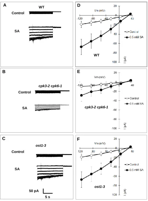
Fig. 2.5 SA activation of S-type anion currents in wild-type (WT), cpk3-2 cpk-6-1, and ost1-3 GCPs. A-C, Representative whole-cell S-type anion currents of wild-type GCPs (A), cpk3-2 cpk6-1 GCPs (B), and ost1-3 GCPs (C) in the absence (top trace) or presence of 0.5 mM SA (bottom trace). D-F, Average current-voltage curves of wild-type GCPs (D) (n=6, Control; n=6, SA), cpk3-2 cpk6-1 GCPs (E) (n=6, Control; n=6, SA), and ost1-3 (F) (n=4, Control; n=6, SA). Open and closed symbols indicate absence and presence of 0.5 mM SA, respectively. GCPs were pretreated with 0.5 mM SA for 10 min before starting patch clamp analysis. Error bars represent SE.
WT
ost1-3 D
E Control
SA A WT
Control
ost1-3
SA C B
cpk3-2 cpk6-1 Control
SA
50 pA 5 s
cpk3-2 cpk6-1
F
28
2.4.5 SA-induced stomatal closure is impaired in S59/S120 double disruption SLAC1 mutants but not in S59 and S120 single disruption SLAC1 mutants
It has been reported that the amino acid residues S59 and S120 are the key phosphorylation sites of SLAC1 and are required for the activation of SLAC1 by CPKs and OST1 protein kinases, respectively (Geiger et al., 2009, 2010;
Brandt et al., 2012). Brandt et al. (2015) recently reported that ABA-induced stomatal closure was impaired in the S59 and S120 double-disruption mutant SLAC1-S59A/S120A (serine is mutated to alanine), but not in S59A or S120A single phospho-site mutants, proving the in-planta function of both of the phosphorylation sites. Next to investigate the function of the two phosphorylation sites S59 and S120 of SLAC1 in guard cell SA signaling, we performed stomatal bioassay analysis using slac1-1 complementation lines with phospho-site-mutated SLAC1, SLAC1-S59A, SLAC1-S120A, and SLAC1-S59A/S120A (Brandt et al., 2015). SLAC1-WT complementation lines showed SA-sensitive stomatal phenotype similar to that of wild-type plants. We found that SA induced stomatal closure in the SLAC1-S59A and SLAC1-S120A single mutants but not in the SLAC1-S59A/S120A double mutant (Fig. 2.6). The impairment of SA-induced stomatal closure in the SLAC1-S59A/S120A double mutant suggests that both S59 and S120 sites are required for guard cell SA signaling.
29 1
2 3 4
Stomatal aperture (µm)
Control SA
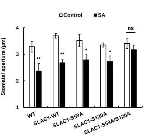
Fig. 2.6 SA-induced responses of stomatal apertures in slac1-1 complementation lines SLAC1-WT, SLAC1-S59A, SLAC1-S120A, and SLAC1-S59A/S120A.
Averages of stomatal apertures from at least three independent experiments (total stomata per bar ≥ 60) are shown. Error bars represent SE. ** and * indicate p <
0.01 and p < 0.05, respectively.
2.4.6 SA elicited ROS production in the cpk3 cpk6 mutant as well as in the wild-type plants
ROS such as superoxide anion (O2-) and hydrogen peroxide (H2O2) are accumulated in the guard cells after perception of SA and subsequently functions as second messenger in the promotion of stomatal closure by SA (Lee et al., 1999; Mori et al., 2001; Khokon et al, 2011, 2017). It is reported that SA elicits SHAM-sensitive peroxidase-mediated ROS burst that triggers activation of
** ** * *
ns
30
downstream SA signaling components in guard cells (Mori et al., 2001; Khokon et al., 2011). As shown above, we found CPK3 and CPK6 are required for SA- induced stomatal closure. Here we examined the effects of SA on ROS production in the cpk3-2 cpk6-1 mutants to investigate the position of ROS production and the two CPK in the SA signal pathway in guard cells.
We measured the O2- production in guard cell enriched-epidermal tissues using CLA (Cripridina lucigenin-derived chemiluminescent agent: 2-methyl-6- phenyl-3,7- dihydroimidazo[1,2-a]pyrazin-3-one) (Kawano et al. 1998; Mori et al 2001). Application of 0.5 mM SA evoked O2- production in wild-type epidermis (Fig.
2.7), and the O2- production was inhibited by SHAM, but not by a NADPH oxidase inhibitor DPI, confirming that the SA-triggered O2- production is mediated by SHAM-sensitive peroxidases (Mori et al., 2001; Khokon et al., 2011), but not DPI- sensitive NADPH oxidases such as RBOHD/F (Kwak et al., 2003). Application of 0.5 mM SA also evoked O2- production in the cpk3-2 cpk6-1 epidermis (Fig. 2.8).
In the previous studies, 3, 3′-diaminobenzidine (DAB)-based apoplastic H2O2
measurement in whole leaves was also employed to dissect guard cell SA signaling (Khokon et al., 2011; Prodhan et al., 2017). The DAB staining analysis revealed that disruption of CPK3 and CPK6 did not affect SA-induced H2O2
production in the whole leaves (Fig. 2.9). Together, these results indicate that CPK3 and CPK6 protein kinases function downstream of ROS production in guard cell SA signaling.
31
5000 RLU
Fig. 2.7 Effects of SHAM and DPI on SA-elicited O2- production in guard cell- enriched epidermal peels of Arabidopsis wild-type plants. A, Typical traces of the CLA-chemiluminescence reflecting the production of O2- in the presence and absence of 2 mM SHAM (a peroxidase inhibitor) or 20 μM DPI (an NADPH oxidase inhibitor). SA at 0.5 mM was added at the time showed by an arrow head.
B, Total CLA-chemiluminescence reflecting the production of O2- as recorded in A.
Total RLU (relative luminescence unit) is the sum of luminescence between 1 and 15 minutes. Each data point reflects the mean (n = 3). The error bars represent SE. Values indicated by the same letter do not differ significantly at the 5% level, as determined by Tukey’s test. SHAM, Salicylhydroxamic acid; DPI, Diphenyleneiodonium chloride
Control
SA
SA + SHAM
SA + DPI Time (min)
3 6 9 12 15
100 400 700 1000 1300 1600
Control SA SA + SHAM SA + DPI CLA chemiluminescence Total RLU X 100
60 s A
B
b
a a
b SA
WT
WT
32
100 400 700 1000 1300 1600
WT cpk3-2 cpk6-1
CLA chemiluminescence Total RLU (X 100)
Control SA
Fig. 2.8 SA-elicited O2- production in wild-type (WT) plant and cpk3-2 cpk6-1 mutant. A, Typical kinetics of the CLA chemiluminescence reflecting the O2-
production in the presence or absence of SA in WT and cpk3-2 cpk6-1 mutant. B, Total CLA-chemiluminescence reflecting the production of O2- as recorded in A.
Total RLU (relative luminescence unit) is the sum of luminescence between 1 and 15 minutes. Each data point reflects the mean (n=5). * differs significantly (p <
0.05) from controls as assessed by t-test. The error bars represent SE.
Control
SA
Control
SA
WT
Time (min) A
SA
3 6 9 12 15
60 s 5000 RLU
cpk3-2 cpk6-1
* B
WT
cpk3-2 cpk6-1
cpk3-2 cpk6-1
*
33 20
60 100 140 180 220
WT cpk3-2 cpk6-1
DAB staining (arb units)
Control 0.5 mM SA 1 mM SA
Fig. 2.9 Concentration-dependent SA-elicited apoplastic H2O2 production in the WT and cpk3-2 cpk6-1 mutants. The vertical scale represents the relative values of pixel intensity of DAB brown color (ROS) when the values of SA treated leaves are normalized to control value taken as 100 for each experiment. Averages of H2O2 production from four independent experiments (total leaves per bar = 16) are represented. Values indicated by the same letter do not differ significantly at the 5% level, as determined by Tukey’s test.
2.5 DISCUSSION
SA and ABA induce stomatal closure in plants and thus play a crucial role in adaptation to stress conditions. Significant efforts have been devoted to identifying signaling components in guard cell responses to ABA. In comparison, very few studies have focused on SA signaling in guard cells. Although the roles of CPKs, OST1, and SLAC1 in ABA signaling in guard cells are elucidated, those in SA signaling are to be examined. In this study, we present the involvement of
a a
b b c c
34
CPKs and SLAC1 in guard cell SA signaling and elucidate the integration of SA signaling and ABA signaling in guard cells.
2.5.1 SA requires CPK3 and CPK6 protein kinases but not OST1 for induction of stomatal closure
Several CPKs are important mediators of Ca2+-dependent stomatal closure and S-type anion channel activation and play roles in ABA signaling in guard cells (Mori et al., 2006; Geiger et al., 2010; Brandt et al., 2015). Mori et al (2006) have reported that CPK3 and CPK6 positively regulate ABA-induced stomatal closure in Arabidopsis. In this study, we found that the disruption of CPK3 and CPK6 also impaired the SA-induced stomatal closure (Fig. 2.2). It has been reported that SA regulates gene expression involved in innate response via Ca2+-dependent pathway (Du et al., 2009; Wang et al., 2009; Coca and San Segunda, 2010). As well, previous pharmacological experiments suggest that guard cell SA signaling is Ca2+-dependent. Our group previously reported that both Ca2+ chelator EGTA (Ethylene glycol-bis(2-aminoethylether)-N,N,N′,N′-tetraacetic acid), and Ca2+
channel blocker LaCl3 significantly suppressed the SA-induced stomatal closure, suggesting the involvement of extracellular free Ca2+ in the modulation of SA- induced stomatal closure (Khokon et al., 2011). However, molecular identities of the Ca2+ sensors that are responsible for guard cell SA signaling were unknown. It is reported that CPKs play a role as [Ca2+]cyt sensors in many aspects of plant physiological processes (Klimecka et al. 2007) and that CPK3 and CPK6 function as Ca2+ sensors in stomatal ABA signaling (Mori et al. 2006). Therefore, our findings with the previous reports suggested that CPK3 and CPK6 function as important Ca2+ sensors in Ca2+-dependent SA signaling in guard cells. The CPK-
35
dependent Ca2+ recognition could be key for an integration mechanism between SA signaling and ABA signaling in guard cells.
The OST1 protein kinase is reported to play a key role in the ABA responses of Arabidopsis guard cells (Mustilli et al., 2002; Yoshida et al., 2002;
Yin et al., 2013; Imes et al., 2013; Acharya et al., 2013). In addition to ABA, microbe-associated molecular-patterns (MAMPs) such as yeast elicitor (YEL, Ye et al., 2015) requires OST1 kinase to induce stomatal closure. It has been suggested that SA acts downstream of MAMP perception in MAMP-induced stomatal closure (Montillet et al., 2013; Montillet and Hirt., 2013). Our results revealed that different from the MAMPs, SA-induced stomatal closure does not require OST1 (Fig. 2.3), suggesting that MAMPs activate the OST1-dependent pathway via a SA-independent manner in guard cells. The detail crosstalk mechanism for SA-dependent pathway and OST1-dependent pathway in guard cell MAMP signaling needs to be further analyzed.
2.5.2 SLAC1 and its activation by phosphorylation of S59 and S120 are required for SA signaling in guard cells
SLAC1 mediates S-type anion channel activity and stomatal closure (Negi et al., 2008; Vahisalu et al., 2008). In this study, we also found that SLAC1 S-type anion channel is required for SA-induced stomatal closure (Fig 2.4) and the SA activation of S-type anion channels was impaired in the cpk3-2 cpk6-1 GCPs (Fig 2.5 B, E) but not in ost1-3 (Fig. 2.5 C,F), suggesting that CPK3 and CPK6, but not OST1 regulates SA signals for the SLAC1 activation. CPK3 and CPK6 are also involved in the ABA activation of S-type anion channels (Mori et al., 2006).
36
Two-electrode voltage clamp analysis using Xenopus oocytes revealed that CPKs phosphorylate S59, and OST1 phosphorylate S120 of SLAC1, resulting in activation of the channel (Brandt et al., 2012; Geiger et al., 2009, 2010;
Maierhofer et al., 2014). Conversely, in in vitro protein kinase assay using recombinant proteins demonstrated that CPKs phosphorylate S120, and OST1 phosphorylate S59 for the activation (Vahisalu et al., 2010; Brandt et al., 2015).
Moreover, in planta analysis concludes that the both phosphorylation sites S59 and S120 of SLAC1 are required for ABA-induced stomatal closure in Arabidopsis (Brandt et al., 2015). Similar to ABA (Brandt et al., 2015), SA-induced stomatal closure was impaired in the S59A/S120A double disruption SLAC1 mutant but not in the S59A or S120A single disruption SLAC1 mutants (Fig. 2.6). These results demonstrated that in guard cell SA signaling, although OST1 kinase is absent (Fig.
2.3), CPKs and other protein kinases (e.g. CIPKs, Mainhofer et al., 2014) can phosphorylate S59 and S120 of SLAC1 to activate S-type anion channel, resulting in stomatal closure.
2.5.3 CPKs function downstream of ROS production in guard cell SA signal cascade
It has been shown that ROS functions as an important second messenger in guard cells during stomatal closure (Pei et al., 2000; Zhang et al., 2001; Suhita et al., 2004; Khokon et al., 2011). It was reported that ROS production in ABA signaling and MeJA signaling in guard cells is mediated by plasma membrane NAD(P)H-oxidases (Kwak et al., 2003; Suhita et al., 2004), while ROS production in guard cell SA signaling is mediated by SHAM-sensitive peroxidases (Mori et al., 2001; Khokon et al., 2011).
37
It was suggested that CPK3 and CPK6 function downstream of ROS production in guard cell ABA signaling (Mori et al., 2006) and that CPK6 also functions downstream of ROS production in guard cell MeJA signaling (Munemasa et al., 2011). In this study, we found that SA elicited ROS production in cpk3 cpk6 guard cell-enriched epidermis as well as wild-type guard cell- enriched epidermis (Fig. 2.8). This result with the previous reports indicates that the CPKs function downstream of SHAM-sensitive peroxidase-mediated ROS production as well as NAD(P)H oxidase-mediated ROS production in guard cell signaling. Therefore, the presented results, as well as previous reports (Mori et al., 2006; Munemasa et al., 2011), provide evidence that the hormone-triggered ROS signals activate CPKs that function as signal integrators in guard cells. The mechanism by which SA-elicited ROS signal is transduced into activation of guard cell CPKs remains unclear and needs to be investigated in future.
2.5.4 Integration of SA and ABA signaling pathways in Arabidopsis guard cells
Based on the results discussed here, we propose a model (Fig. 2.10) showing the overlapping between SA signaling and ABA signaling in guard cells.
Briefly, SA-triggered ROS signal activates not OST1, but CPKs and other protein kinases that phosphorylate S59 and S120 of SLAC1 and activate it, whereas ABA needs both CPKs and OST1 for full activation of SLAC1 (Brandt et al., 2015).
Thus the Ca2+/CPK-dependent signaling branch is key for integration of SA signaling with ABA signaling in guard cells. It has been reported that SA, as well as ABA, functions as a key mediator for biotic stress-triggered stomatal closure (Melotto et al., 2006; Zeng et al., 2010; Montillet et al., 2013). In addition, a
38
previous research reported a SA-independent biotic signaling that antagonizes Ca2+-dependent, but OST1-independent ABA signaling in guard cells (Kim et al., 2011). The Ca2+/CPK-dependent integration mechanism of SA signaling with ABA signaling described here might be one of the innate immune responses for overcoming pathogen-induced inhibition of Ca2+-dependent ABA-induced stomatal closure.
Fig. 2.10 Proposed model of integration between SA signaling and ABA signaling in Arabidopsis guard cells. SA triggers SHAM-sensitive peroxidase-mediated ROS signal that activates CPKs, but not OST1 protein kinase. Then the CPKs together with other unknown protein kinases (e.g. CIPKs) phosphorylate S59 and S120 of SLAC1 and activate it, while ABA requires OST1 as well as CPKs for the activation of SLAC1. These suggest that the SA signaling integrates with ABA signaling via Ca2+/CPK-dependent signaling pathway in guard cells.
SA
Stomatal closure
SHAM-sensitive peroxidase
CPK3 CPK6
ABA
OST1
P S59 + S120 P
SLAC1 ROS
39
CHAPTER 3
Chitosan signaling in guard cells requires endogenous salicylic acid
3.1 Abstract
An elicitor chitosan (CHT) induces stomatal closure but the mechanism remains to be clarified. A phytohormone salicylic acid (SA) is crucial for elicitor-induced defense signaling in plants. Here we investigated whether endogenous SA is required for CHT signaling in guard cells. In the SA-deficient nahG mutant, treatment of CHT did not induce either apoplastic reactive oxygen species (ROS) production or stomatal closure but co-treatment of CHT and SA induced both apoplastic ROS production and stomatal closure, indicating the involvement of endogenous SA in CHT-induced apoplastic ROS production and CHT-induced stomatal closure. Furthermore, CHT induced transient cytosolic free calcium concentration increments in the nahG mutant in the presence of exogenous SA but not in the absence of exogenous SA. These results provide evidence that endogenous SA is a crucial element in CHT-induced stomatal closure.
3.2 Introduction
A pair of guard cells forms a stomatal pore, which is the most important port of entry for pathogens, gaseous exchange and transpiration. Guard cells respond to a variety of biotic and abiotic stimuli such as elicitors, salicylic acid (SA), and abscisic acid (ABA) and consequently control innate immunity, exchange of CO2 and O2, and transpirational loss of water (Murata et al., 2015).
40
Elicitor is a chemical or biological molecule that mimics microbial attack and causes physiological changes in plants (Zhao et al., 2005). Chitosan (CHT), a fungal elicitor, is a deacylated derivative of chitin which is present mainly in the exoskeleton of insects, crustaceans and cell wall of fungi (Bartnicki-Garcia, 1970).
CHT induces stomatal closure in plants (Lee et al., 1999; Khokon et al., 2010;
Salam et al., 2012), but the mechanism remains unclear.
Previous studies have suggested that SA is important for CHT signaling in plants. For instances, CHT oligosaccharide induces defense responses via the SA-mediated signaling pathway (Jia et al., 2016), and induces methyl-salicylate accumulation in plants (Obara et al., 2002). Hossain et al. (2011) reported that methyl jasmonate (MeJA)-induced stomatal closure requires endogenous ABA whereas Issak et al. (Issak et al., 2013) reported that SA-, yeast elicitor (YEL)- or CHT-induced stomatal closure requires neither endogenous ABA nor endogenous JA in Arabidopsis. However, whether CHT signaling in guard cells requires endogenous SA are to be investigated.
It is well-known that reactive oxygen species (ROS) play a role as a second messenger in guard cell signaling (Pei et al., 2000; Khokon et al., 2011) and that ROS production is crucial for signal integration in guard cells (Murata et al., 2015;
Mori et al., 2009; Song et al., 2014). CHT signaling in guard cells is accompanied by ROS production which is mediated by salicylhydroxamic acid (SHAM)-sensitive peroxidases (Lee et al., 1999; Khokon et al., 2010; Salam et al., 2012).
Calcium (Ca2+) functions as an important second messenger in signaling in guard cells (Pei et al., 2000; Grabov et al., 1999; Roelfsama et al., 2010). Elicitors trigger ROS production which activates non-selective Ca2+ channels leading to elevation of cytosolic free Ca2+ concentration ([Ca2+]cyt) (Khokon et al., 2010;
41
Salam et al., 2012; Klusener et al., 2002; Ye et al., 2013). CHT elicits [Ca2+]cyt
oscillations for the induction of stomatal closure in Arabidopsis (Salam et al., 2012).
To elucidate the roles of endogenous SA in CHT signaling in guard cells, here we analyzed CHT-induced apoplastic ROS production, [Ca2+]cyt oscillations, and stomatal closure in the SA-deficient nahG mutant. We found that treatment of CHT did not induce either apoplastic ROS production or stomatal closure but co- treatment of CHT and SA induced both apoplastic ROS production and stomatal closure. In addition, CHT elicited transient [Ca2+]cyt increments in the presence of SA but not in the absence of SA in the nahG mutants. Based on the results, we concluded that endogenous SA is an important signal transducer in CHT signaling in Arabidopsis guard cells.
3.3 Materials and Methods
3.3.1 Plants and Their Growth Conditions
Arabidopsis ecotype Columbia-0 (Col-0) was used as wild-type plant. The nahG (Delaney et al., 1994) and sid2-2(Wildermuth et al., 2001) plants were used as SA-deficient mutants. Plants were grown in vermiculite:soil (v/v) 1:1 mixture in controlled growth conditions under a photoperiod of 16-h-light/8-h-dark with photon flux density of 80 µmolm-2s-1. The relative humidity and temperature were 70% and 21°C. Hyponex (0.1%) nutrient solution was provided to the plants twice per week. Four-week-aged plants were used for the experiments.
42 3.3.2 Analysis of Stomatal Apertures
Stomatal apertures were measured as described previously (Munemasa et al., 2007). Young fully expanded leaves were floated on assay buffer (5 mM KCl, 50 µM CaCl2, and 10 mM MES, pH 6.15) for 2 h in the light to induce stomatal opening. The solutions of chemicals such as CHT (50 µg/mL), SA (50 µM), and catechol (50 to 500 µM) were directly added to the buffer solution either individual or in combination. After again 2 h incubation in the light, the leaves were blended in tap water in a commercial blender for 25 to 30 s. The blended-epidermal tissues were collected using a nylon mesh (pore size 100 µm) and were mounted on glass slides for microscopic observation. Twenty stomatal apertures were measured for each individual experiment.
3.3.3 Measurement of apoplastic ROS Production
The generation of ROS was measured by the Thordal-Christensen et al.
(1997) method. The leaves were floated on assay buffer containing 5 mM KCl, 50 µM CaCl2, 10 mM MES, and 0.1% Tween 20, pH 6.15 in the light for 3 h. Then 3,3′-diaminobenzidine (DAB) at 1 mg/mL was added to the assay buffer and gently infiltrated for 2 h in a vacuum. In the presence or absence of SA, CHT was then added to the buffer solution and again incubated for 2 h. The leaves were depigmented by boiling in ethanol. Hydrogen peroxide (H2O2) was visualized as reddish-brown color which was quantified using Image-J software (National Institutes of Health, USA).
43
3.3.4 Measurement of [Ca2+]cyt Oscillations in Guard Cells
Four- to six-week-old wild-type and nahG plants expressing a Ca2+-reporter fluorescence protein, Yellow Cameleon 3.6 (YC3.6) were used for the observation of guard cell [Ca2+]cyt oscillations as described previously (Hossain et al., 2011;
Islam et al., 2010). The lower surface of the excised leaf was attached on a glass slide using medical adhesive (Stock No. 7730; Hollister). Then the upper surface and mesophyll tissues of the leaves were removed. Isolated lower-epidermal peels were incubated in the solution containing 5 mM KCl, 50 µM CaCl2, 10 mM MES (pH 6.15-Tris) under light for 2 h to enhance stomatal opening. The [Ca2+]cyt
oscillations were monitored in the turgid guard cells. Guard cells were treated with 50 mg/mL CHT solution by using a peristaltic pump in the 5 min after monitoring and 50 μM SA was added just after CHT application. For dual-emission ratio imaging of YC3.6, a 445DRLP dichroic mirror, a 440AF21 excitation filter, and two emission filters, 480DF30 for cyan fluorescent protein (CFP) and 535DF25 for yellow fluorescent protein (YFP) were used. The CFP and YFP fluorescence intensities of guard cells were imaged and analyzed using AQUA COSMOS software (Hamamatsu Photonics). Similar exposure time was applied for both CFP and YFP.
3.3.5 Statistical Analysis
Two-way ANOVA was performed to compare the significance of differences of mean values of stomatal apertures and ROS production between the groups (e.g., between wild-type and nahG or sid2-2 plants) and t-test was performed to compare that within the group. The χ2 test was performed to assess the