Multipoint Molecular Recognition through
Non-Werner Type Coordination Bonding by Ag(I)-Macrocycles
(Ag(I)
)
27
12
(
)
Abstract
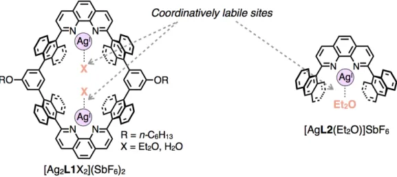
Macrocycles provide nano-spaces for molecular recognition and chemical reactions depending on the size, shape, and chemical property of their cyclic skeletons. To construct functional macrocycles, it is important to precisely arrange functional groups within their inner surfaces. Herein, a dinuclear Ag(I)-macrocycle and a mononuclear Ag(I)-half-macrocycle which have anthracene-based nano-spaces functionalized with coordinatively labile sites of Ag(I) ions have been newly synthesized, and their guest binding abilities were examined in detail focusing on non-Werner type coordination properties of Ag(I) ions (Figure 1).
A dinuclear Ag(I)-macrocycle [Ag2L1X2](SbF6)2 (X = Et2O or H2O) and its CF3SO3– salt formed host-guest
complexes with several kinds of aromatic molecules such as p-xylenes, anthracene, [2.2]paracyclophane (pCp), and ferrocene (FeCp2) derivatives in solution and/or in the solid state through Ag–π interactions (Figure 1a,b). Notably,
the affinities of [Ag2L1X2](SbF6)2 to anthracene, pCp, and FeCp2 were particularly high as the binding constants
(Ka = [Guest⊂Host]/([Guest][Host]) M–1) were estimated to be as large as Ka = 104–109 M–1 in CDCl3 at 300 K.
Single crystal X-ray analyses of the resulting complexes revealed that the structures of these aromatic guest molecules were well fitted to form Ag–π interactions at both sides of Ag(I) ions on the nano-space of [Ag2L1X2](SbF6)2. These results suggest that multipoint Ag–π interactions at the inner surface of the dinuclear
Ag(I)-macrocycles work as effective driving forces to bind aromatic molecules. Moreover, electrochemical measurements revealed that the redox reactivity of an included ferrocene within [Ag2L1X2](SbF6)2 was markedly
changed due to the cationic character of the neighboring Ag(I) ions.
A mononuclear Ag(I)-half-macrocycle [AgL2(Et2O)]SbF6 can effectively bind one molecule of ruthenocene
(RuCp2) in CD2Cl2 with a binding constant Ka > 104 M–1 at 300 K (Figure 1c). The single crystal X-ray analysis of
the resulting complex revealed the formation of a Ru–Ag type metal-to-metal dative bonding between a Lewis basic metal center of RuCp2 and a Lewis acidic Ag(I) ions on [AgL2(Et2O)]SbF6. These results suggest that a Ru–
Ag type metal-to-metal dative bonding works as an effective driving force for host-guest binding.
The present results suggest that metallo-macrocycles and half-macrocycles equipped with non-Werner type coordination centers provide novel binding motifs for host-guest complexation utilizing non-Werner type coordination: metal–arene interactions and metal–metal interactions as driving forces. Such metallo-hosts would provide novel functions, such as guest separation and activation, taking advantage of specific coordination properties of well-arranged non-Werner type coordination centers.
Figure 1. Host-guest complexation between a) a dinuclear Ag(I)-macrocycle [Ag2L1X2](SbF6)2 or b) its CF3SO3– salt with aromatic
molecules via Ag–π interactions. c) Host-guest complexation between a mononuclear Ag(I)-half-macrocycle [AgL2(Et2O)]SbF6 and
Abbreviations
A adenine
ATP adenosine triphosphate
ATR attenuated total reflection
a.u. arbitrary unit
bpy 2,2’-bipyridine
C cytosine
Cp cyclopentadienyl
COSY correlation spectroscopy
DHAP dihydroxyacetone phosphate
DMF N,N-dimethylformamide
DMSO dimethyl sulfoxide
DNA deoxyribonucleic acid
ESI electrospray ionization
Et2O diethyl ether
FeCp2 ferrocene
FeCp2’ hydroxymethyl ferrocene
G guanine
GPC gel permeation chromatography
HPLC high performance liquid chromatography
HRMS high resolution mass spectrometry
IR infrared spectroscopy
J coupling constant
M molar
M.p. melting point
NMR nuclear magnetic resonance
Nu nucleophile
ORTEP Oak Ridge thermal-ellipsoid plot
ROE rotating frame Overhauser effect
ROESY rotating frame Overhauser effect spectroscopy
RuCp2 ruthenocene pCp [2.2]paracyclophane Sol. solvent T thymine TBA n-tetrabutylammonium THF tetrahydrofuran TMS trimethylsilane
TOF time of flight
UV-Vis ultraviolet-visible
Contents
Abstract
··· i
Abbreviations
··· ii
Contents
··· iii
1. General Introduction
··· 1
1–1. Molecular Recognition within Biology
··· 2
1–2. Molecular Recognition by Macrocycles
··· 5
1–3. Metallo-macrocycles
··· 7
1–4. Molecular Architectures using Non-Werner Type Coordination ··· 10
1–5. The Aim of This Research
··· 14
1–6. References
··· 16
2. Inclusion of Aromatic Guest Molecules within a Dinuclear Ag(I)-Macrocycle via Multipoint
Ag–π Interactions
··· 19
2–1. Introduction
··· 20
2–2. Design and Synthesis of a Dinuclear Ag(I)-Macrocycle
··· 23
2–3. Host-Guest Interactions between a Dinuclear Ag(I)-Macrocycle
and Aromatic Guest Molecules via Ag–π Interactions
··· 30
2–4. Conclusion
··· 58
2–5. Experimental
··· 60
3. Host-Guest Interaction via Metal-to-Metal Dative Bonding between Mononuclear
Ag(I)-Half-Macrocycle and Ruthenocene
··· 79
3–1. Introduction
··· 80
3–2. Design and Synthesis of a Mononuclear Ag(I)-Half-Macrocycle ··· 83
3–3. Binding of an Organometallic Molecule via Metal-to-Metal
Dative Bonding
··· 87
3–4. Conclusion
··· 95
3–5. Experimental
··· 96
3–6. References
··· 102
4. Concluding Remarks
··· 105
A list of publications
··· 111
Acknowledgement
··· 112
Chapter 1.
1–1. Molecular Recognition within Biology
All of our living bodies are constructed as mixtures of various kinds of molecules. Almost all of physiological activities, such as replication, translation, transcription of genetic codes, metabolism, signalization, are conducted at the same time within highly complicated mixtures of molecules. In spite of such intricate conditions, most of chemical reactions in biological systems are conducted in highly organized manners without remarkable side reactions to establish highly sophisticated systems as lives. One of the most important mechanisms to
organize such marvelous systems is molecular recognition[1] which means “the selective
binding of a substrate by a molecular receptor to form a supramolecular species”.[1b] Molecular
recognition plays fundamental roles in managing our living activities such as hybridization of
DNA, antibody-antigen bonding, and enzymatic reactions.[2] In these processes, receptor
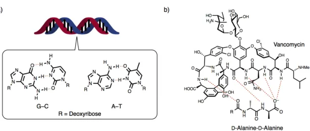
molecules (hosts) bind target substrates (guests) with high selectivity via multipoint and complementally intermolecular and intracomplex interactions such as coordination bonding, hydrogen bonding, hydrophobic effect, and van der Waals interaction (Figure 1–1).
Figure 1–1. Schematic image of molecular recognition.
Molecular recognition of a target substrate usually takes place at specific positions of a
receptor molecule: molecular binding sites (Figure 1–1 left).[1] In general, molecular binding
sites have complement shapes and spaces to their target substrates. On the surfaces of molecular binding sites, multiple functional groups are precisely arranged in a molecular to atomic scale so as to create complementally and multipoint intermolecular interactions and contacts with target substrates. For example, in the hybridization of a DNA double helix, a DNA single strand binds its own complementally strand at its arrangement of nucleobases via multivalent intermolecular interactions such as hydrogen bonding, π–π interaction, and hydrophobic effect
(Figure 1–2a).[2] The selectivity of hybridization is precisely programmed by the sequence of
nucleobases (A, C, G, and T) in a molecular to atomic scale, where A–T and C–G can create complementally hydrogen bonding, so called Watson-Crick base pairs. Vancomycin is a kind of
glycopeptides which works as an antibiotic reagent (Figure 1–2b).[3] Vancomycin selectively
binds to the D-alanine-D-alanine sequence of cell wall synthesis enzymes of eubacteria to inhibit
its propagation. Well-defined multipoint hydrogen bonding and hydrophobic contacts between
the surface of vancomycin and the D-alanine-D-alanine sequence works as a driving force for
the enzyme selective binding.
Figure 1–2. a) Hybridization of a DNA double strand and Watson-Crick base pairs, b) recognition of a
D-alanine-D-alanine sequence by vancomycin.
Some receptor molecules provide confined nano-spaces of specific size and shape surrounded by their skeletons (Figure 1–3). Such nano-spaces often work as effective molecular binding sites, because their inner-surfaces are suitable to form multiple intermolecular interactions and contacts in large areas with their target substrates. Moreover, structural and chemical/physical properties of the inner surfaces can significantly affect conformations or properties of bound substrates to induce specific reactivities or properties. For instance,
valinomicyn is a kind of macrocyclic transmembrane K+ transporters, which provides a
confined nano-space surrounded by a covalently linked dodecadepsipeptide skeleton with multi
O-atoms (Figure 1–3a).[4] Valinomicyn can selectively include a K+ ion within its nano-space
via multipoint ion-dipole interactions at inward O-atoms. As another example, enzymes provide hydrophobic nano-pockets as active centers of catalytic reactions, which are constructed by folding of polypeptides. On their inner surfaces, multiple functional groups such as amino-acid
residues and metal ions are precisely arranged.[2] Multipoint intermolecular interactions and
target substrates. For instance, type-II aldolase, which catalyzes asymmetric aldol reactions between dihydroxyacetone phosphate (DHAP) and various aldehydes, possesses a nano-space
arranged with various amino acid residues and a Zn(II) ion (Figure 1–3b).[5] In the proposed
transition state, DHAP and aldehyde are bound in a specific configuration via hydrogen bonding and coordination bonding with amino acid residues and the Zn(II) ion. In this structure,
Brønsted or Lewis acidic/basic characters of these functional groups activate bound substrates
to enhance asymmetric aldol reactions under ambient conditions.
Figure 1–3. a) Recognition of K+ by valinomycin, b) a schematic illustration of an active center of type-II aldolase.
In summary, chemical phenomena in biological systems are well organized by molecular recognition processes which utilize well designed intermolecular and intracomplex interactions and contacts between specific pairs of molecules. To realize sophisticated molecular recognition, it is important to design receptor molecules which have molecular binding sites with specific sizes, shapes and precise arrangements of multi functional groups.
1–2. Molecular Recognition by Macrocycles
As described in the previous section, to realize functional molecular recognition processes, it is very important to create host molecules with molecular binding sites which can produce multipoint intermolecular interactions with target guest molecules. In particular, host molecules possessing a confined nano-space show promise for multipoint intermolecular interactions in large areas with guest molecules.
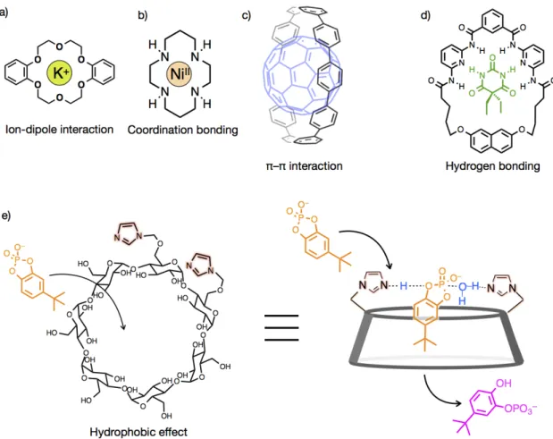
Macrocycles such as crown ether,[6] cyclodextrin,[7] and cyclam[8] have a hollow nano-space
surrounded by their covalently-linked cyclic skeleton (Figure 1–4). Within their nano-spaces, macrocycles can effectively encapsulate guest molecules of specific size and shape through
multipoint intermolecular interactions and contacts at their inner surface.[1d]
Figure 1–4. Representative examples for host-guest complexes of macrocycles and guest molecules. a)
Inclusion of K+ within dibenzo-18-crown-6 through ion-dipole interaction,[6] b) inclusion of Ni(II) within
cyclam through coordination bonding,[8a] c) inclusion of C60 by [10]cycloparaphenylene through π–π
interaction,[10] d) inclusion of barbiturate through hydrogen bonding,[11] and e) activation of hydrolysis of a cyclic phosphoester within the a hydrophobic nano-cavity of a β-cyclodextrin functionalized with two imidazole moieties.[12]
within macrocycles, which is an important factor to determine the type of guest molecules.
Ion-dipole interaction and coordination bonding are often utilized as powerful driving forces to
encapsulate metal ion(s) within a macrocycle.[9] Crown ethers are one of earlier examples of
synthetic macrocycles investigated by Pedersen and co-workers (Figure 1–4a).[6] Within their
cyclic poly-ether frameworks, alkali or alkaline earth metal ions such as Na+, K+, or Ca2+ can be
selectively encapsulated depending on the cavity sizes of the cyclic structures through multipoint ion-dipole interactions arising from radially-inwardly projecting C–O bonds. Similarly, macrocyclic polyamines, like cyclam, can bind various kinds of transition metals within their nano-cavity normally using three to six coordination bonds between metals and
N-atoms (Figure 1–4b).[8a] For the inclusion of aromatic molecules, π–π interaction is often used
as a central driving force. [10]Cycloparaphenylene is a kind of macrocycles composed of
covalently linked ten p-phenylene moieties (Figure 1–4c).[10] Within its nano-space surrounded
by aromatic π-planes, this macrocycle accommodates a C60 molecule in CD2Cl2 by multipoint
interactions with aromatic π-planes.
As an important advantage of macrocycles, they have robust and covalently-linked skeletons. Therefore, we can precisely arrange functional groups pre- or post-synthetically on their skeletons so as to control guest binding abilities and functionality. Hamilton and
co-workers prepared a macrocycle shown on Figure 1–4d.[11] This macrocycle has an alternate
arrangement of multiple NH and N moieties as hydrogen bonding donors and acceptors. This macrocycle can strongly bind to one molecule of barbiturate within its nano-cavity through multipoint and complementally hydrogen bonding with its well designed inner surface (Figure 1–4d). Cyclodextrins are kinds of macrocycles which can encapsulate many kinds of organic
molecules in aqueous media due to hydrophobic effect.[7] To the skeleton of β-cyclodextrin,
Breslow and co-workers post-synthetically added two imidazole moieties (Figure 1–4e).[12]
Using an arranged imidazole pair as a concerted acid base catalyst center, this macrocycle catalyzed site-selective hydrolysis of a cyclic phosphoester bound within its nano-space.
In summary, macrocycles provide a confined nano-space for guest binding and activation using multiple intermolecular interactions and contacts between their inner surface and guest molecules. Furthermore, by arranging functional groups on their covalently-linked skeletons, we can design their guest binding ability and functions.
1–3. Metallo-macrocycles
As discussed in the previous section, macrocycles provide excellent nano-spaces for molecular recognition and guest activation depending on the shape, size, and chemical/physical property of their inner surfaces. To construct functional nano-spaces, it is important to precisely design arrangement styles of functional groups on their covalently-linked skeletons.
Metal ions or complexes exhibit specific properties that cannot be achieved by ordinary organic molecules, for instance, coordinating property, redox reactivity, magnetism, Lewis acidity, and electrostatic natures. Focusing on such specific properties of metal ions, various kinds of metallo-macrocycles which contain metal ions on their cyclic skeletons as function-centers have been investigated. In such macrocycles, guests and metal ions can interact with each other to exhibit unique guest binding behaviors or metal dependent functions as follows.
Figure 1–5. Representative examples of metallo-macrocycles which can work as sensors to detect guest
molecules. a) A cyclam derivative possessing four Ru(bpy)32+,[13a] b) a ferrocene-viologen based
macrocycle,[14b] and c) a metallo-macrocycle with two Cu(II) porphyrin complexes.[15] Figure 1–5c is
reproduced with permission from J. Am. Chem. Soc. 2011, 133, 9290–9292,[15] Copyright 2011 American Chemical Society.
Metal complexes with unique emission,[13] redox,[14] or magnetic properties[15] are often
incorporated into cyclic frameworks of macrocycles (Figure 1–5). Upon host-guest complexation, properties of metal ions can be significantly affected by the neighboring guest molecules to give off modified chemical/physical properties as responses. Therefore, such macrocycles can work as sensors to detect specific guest molecules. For instance, a cyclam
derivative possessing four Ru(bpy)32+ chromophore centers can work as an emission sensor for
metal ions (Figure 1–5a).[13a] Upon Ni(II) binding with its cyclam unit, the emission of
Ru(bpy)32+ centers was greatly reduced due to the heavy atom effect arising from bound metal
receptor for ATP2–.[14b] The binding behavior of ATP2– to this macrocycle can be detected from
a negative shift of the ferrocene-centered redox potential because of an anionic character of the
included guest.Aida and co-workers recently prepared a metallo-macrocycle with two Cu(II)
porphyrin complexes on its framework, which can bind one molecule of paramagnetic La@C82
(Figure 1–5c).[15] The resulting host-guest complex shows a specific ferromagnetic character
derived from a spin-spin coupling among two Cu(II) centers and the included guest molecule. Metallo-macrocycles which have coordinatively labile sites of transition metal centers directing toward the inwards of their nano-spaces exhibit guest binding behaviors through direct
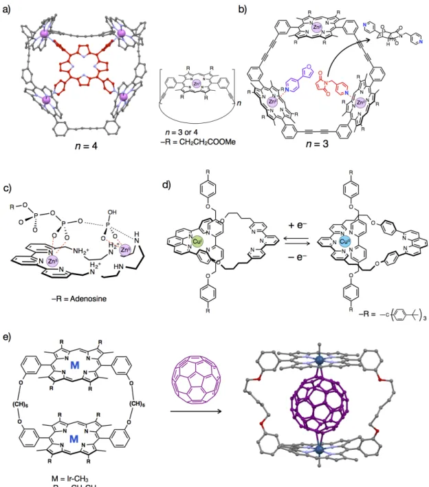
coordination bonding between guests and metals as driving forces (Figure 1–6).[16] Such
metallo-macrocycles potentially have several advantages based on specific properties of metal
coordination.[17] Firstly, coordination bonding is generally stronger and has higher directionality
than other non-covalent interactions. Therefore, strong host-guest interactions can be achieved using such metallo-macrocycles. Notably, the modes of metal arrangement significantly affect the affinity and binding pattern of included guests. Secondly, metal coordination can modify electronic properties of bound guests due to their cationic and Lewis acidic characters of metal centers, which have great potential to induce specific chemical reactions. Furthermore, coordination structures and metal-ligand affinity highly depend on the type and oxidation states of metal ions. Thus, the guest selectivity is possibly modified from many aspects.
Followings are excellent examples of metallo-macrocycles which utilize coordination bonding as driving forces for guest binding and/or activation (Figure 1–6). Sanders and co-workers reported a series of metallo-macrocycles which possess tetra- or trinuclear
Zn(II)-porphylin scaffolds (Figure 1–6a,b).[16a,18] A tetranuclear Zn(II)-macrocycle strongly
encapsulates one molecule of meso-tetra(pyridyl)porphyrin through multipoint coordination
bonding between Zn(II) and pyridyl N-atoms as driving forces (Figure 1–6a).[16a] Furthermore,
they also found that a trinuclear Zn(II)-macrocycle can accelerate a stereochemically selective
Diels-Alder reaction between a pyridine-substituted diene and a dienophile (Figure 1–6b).[18]
Such a specific reactivity is based on a condensation effect and controlled conformations of the substrates bound within the macrocycle through coordination bonding. Valtancoli and
co-workers reported that a Zn(II)-macrocycle catalyzed ATP hydrolysis (Figure 1–6c).[19a] In
this reaction, ATP is supposed to be activated by a Lewis acidic character of Zn(II) ions. Sauvage and co-workers reported that a polypyridyl-based metallo-macrocycle with a Cu(I)/(II) center can bind a 2,2’-bipyridine-based axle molecule via coordination bonding to create a
pseudo-rotaxane (Figure 1–6d).[20] This pseudo-rotaxane exhibits pirouetting motions upon
oxidation/reduction of the Cu(I)/(II) center based on valence-dependent coordination structures of the Cu(I)/(II) center. In 2007, Aida and co-workers reported that a metallo-macrocycles with
two Ir-porphyrins can effectively bind a C60 molecule in organic solvents (Figure 1–6e).[16d] The
crystal structure of the resulting complex suggests C60 was included via multipoint η2-type Ir–π
interactions. This is one of a few examples of metallo-macrocycles which utilize non-Werner
type coordination[21c] as a driving force for guest binding.
Figure 1–6. Representative examples of metallo-macrocycles which have coordinatively labile sites of
metal ions on their frameworks. a) Crystal structure of the host-guest complex of meso-tetra(pyridyl)porphyrin and a tetranuclear Zn(II)-macrocycle,[16a] b) activation of a stereoselective Diels-Alder reaction within a trinuclear Zn(II)-macrocycle,[18] c) activation of ATP hydrolysis using a
Zn(II)-macrocycle,[19a] d) pirouetting motions of a pseudo-rotaxane possessing a mononuclear Cu(I)/(II)-macrocycle,[20] and e) inclusion of C60 within a dinuclear Ir-porphyrin macrocycle via
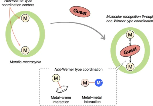
1–4. Molecular Architectures using Non-Werner Type Coordination
Coordination bonding (and complexes) are generally categorized into the following two
types: Werner type coordination (complexes) and non-Werner type coordination (complexes).[21]
Although there is no clear boundary between them, coordination bonding generated between metals and coordinating hetero atoms with lone-pair electrons (such as N-, O-, and halogen atoms) are usually categorized as Werner type coordination which is named after Alfred Werner
who developed the basis of coordination chemistry (Figure 1–7a).[21d] Whereas other types of
coordination bonding, such as metal–arene bonds, metal–CO bonds, or metal–metal bonds, are
often categorized as non-Werner type coordination (Figure 1–7b).[21] As exemplified in the
previous sections, Werner type coordination has been widely utilized as building blocks of metallo-supramolecular complexes or host-guest complexes, because the sites and directions of
bonds are predictable.[22] Whereas non-Werner type coordination, in particular metal–arene
interaction[23] and metal–metal interaction,[24] are also attractive, because their coordination
geometry and reactivity are quite distinct from Werner type coordination. Such unique coordination properties of non-Werner type coordination often provide efficient methods to create novel molecular architectures.
This section describes several representative examples of molecular architectures which utilize metal–arene interactions and metal-to-metal dative bonding: ‘metal-only Lewis pairs
with transition metal Lewis bases’[24h] as non-Werner type coordination.
Figure 1–7. Representative examples of a) Werner type complexes and b) non-Werner type complexes.
1–4–1. Metal–arene interaction
Metal ions can vertically bind to the aromatic π-plane to create metal–arene complexes.[23]
Some of metal–arene complexes play fundamental roles in organometallic chemistry, because
they can induce specific chemical reactions to the bound aromatic rings.[23c] For instance,
rings and Cr(CO)6, represent η6-type Cr–arene interactions (Figure 1–8a). In these complexes,
electron withdrawing effects of Cr(CO)3 units facilitate nucleophilic substitution reactions of the
bound aromatic rings.[25]
Notably, the thermodynamic stabilities and binding modes (hapticities) of metal–arene interactions vary with the type of metals and aromatic molecules. Such diverse coordination structures make it possible to create unique molecular architectures. Murahashi and co-workers succeeded in preparing a series of multinuclear Pd-complexes taking advantages of vast
π-surfaces of polycyclic aromatic hydrocarbons as templates (Figure 1–8b).[26] Metal–arene
interactions are sometimes utilized to create specific supramolecular structures. Ag(I) ions are well known to weakly bind to the peripheries of neutral aromatic hydrocarbons via Ag–π
interactions in usually η1 to η2 manners.[27] Taking advantage of Ag–π interactions as linkers,
various kinds of coordination polymers have been reported by Amma and Munakata, some of
which possess specific multilayer structures (Figure 1–8c).[27a,c] Notably, Ag–π interaction is a
weak interaction,[27e] therefore studies in solution have been mainly limited to the systems which
utilize Ag–π interactions in multipoint manners.[28] For instance, [2.2.2]paracyclophane which
possess a macrocyclic structure with three aromatic rings can strongly bind Ag(I) ions in
chloroform through multipoint Ag–π interactions (Figure 1–8d).[28a]
Figure 1–8. Representative examples of metal–arene complexes. a) A nucleophilic substitution reaction
of a (arene)tricarbonylchromium derivative (Nu: nucleophile),[23c,25] b) a multinuclear Pd-complex using tetracenes as templates,[26] c) a multilayer coordination polymer composed of Ag(I) ions and pyrenes
1–4–2. Metal-to-metal dative bonding
Metal–metal bonding is an attractive interaction which enables to create
electronically-coupled supramolecules with specific structural characteristics.[24] In particular,
metal assembled complexes composed of closed-shell group 11 metals (Cu(I), Ag(I), and Au(I)) and planar Pt(II) complexes have been widely investigated because of their specific emission
and absorption properties.[24a-f,i,j] In these cases, each metal ion shares electrons more or less
equally to form metal–metal bonding, which is often called covalent bonding or non-dative
bonding.[24c,h] While there is another category of metal–metal interactions: metal-to-metal dative
bonding, in which one metal ions work as Lewis base to provide electrons to the other Lewis
acidic metal center.[24c,g,h] The distinction between these two categories of metal–metal bonding
is not always clear-cut and often considered empirically. However, in the cases of latter, proper choices of Lewis acidic and basic metals possibly provide strategies to design metal assembled
architectures with alternative arrangements of hetero metals.[24c,g,h] For instance, an electron rich
Pt(II) center containing a cyclo-metalated ligand can work as an electron donor to create
Pt(II)→Ag(I) dative bonding.[24g,h] Taking its advantage, Ito and co-workers succeeded to
prepare a metal assembled complex with an alternative linear arrangement of multinuclear Pt(II)
and Ag(I) ions in the crystalline state (Figure 1–9a).[29] Also, transition metal centers of group 8
metallocenes are known to act as Lewis bases due to the donation property of occupied e2g
orbital.[30,31] Based on such Lewis basic characteristics of these metal centers, ruthenocene,
osmocene, and ferroceneophane derivatives which have sterically accessible metal centers can bind to Lewis acidic metal ions such as Hg(II) and Sn(IV) via metal-to-metal dative bonding to
create multinuclear metal complexes (Figure 1–9b).[31] Moreover, some of metal-to-metal
bonded complexes show unique reactivity based on cooperative effects of multi metal
centers.[32] For instance, hetero dinuclear M→M’ complexes (M = Fe(0), Ru(0); M’ = Cu(I),
Ag(I)) activate the cleavage of H2 molecule to catalyze semi-hydrogenation of alkynes (Figure
Figure 1–9. Representative examples of heteronuclear complexes through metal-to-metal dative bonding.
a) A crystal structure of a metal assembled complex with an alternative linear arrangement of Pt(II) and Ag(I) ions,[29] b) a crystal structure of a reported Hg(II)-complex of osmocene,[31a] c) molecular structures of hetero dinuclear M→M’ complexes (M = Fe(0), Ru(0); M’ = Cu(I), Ag(I)) which catalyze semi-hydrogenation of alkynes, and d) hypothetical mechanism for the catalytic reactions.[32c]
1–5. The Aim of This Research
As shown in sections 1–2 and 1–3, nano-spaces of macrocycles exhibit special functionalities for guest binding and activation. In order to control guests binding and activation abilities, it is significantly important to design functional groups in terms of shape, size, and mode of arrangement on the inner surface of macrocycles. Metal coordination is one of the powerful tools to bind guest molecules and develop further functionalities. However, in the cases of conventional metallo-macrocycles, utilization of metal coordination is limited to Werner type coordination. Metallo-macrocycles, which utilize non-Werner type coordination, such as matal–arene and metal–metal interactions for space-functions, have not been
extensively investigated so far, except a few examples,[16d] in spite of their attractive
coordination structures and reactivities quite distinct from classical Werner type coordination. In this research, I aim to develop metallo-macrocycles possessing a non-Werner type coordination center as a functional platform with a view to developing host-guest systems based on non-Werner type coordination (Figure 1–10). Nano-spaces arranged with coordinatively labile sites of non-Werner type coordination centers provide novel platforms for host-guest complexiation with unique structures, selectivity, and properties. For instance, binding and activation of guest molecules without coordinating hetero-atoms would be achieved. Furthermore, specific chemical properties of non-Werner type coordination centers would affect structures, reactivities, and properties of guest molecules included within nano-spaces.
Figure 1–10. The concept of this study: molecular recognition through non-Werner type coordination
In this work, a dinuclear Ag(I)-macrocycle [Ag2L1X2](SbF6)2 (X = Et2O or H2O) and a
mono-nuclear Ag(I)-half-macrocycle [AgL2(Et2O)]SbF6 were designed and synthesized, which
have anthracene-based nano-spaces arranged with coordinatively labile sites of Ag(I) ions as
non-Werner type coordination centers (Figure 1–11). Moreover, their guest binding ability was
examined in detail focusing on the non-Werner type coordination behaviors of Ag(I) ions: Ag–π interaction and metal-to-metal dative bonding.
Chapter 2 describes host-guest complexation behaviors between a dinuclear Ag(I)-macrocycles and pristine aromatic molecules using multipoint Ag–π interactions as driving forces. Chapter 3 describes host-guest complexation behaviors between a mononuclear Ag(I)-half-macrocycle and ruthenocene as a pristine organometallic molecule using Ru–Ag type metal-to-metal dative bonding.
Figure 1–11. Molecular structures of a dinuclear Ag(I)-macrocycle and a mono-nuclear
1–6. References
[1] a) Lehn, J.-M. Angew. Chem. Int. Ed. Engl. 1988, 27, 89–112. b) Lehn, J.-M. Angew. Chem. Int. Ed. Engl. 1990, 29, 1304–1319. c) Ariga, K.; Ito, H.; Hill, J. P.; Tsukube, H. Chem. Soc. Rev. 2012, 41, 5800–5835. d) Supramolecular Chemistry, 2nd Edition; Steed, J. W., Atwood, J. L., Eds.; WILEY, 2009.
[2] ; ; , , 2004.
[3] Knox. J.; Pratt, R. F. Anti. Agents. Chem. 1990, 34, 1343–1347. [4] Neupert-Laves, K.; Dobler, M. Helv. Chim. Acta 1975, 58, 432–442.
[5] Machajewski, T. D.; Wong, C.-H. Angew. Chem. Int. Ed. 2000, 39, 1352–1374. [6] Pedersen, C. J. Angew. Chem. Int. Ed. Engl. 1988, 27, 1021–1027.
[7] Breslow, R.; Dong, S. D. Chem. Rev. 1998, 98, 1997–2011.
[8] a) Barefield, E. K.; Mocella, M. T. J. Am. Chem. Soc. 1975, 97, 4238–4246. b) Kimura, E. Tetrahedron 1992, 48, 6175–6217. c) Wainwright, K. P. Coord. Chem. Rev. 1997, 166, 35–90.
[9] Christensen, J. J.; Eatough, D. J.; Izatt, R. Chem. Rev. 1974, 74, 351–384.
[10] Iwamoto, T.; Watanabe, Y.; Sadagiro, T.; Haino, T.; Yamago, S. Angew. Chem. Int. Ed. 2011, 50, 8342–8344.
[11] Chang, S.-K.; Engen, D. V.; Fan, E.; Hamilton, A. D. J. Am. Chem. Soc. 1991, 113, 7640–7645. [12] Breslow, R. Acc. Chem. Res. 1995, 28, 146–153.
[13] a) Josceanu, A. M.; Moore, P.; Rawle, S. C.; Sheldon, P.; Smith, S. M. Inorg. Chim. Acta 1995, 240, 159–168. b) Keefe, M. H.; Benkstein, K. D.; Hupp, J. T. Coord. Chem. Rev. 2000, 205, 201–228.
[14] a) Beer, P. D.; Gale, P. A.; Chen, G. Z. J. Chem. Soc., Dalton Trans. 1999, 1897–1909. b) Reynes, O.; Bucher, C.; Moutet, J.-C.; Royal, G.; Saint-Aman, E. Chem. Commun. 2004, 40, 428–429.
[15] Hajjaj, F.; Tashiro, K.; Nikawa, H.; Mizorogi, N.; Akasaka, T.; Nagase, S.; Furukawa, K.; Kato, T.; Aida, T. J. Am. Chem. Soc. 2011, 133, 9290–9292.
[16] a) Anderson, S.; Anderson, H. L.; Bashall, A.; McPartlin, M.; Sanders, J. K. M. Angew. Chem. Int. Ed. 1995, 34, 1096–1099. b) Amendola, V.; Fabbrizzi, L.; Mangano, C.; Pallavicini, P.; Poggi, A.; Taglietti, A. Coord. Chem. Rev. 2001, 219–221, 821–837. c) Fabbrizzi, L.; Foti, F.; Patroni, S.; Pallavicini, P.; Taglietti, A. Angew. Chem. Int. Ed. 2004, 43, 5073–5077. d) Yanagisawa, M.; Tashiro, K.; Yamasaki, M.; Aida, T. J. Am. Chem. Soc. 2007, 129, 11912–11913. e) Devoille, A. M. J.; Richardson, P.; Bill, N. L.; Sessler, J. L.; Love, J. B. Inorg. Chem. 2011, 50, 3116–3126.
[17] ; , , 2009.
[18] Clyde-Watson, Z.; Vidal–Ferran, A.; Twyman, L. J.; Walter, C. J.; McCallien, D. W. J.; Fanni, S.; Bampos, N.; Wylie, R. S.; Sanders, J. K. M. New J. Chem. 1998, 22, 493–502.
[19] a) Bazzicalupi, C.; Bencini, A.; Bianchi, A.; Danesi, A.; Giorgi, C.; Lodeiro, C.; Pina, F.; Santarelli, S.; Valtancoli, B. Chem. Commun. 2005, 2630–2632. b) Bencini, A.; Lippolis, V.; Valtancoli, B. Inorg.
Chim. Acta 2014, 417, 38–58.
[20] Poleschak, I.; Kern, J.-M.; Sauvage, J.-P. Chem. Commun. 2004, 474–476.
[21] a) , , 1982. b) ; ;
, , 1995. c)
, , 2006. d) Werner, H. Angew. Chem. Int.
Ed. 2013, 52, 6146–6153.
[22] a) Chakrabarty, R.; Mukherjoo, P. S.; Stang, P. J. Chem. Rev. 2011, 111, 6810–6918. b) Nakamura, T.; Ube, H.; Shionoya, M. Chem. Lett. 2013, 42, 328–334.
[23] a) Muetterties, E. L.; Bleeke, J. R.; Wucherer, E. J. Chem. Rev. 1982, 82, 499–525. b) Fagan, P. J.; Ward, M. D.; Calabrese, J. C. J. Am. Chem. Soc. 1989, 111, 1698–1719. c) Modern Arene Chemistry; Astruc, D., Eds.; WILEY-VCH Verlag GmbH & Co. KGaA: Weinheim, 2002.
[24] a) Krogmann, K. Angew. Chem. Int. Ed. 1969, 8, 35–42. b) Pyykkö, P. Chem. Rev. 1997, 97, 597– 636. c) Ito, T.; Kajiwara, T.; in Metal Assembled Complexes; Okawa, H.; Ito T. Eds.; Kagaku Dojin,
Kyoto, 2003, p.3–10 (Japanese). d) ; ,
, 2007. e) Katz, M. J.; Sakai, K.; Leznoff, D. B. Chem. Soc. Rev. 2008, 37, 1884–1895. f) Sculfort, S.; Braunstein, P. Chem. Soc. Rev. 2011, 40, 2741–2760. g) Díez, Á.; Lalinde, E.; Moreno, M. T. Coord. Chem. Rev. 2011, 255, 2426–2447. h) Bauer, J.; Braunschweig, H.; Dewhurst, R. D. Chem. Rev. 2012, 112, 4329–4346. i) Krogman, J. P.; Thomas, C. M. Chem. Commun. 2014, 50, 5115–5127. j) Schmidbaur, H.; Schier, A. Angew. Chem. Int. Ed. 2015, 54, 746–784.
[25] Brown, D. A.; Raju, J. R. J. Chem. Soc. A 1966, 40–43.
[26] Murahashi, T.; Fujimoto, M.; Oka, M.; Hashimoto, Y.; Umemura, T.; Tatsumi, Y.; Nakao, Y.; Ikeda, A.; Sasaki, S. Kurosawa, H. Science 2006, 313, 1104–1107.
[27] a) Griffith, E. A. H.; Amma, E. L. J. Am. Chem. Soc. 1974, 96, 5407–5413. b) Dias, H. V. R.; Wang, Z.; Jin, W. Inorg. Chem. 1997, 36, 6205–6215. c) Munakata, M.; Wu, L. P.; Ning, G. L. Coord. Chem. Rev. 2000, 198, 171–203. d) Lindeman, S. V.; Rathore, R.; Kochi, J. K. Inorg. Chem. 2000, 39, 5707– 5716. e) Maier, J. M.; Jungwun Hwang, P. L.; Smith, M. D.; Shimizu, K. D. J. Am. Chem. Soc. 2015, 137, 8014–8017.
[28] a) Pierre, J.-L.; Baret, P.; Chautemps, P.; Armand, M. J. Am. Chem. Soc. 1981, 103, 2986–1988. b) Ikeda, M.; Takeuchi, M.; Shinkai, S. Tani, F.; Naruta, Y.; Sakamoto, S.; Yamaguchi, K. Chem. Eur. J.
2002, 8, 5542–5550.
[29] Yamaguchi, T.; Yamazaki, F.; Ito, T. J. Am. Chem. Soc. 2001, 123, 743–744. [30] Green, J. C. Chem. Soc. Rev. 1998, 27, 263–271.
[31] a) Watanabe, M.; Sano, H. Bull. Chem. Soc. Jpn. 1990, 63, 1455–1461. b) Watanabe, M.; Nagasawa, A.; Sato, M.; Motoyama, I.; Takayama, T. Bull. Chem. Soc. Jpn. 1998, 71, 1071–1079. c) Enders, M.; Kohl, G.; Pritzkow, H. Organometallics 2002, 21, 1111–1117. d) Gramigna, K. M.; Oria, J. V.; Mandell,
C. L.; Tiedemann, M. A.; Dougherty, W. G.; Piro, N. A.; Kassel, W. S.; Chan, B. C.; Diaconescu, P. L.; Nataro, C. Organometallics 2013, 32, 5966−5979.
[32] a) Jayarathne, U.; Mazzacano, T. J.; Bagherzadeh. S.; Mankad, N. P. Organimetallics 2013, 32, 3986–3992. b) Mazzacano, T. J.; Mankad, N. P. J. Am. Chem. Soc. 2013, 135, 17258–17261. c) Karunananda, M. K.; Mankad, N. P. J. Am. Chem. Soc. 2015, 137, 14598–14601.
Chapter 2.
Inclusion of Aromatic Guest Molecules within
2–1. Introduction
Aromatic molecules have attracted attention from many researchers because of their simple
but rigid planar structures, electronic properties, reactivity, and supramolecular behaviors.[1]
They are also considered as attractive host molecules in the field of host-guest chemistry, because host molecules provide nano-spaces to recognize, isolate, and control chemical or
physical properties of included aromatic molecules.[2] As the stabilities and properties of the
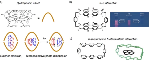
resulting host-guest complexes are significantly affected by close and multipoint interactions or contacts between host and aromatic guest molecules, investigation of binding modes and motifs of aromatic molecules is one of the most important issues particularly in the field of supramolecular chemistry. For the inclusion of aromatic molecules, hydrophobic effect is often used in aqueous media. For instance, Gibb and Ramamurthy reported that extended cavitand can
include two molecules of aromatic hydrocarbon at the same time in water (Figure 2–1a).[2a,b] In
these cases, included aromatic guests exhibit specific excimer emission or a stereo-selective photo-dimerization reaction due to the condensation effect and pre-organization of the aromatic molecules within the nano-space. π–π interaction or electrostatic interaction are also utilized as driving forces to include aromatic molecules. Wrüthner recently reported that a perylene bisimide cyclophane possessing a nano-space with electron deficient π-planes can effectively bind various kinds of aromatic hydrocarbons in organic solvent using multiple π–π interactions
(Figure 2–1b).[2d] In this case, the stability constants of the complexation tend to increase as the
face-to-face π–π overlaps between host and guest become larger. Moreover, this cyclophane works as a fluorescent probe to detect aromatic guest molecules based on specific emission properties depending on the electron transfer between guests and electron deficient π-planes of the host. A cationic macrocycle by Stoddart can also bind to various kinds of polycyclic aromatic hydrocarbons using π–π interaction and electrostatic interaction, which has a potential
to extract polycyclic aromatic hydrocarbons from crude oil from Saudi Arabia (Figure 2–1c).[2c]
As an important category of host molecules, metallo-macrocycles have been widely studied to create functional host-guest complexes due to specific chemical and physical properties of metal ions which depend on the type, number and mode of arrangement of metal ions on their
skeletons.[3] In particular, metallo-macrocycles that possess coordinatively labile sites of metal
ions on their inner surfaces can utilize coordination bonding as powerful driving forces for guest binding and activation. As mentioned in the previous chapter, several metal ions can bind to the aromatic π-planes through metal–arene interactions, which has developed specific molecular
architectures or chemical reactions involving aromatic molecules.[4,5] Herein, I envisioned that
ions would provide novel and effective binding motifs to recognize and activate aromatic guest molecules of suitable sizes and shapes utilizing multiple metal–arene interactions as driving forces (Figure 2–2). Such host-guest complexes mediated by multipoint metal–arene interactions are expected to exhibit specific physical or chemical properties to included aromatic guest molecules based on metal coordination.
Figure 2–1. Representative examples of the inclusion complexes of aromatic guest molecules. a)
Inclusion of two anthracenes and acenaphthylenes within an extended cavitand and their specific photo-chemical properties.[2a,b] b) The molecular structure of a perylene bisimide cyclophane and its
specific emission properties upon guest binding.[2d] c) The molecular structure of a cationic macrocycle and its inclusion complex of pyrene.[2c] Figures 2–1a–c are reproduced with permission from Chem. Commun. 2007, 1062–1064,[2b] Copyright 2007 The Royal Society of Chemistry, Angew. Chem. Int. Ed.
2015, 54, 10165–10168,[2d] Copyright 2015 Wiely-VCH Verlag GmbH, Weinheim, and from J. Am. Chem. Soc. 2013, 135, 183–192,[2c] Copyright 2013 American Chemical Society, respectively.
Figure 2–2. A schematic drawing of the inclusion of aromatic molecules via multipoint Ag–π interactions
within a nano-space of a dinuclear Ag(I)-macrocycle [Ag2L1X2](SbF6)2.
In this chapter, I describe the synthesis and guest binding behaviors of a dinuclear
Ag(I)-macrocycle [Ag2L1X2](SbF6)2 (X = Et2O or H2O) (Figure 2–2). This complex possesses a
surface. Herein, I revealed that [Ag2L1X2](SbF6)2 provides a novel and effective motif to bind
aromatic guest molecules utilizing multipoint Ag–π interactions as driving force.[5] Furthermore,
I found that the cationic character of Ag(I) ions can modify the redox potentials of included aromatic molecules.
2–2. Design and Synthesis of a Dinuclear Ag(I)-Macrocycle
A cyclophane-type macrocyclic ligand L1 was newly designed as a platform of a metallo-macrocycle with a rigid and well-defined aromatic framework (Figure 2–3). Two phenanthrolines, placed in about 1 Å apart from each other, can work as bidentate metal binding
sites to immobilize two metal ions on the inner surface of the nano-space in a certain distance.[6]
Four anthracenes placed as main building brocks are supposed to stand orthogonal to the cyclic framework of L1 due to the steric repulsion among H-atoms of the neighboring aromatic
skeletons to provide a three dimensional thick cavity.[7]
Ag(I) ion was selected here, because Ag(I) ion is well known to bind to the periphery of
various types of aromatic π-planes through Ag–π interactions.[5] Besides, the coordinatively
labile character of Ag(I) ion would provide an effective platform as a guest binding site to
create thermodynamically stable host-guest assemblies.[8]
Due to the above-mentioned characteristics, L1 can provide a well defined three dimensional nano-space with a precise arrangement of coordinatively labile sites of Ag(I) ions upon metal complexation. This would work as an effective binding site for aromatic molecules through multipoint Ag–π interactions.
Figure 2–3. Molecular design of macrocyclic ligand L1.
The macrocyclic ligand L1 was prepared by sequentially connecting aromatic fragments by
Pd-catalyzed coupling reactions (Scheme 2–1).
9,10-Bis(4,4,5,5-tetramethyl-1,3,2-dioxaborolan-2-yl)anthracene (3),
2,9-dichrolo-1,10-phenanthroline (4), and 1,3-dibromo-5-(hexyloxy)benzene (5) were connected to afford V-shaped precursors 6 and 7. The final cyclization reaction between 6 and 7 afforded L1 in 5% yield. Although, L1 was hardly soluble in common organic solvents (acetone,
n-hexane, benzene etc), it can be dissolved in CHCl3 at a concentration about 100 µM. L1 was
characterized by NMR (1H, COSY, and ROESY), HRMS (ESI), and the following crystal
structures of its derivatives (Figure 2–4–5, 2–7–8). The UV-Vis spectrum of L1 in CHCl3
showed an absorption band around 350–450 nm, which is typical to π-π* transition of
anthracene moieties (Figure 2–6).[7b] Emission spectrum of L1 showed broad band around 470
nm in CHCl3.
Scheme 2–1. Synthesis of macrocyclic ligand L1.
Figure 2–4. 1H NMR spectra of L1 (500 MHz, CDCl
Figure 2–5. ESI-TOF mass spectrum of L1 in CHCl3/CH3CN.
Figure 2–6. a) UV-Vis (blue line, 5 µM, l = 1.0 cm, 293 K in CHCl3) and fluorescence (red line, 5 µM,
293 K, λex = 365 nm in CHCl3) spectra of L1 and b) a photo of the emissive L1 in CDCl3 under UV
irradiation (room temperature, λex = 365 nm).
A dinuclear Ag(I)-macrocycle [Ag2L1X2](SbF6)2 (X = Et2O or H2O) was prepared by the
complexation between L1 and Ag(I) salt (Scheme 2–2). As an Ag(I) source, AgSbF6 was
selected because of the weak Lewis basic character of SbF6–, which would not inhibit
interaction between guest and Ag(I) centers as a competing coordinative species. Upon reaction
between L1 and 4.0 eq of AgSbF6 in CHCl3/(CH3)2CO followed by crystallization by Et2O
vapor diffusion in the dark, the dinuclear Ag(I)-macrocycle [Ag2L1X2](SbF6)2 was isolated in
63% yield as yellow crystals. The [Ag2L1X2](SbF6)2 complex was fully characterized by NMR,
Scheme 2–2. Synthesis of [Ag2L1X2](SbF6)2.
In the resulting crystal structure, the dinuclear Ag(I)-macrocycle exists as a 1:1 co-crystal
of [Ag2L1(Et2O)2](SbF6)2 and [Ag2L1(H2O)2](SbF6)2 (Figure 2–7–8). In each complex, two
Ag(I) ions are bound by two phenanthroline ligands in 9.433(1) Å and 9.181(1) Å apart from each other, respectively. In addition to two N-atoms of phenanthrolines, each Ag(I) ion is
coordinated by one O-atom of Et2O or H2O as a coordinating solvent, respectively, in a trigonal
planar coordination geometry. As expected, the dihedral angles among four anthracenes and adjacent aromatic rings were estimated to be around 80–90°. Thus, three dimensional nano-spaces arranged with a face to face arrangement of solvated Ag(I) ions have been created (Figure 2–8).
Figure 2–7. ORTEP views (50% probability level) of a) [Ag2L1(Et2O)2](SbF6)2 and b)
[Ag2L1(H2O)2](SbF6)2. Side alkyl-chains are omitted for clarity. (Ag: magenta, C: grey, C of Et2O: light
Figure 2–8. Space filling models of a–b) [Ag2L1(Et2O)2](SbF6)2 and c–d) [Ag2L1(H2O)2](SbF6)2. Side
alkyl-chains are omitted for clarity. (Ag: magenta, C: grey, C of Et2O: light blue, F: yellow, H: white, N:
blue, O: red, Sb: pink)
The 1H NMR spectrum of the resulting complex [Ag
2L1X2](SbF6)2 (X = Et2O or H2O) in
CDCl3 at 300 K showed phenanthroline’s signals (Ha–c) which were approximately 0.2 ppm
downfield shifted from those of the original macrocyclic ligand L1 due to the effect of the
coordination with Ag(I) ions (Figure 2–9).[6] Observation of only one single set of
phenanthroline’s signals suggests that the exchange reaction of bound and free coordinating
solvents (Et2O or H2O) is faster than the timescale of 1H NMR measurement at 300 K due to the
labile nature of the coordination bond between Ag(I) ions and solvent molecules.
Figure 2–9. 1H NMR spectra of [Ag2L1X2](SbF6)2 (500 MHz, CDCl3, 300 K). Acetone was included
during the processes of crystallization. b a c g d f e h i CHCl3 TMS H2O Et2O Et2O j k m,n o Acetone l CHCl3 a b c d e f g h i j k l m n o N N O AgI X X = Et2O or H2O
Figure 2–10. ESI-TOF mass spectrum of [Ag2L1X2](SbF6)2 in CHCl3.
To estimate the stability of the coordination bond between Ag(I) ions and two
phenanthrolines, 1H NMR study of a mixtures of isolated [Ag
2L1X2](SbF6)2 and different
amounts of L1 were performed (Figure 2–11). With an increase in the net equivalence of Ag(I)
ion, signals of D2h-symmetrical metal-free macrocyclic ligand L1 were firstly replaced by more
complicated signals corresponding to C2v-symmetrical mononuclear Ag(I)-macrocycle
[AgL1X]SbF6 (Figure 2–11d,e). Then, the signals were changed into simple D2h-symmetrical
signal patterns ascribable to the dinuclear Ag(I)-macrocycle [Ag2L1X2](SbF6)2 (Figure 2–11a–
c). These results suggest that the signals of L1, [AgL1X]SbF6, and [Ag2L1X2](SbF6)2 were
observed separately in CDCl3 at 300 K. Notably, 1H NMR spectrum of isolated
[Ag2L1X2](SbF6)2 showed only one set of signals in highly diluted solution (30 µM) in CDCl3
(Figure 2–11a), indicating that coordination bonding between Ag(I) ion and phenanthroline was
stable and the dissociation of Ag(I) ions from [Ag2L1X2](SbF6)2 was ignorable even in such a
low concentration condition.
200 400 600 800 1000 1200 1400 1600 1800 2000 2200 2400 2600 2800 m/z 812 814 816 820 812 814 816 820 Experimental Theoretical [Ag22]2+ 814.10 814.20 814.10
Figure SX. ESI-TOF mass spectrum of the CDCl3 solution of [Ag22](SbF6)2.
Experimental Theoretical 917 919 921 921 919 917 918.80 918.77 [pCp⊂Ag22]2+ 200 400 600 800 1000 1200 1400 1600 1800 2000 2200 2400 2600 2800 m/z 918.80
Figure SX. ESI-TOF mass spectrum of the CDCl3 solution of [pCp⊂Ag22](SbF6)2. 813 815 817 815 817 813 814.19 814.20 Experimental Theoretical [Ag2L1]2+ 814.20
Figure 2–11. Partial 1H NMR spectra of the mixtures of [Ag
2L1X2](SbF6)2 and different amounts of L1
(500 MHz, CDCl3, 300 K). a) [[Ag2L1X2](SbF6)2]0 = 30 µM, [L1]0 = 0 µM, b) [[Ag2L1X2](SbF6)2]0 = 25
µM, [L1]0 = 17 µM, c) [[Ag2L1X2](SbF6)2]0 = 20 µM, [L1]0 = 27 µM, d) [[Ag2L1X2](SbF6)2]0 = 17 µM,
[L1]0 = 35 µM, and e) [[Ag2L1X2](SbF6)2]0 = 0 µM, [L1]0 = 80 µM. [L1]0 and [[Ag2L1X2](SbF6)2]0
indicate the initial concentrations of L1 and [Ag2L1X2](SbF6)2, respectively. The net equivalence
Ag(I)/L1 was calculated as follows:
2–3. Host-Guest Interactions between a Dinuclear Ag(I)-Macrocycle
and Aromatic Guest Molecules via Ag–π Interactions
As the dinuclear Ag(I)-macrocycle [Ag2L1X2](SbF6)2 possesses a nano-space equipped
with solvated two Ag(I) ions, the cavity would provide a suitable space to include guest molecule(s) through coordination bonding. In this section, guest binding behaviors of
[Ag2L1X2](SbF6)2 were investigated in terms of guest selectivity and stability of inclusion
complexes. I found that [Ag2L1X2](SbF6)2 can bind several kinds of pristine aromatic molecules
using Ag–π interaction, by the ligand exchange reaction with coordinating solvents as characterized by NMR, ESI-TOF mass, and single crystal X-ray analyses. Notably, aromatic hydrocarbons, which can form Ag–π interactions with both of the two Ag(I) ions of
[Ag2L1X2](SbF6)2, have relatively high affinity to the dinuclear Ag(I)-macrocycle .
2–3–1. Binding of pristine aromatic hydrocarbons
2–3–1.
本節については3年以内に雑誌等で刊行予定のため、非公開
2–4.
本節については3年以内に雑誌等で一部刊行予定のため、非公開
本記述については3年以内に雑誌等で刊行予定のため、非公開
本記述については3年以内に雑誌等で刊行予定のため、非公開
2–3–2. Binding of a sandwich-shaped aromatic molecule
From the experimental results described in the previous section, multipoint Ag–π
interactions between [Ag2L1X2](SbF6)2 and π-surfaces of aromatic rings work as an effective
driving force to bind pristine aromatic molecules. The above-mentioned crystal structure of
p-xylene inclusion complex (p-Xylene)2⊂[Ag2L1(CF3SO3)2] (Figure 2–20) encouraged me to
incorporate [2.2]paracyclophane (pCp) as a sandwich shaped aromatic molecule, which has two stacked p-phenylene rings covalently connected by two alkyl chains in a π–π distance of 3.1 Å. I
then found that [Ag2L1X2](SbF6)2 can effectively bind to one pCp molecule with a significantly
high binding constant (Ka > 109 M–1 in CDCl3 at 300 K), as revealed by NMR, ESI-TOF mass
and single crystal X-ray analyses. Control experiments using host molecules which have similar
structures to [Ag2L1X2](SbF6)2 revealed that multipoint Ag–π interactions between pCp and
Ag(I) centers of [Ag2L1X2](SbF6)2 works as a major driving force for pCp binding.
The binding behavior of pCp to Ag(I)-macrocycle was studied by 1H NMR titration
experiment (Figure 2–22). Upon adding pCp to a solution of dinuclear Ag(I)-macrocycle
[Ag2L1X2](SbF6)2 (107 µM) in CDCl3, the signal intensity of the original [Ag2L1X2](SbF6)2
gradually decreased, which were replaced by a new set of signals assignable to the
D2h-symmetrical macrocyclic structure (Figure 2–22b). The signals of [Ag2L1X2](SbF6)2 were
completely replaced by the new signals in the presence of 1.0 eq of pCp (Figure 2–22c). This
result suggests the formation of a 1:1 inclusion complex pCp⊂[Ag2L1](SbF6)2. Considering
from the integral ratios of the NMR signals, the new singlet peaks appeared at 3.82 ppm and
1.17 ppm (Ain and Bin,respectively, in Figure 2–22c) can be assigned as those of the protons of
included pCp. These signals were significantly upfield shifted due to the strong shielding effect
from the anthracene walls (Δδ = –2.6 and –1.9 ppm for Ain and Bin, respectively). This
assignment was strongly supported by the distinct rotating frame Overhauser effect (ROE)
correlation between Ain and the protons (Hi) inside the cavity of [Ag2L1X2](SbF6)2 (Figure 2–
23). Although the host-guest binding was reversible, the intermolecular exchange reaction
between bound and free pCp molecules was slower than the timescale of 1H NMR at 300 K in
CDCl3, because the signals of [Ag2L1X2](SbF6)2 and pCp⊂[Ag2L1](SbF6)2 were observed
separately each other in the presence of less than 1.0 eq of pCp (Figure 2–22b). The formation
of pCp⊂[Ag2L1](SbF6)2 was also supported by ESI-TOF mass spectrometry (m/z = 918.80 for
Figure 2–22. Partial 1H NMR spectra of [Ag2L1X2](SbF6)2 (107 µM) in the presence of a) 0.0, b) 0.5,
and c) 1.0 eq of pCp, and d) pCp only (500 MHz, CDCl3, 300 K). Asterisks represent the signals of
p-dimethoxybenzene used as the internal standard.
Figure 2–24. ESI-TOF mass spectrum of a mixture of L1, AgSbF6 (4.0 eq), and pCp (2.3 eq) in CDCl3.
The structure and the binding mode of this host-guest complex were determined by single
crystal X-ray analysis. Ether vapor diffusion into a mixture of AgSbF6 (4.0 eq), L1 and pCp (1.0
eq) in CHCl3/(CH3)2CO in the dark yielded yellow plate crystals in 71% yield, which were
suitable for single crystal X-ray analysis. In the resulting crystal structure, one molecule of pCp is included within the nano-space of the dinuclear Ag(I)-macrocycle in two substitutionary disordering manners (occupancies: 56% and 44%) (Figure 2–25). The distance between two crystallographically equivalent Ag(I) centers is 8.514(1) Å, which is slightly shorter but approximately identical to those of the original dinuclear Ag(I)-macrocycle (Ag–Ag distances
of [Ag2L1X2](SbF6)2: 9.433(1)–9.181(1) Å). Two Ag(I) ions form a distorted tetrahedral
coordination geometry with two N-atoms of phenanthrolines and two C-atoms of pCp without coordinating solvents or anions (Ag–N1 2.342(6) Å; Ag–N2 2.327(5) Å; Ag–C 2.39(1)–2.56(1)
Å, Figure 2–25b,d). Notably, the π-planes of pCp form η2-type Ag–π interactions with both of
the Ag(I) ions within the nano-space. Although, the direction of these Ag–π bonding are inclined about 30° from the vertical direction of the π-plane of pCp, the resulting Ag–π–π–Ag
structure is quite similar to the case of the (p-Xylene)2⊂[Ag2L1(CF3SO3)2] as described in the
previous section (Figure 2–20). Besides Ag–π interactions, pCp within the nano-space forms multipoint CH–π interactions with anthracene walls (C–π distances: ca. 3.4 Å), which may stabilize this host-guest complex as well (Figure 2–25a,c).
200 400 600 800 1000 1200 1400 1600 1800 2000 2200 2400 2600 2800 m/z 812 814 816 820 812 814 816 820 Experimental Theoretical [Ag22]2+ 814.10 814.20 814.10
Figure SX. ESI-TOF mass spectrum of the CDCl3 solution of [Ag22](SbF6)2.
Experimental Theoretical 917 919 921 921 919 917 918.80 918.77 [pCp⊂Ag22]2+ 200 400 600 800 1000 1200 1400 1600 1800 2000 2200 2400 2600 2800 m/z 918.80
Figure SX. ESI-TOF mass spectrum of the CDCl3 solution of [pCp⊂Ag22](SbF6)2.
Experimental Theoretical 917 919 921 917 919 921 918.77 918.80 Experimental Theoretical pCp⊂[Ag2L1]2+
Figure 2–25. Crystal structure of pCp⊂[Ag2L1](SbF6)2. a,c) Space filling models, and b,d,) ORTEP
views (50% probability level) of a partial structure (some parts of solvent, side-alkyl chains, counter anions, and H-atoms are omitted for clarity). (Ag: magenta, C: grey, C of pCp : green and pale blue, F: yellow, H: white, N: blue, O: red, Sb: pink). pCp is colored based on the disordering patterns (a–b) 56% (green), c–d) 44% (pale blue)).
The 1H NMR spectrum of the isolated single crystal in CDCl
3 showed identical signal
patterns as that of aforementioned titration experiments (Figure 2–26a). This indicates the composition of the host-guest complex in solution was the same as that in the crystalline state.
Notably, in the crystal, pCp within the nano-space is inclined and showed a Ci symmetrical
structure because of the desymmetrization by an η2-type Ag–π bonding (Figure 2–25), whereas
1H NMR signals of the included pCp shows only two singlets (A
in and Bin) corresponding to a
D2h-symmetrical structure (Figure 2–22c). This suggests pCp exhibits fast and fluxional
oscillation or a precession movement within the nano-space of Ag(I)-macrocycle via
haptotropic shifts of Ag(I) ions in the time scale of 1H NMR in CDCl
3 at 300 K, whereas any
rotational movements of pCp in the nano-space were likely to be sterically inhibited.
[pCp⊂[Ag2L1]2+]/([pCp][[Ag2L1]2+]) M–1) was estimated by 1H NMR titration experiments in
CDCl3 at 300 K. Notably, Ka(pCp) was too large to directly determine from 1H NMR titration
experiments at the concentration of about 102 µM, a guest competition experiment in the
presence of ferrocene (FeCp2) as a competing guest was conducted, where, Ka(FeCp2) =
[FeCp2⊂[Ag2L1]2+]/([FeCp2][[Ag2L1]2+]) = 6.2 ± 0.9 × 10⁴ M–1 in CDCl3 at 300 K (the binding
behavior of FeCp2 is described on the section 2–3–3) (Figure 2–26). However, upon addition of
an excess amount (25 eq) of FeCp2 to a solution of pCp⊂[Ag2L1](SbF6)2 in CDCl3 at 300 K, no
guest exchanges were observed in 1H NMR study (Figure 2–26b). This result suggests K
a(pCp)
> 109 M–1 in CDCl3 at 300 K.
Figure 2–26. 1H NMR spectra of pCp⊂[Ag
2L1](SbF6)2 (115 µM) in the presence of a) 0.0 and b) 25 eq
of FeCp2.c) 1H NMR spectra of a mixture of [Ag2L1X2](SbF6)2 (105 µM) and FeCp2 (5.0 eq) (500 MHz,
CDCl3, 300 K). An asterisk represents the signal of p-dimethoxybenzene used as the internal standard. R = n-C6H13 X = Et2O or H2O CDCl3 N N OR RO N N AgI AgI : pCp⊂[Ag2L1](SbF6)2 Fe
FeCp2 No guest exchange
Strong host-guest interaction between [Ag2L1X2](SbF6)2 and pCp was also supported by
titration experiments using UV-Vis spectroscopy (Figure 2–27). Upon adding pCp to the
solution of [Ag2L1X2](SbF6)2 (50 µM) in CHCl3, the spectrum of [Ag2L1X2](SbF6)2 slightly
changed. The absorption change converged in the presence of 1.0 eq of pCp (Figure 2–27b),
which suggests quantitative formation of the host-guest complex pCp⊂[Ag2L1X2](SbF6)2 in
such a highly diluted condition (50 µM) due to strong host-guest interaction. It should be noted
that the slight change in the absorption of [Ag2L1X2](SbF6)2 suggests existence of no
remarkable charge transfer interaction between Ag(I) centers and pCp (Figure 2–27a).
Figure 2–27. a) UV-Vis spectra of the mixtures of [Ag2L1X2](SbF6)2 and different amounts of pCp
([[Ag2L1X2](SbF6)2] = 50 µM, l = 0.1 cm, 293 K in CHCl3), b) absorption change at 390.5 nm vs
equivalence of pCp to [Ag2L1X2](SbF6)2.
Such an extremely strong host-guest interaction is attributed to the multipoint Ag–π
interactions within nano-space of [Ag2L1X2](SbF6)2 as qualitatively evaluated by the following
control experiments.
Firstly, to evaluate the contribution of Ag–π interaction to the stability of the inclusion
complex pCp⊂[Ag2L1](SbF6)2, control experiments using L1 or [M2L1Xm]n+ (M = Hg(II),
Cu(I), and Zn(II); X = solvent or anion) in CDCl3/(CD3)2CO (= 75/1–75/0) at 300 K were
performed (Figure 2–28–29). Even when an excess amount of pCp was added to a solution of each host, the spectral patterns of the host and guest showed almost no changes, suggesting
weak host-guest interactions. It should be noted that the existence of counter anions (CF3SO3–or
BF4–) or (CD3)2CO did not have large effects on the results of these control experiments. Indeed,
the dinuclear Ag(I) complex of L1 with SbF6–, CF3SO3–, or BF4–as counter anions showed high
(Figure 2–30). Above-mentioned results suggest Ag–π interactions work as a major driving
force to include pCp into [Ag2L1X2](SbF6)2.
Figure 2–28. Partial 1H NMR spectra of mixtures of L1 (70 µM), metal sources (3.8 eq), and 0.0–10 eq of pCp (500 MHz, CDCl3/(CD3)2CO = 75/1, 300 K). Metal sources: a) Cu(CH3CN)4BF4, b) Hg(CF3SO3)2,
and c) Zn(CF3SO3)2.
Figure 2–29. Partial 1H NMR spectra of mixtures of L1 (125 µM) and 0.0–7.5 eq of pCp (500 MHz,
Figure 2–30. Partial 1H NMR spectra of mixtures of L1 (70 µM), Ag(I) sources (3.8 eq), and 0.0–1.5eq of pCp (500 MHz, CDCl3/(CD3)2CO = 75/1, 300 K). Ag(I) sources: a) AgSbF6, b) AgBF4, and c)
AgCF3SO3.
Next, to examine the effect of the mode of arrangement of Ag(I) ions within the cyclic
framework of L1, control experiment using a half-macrocycle [AgL2(Et2O)]SbF6, which has a
partial structure of [Ag2L1X2](SbF6)2, as a host was performed in CDCl3/(CD3)2CO at 300 K
(Figure 2–31–32) (preparation and characterization of [AgL2(Et2O)]SbF6 are described in
Chapter 3). Upon adding pCp to a solution of [AgL2(Et2O)]SbF6, gradual shifting of the signals
of [AgL2(Et2O)]SbF6 was observed, suggesting significant host-guest interactions (Figure 2–
31). Distinct from the case of the dinuclear Ag(I)-macrocycle, the intermolecular exchange
reaction of pCp was faster than the timescale of 1H NMR, because the signals gradually shifted
as the amount of pCp increased. The formation of a 1:1 complex pCp⊂[AgL2]+ in solution was
suggested from ESI-TOF mass measurement (m/z = 847.15 for pCp⊂[AgL2]+) (Figure 2–33).
Moreover, binding of pCp by the half-macrocycle via Ag–π interactions was suggested by
single crystal X-ray analysis (Figure 2–34). Upon slow evaporation of a mixture of L2, AgSbF6
(1.5 eq), and pCp (2.0 eq) in CDCl3, yellow block crystals were obtained. In the resulting crystal
structure, one molecule of pCp is bound by two [AgL2]+ from top and bottom via η2-type Ag–π
 2 or b) its CF 3 SO 3 – salt with aromatic molecules via Ag–π interactions](https://thumb-ap.123doks.com/thumbv2/123deta/8494529.922291/3.892.123.783.778.1092/figure-host-complexation-dinuclear-macrocycle-aromatic-molecules-interactions.webp)
![Figure 1–8. Representative examples of metal–arene complexes. a) A nucleophilic substitution reaction of a (arene)tricarbonylchromium derivative (Nu: nucleophile), [23c,25] b) a multinuclear Pd-complex using tetracenes as templates, [26] c) a mul](https://thumb-ap.123doks.com/thumbv2/123deta/8494529.922291/17.892.166.759.623.994/representative-nucleophilic-substitution-tricarbonylchromium-derivative-nucleophile-multinuclear-tetracenes.webp)
 2 and b) [Ag 2 L1(H 2 O) 2 ](SbF 6 ) 2](https://thumb-ap.123doks.com/thumbv2/123deta/8494529.922291/32.892.120.718.137.382/figure-ortep-views-probability-level-ag-sbf-sbf.webp)
 2 (500 MHz, CDCl 3 , 300 K)](https://thumb-ap.123doks.com/thumbv2/123deta/8494529.922291/33.892.171.775.719.1010/figure-h-nmr-spectra-ag-sbf-mhz-cdcl.webp)
