INTRODUCTION
Athletes in the pediatric age group are at risk of developing dis-orders of the lumbar spine caused by mechanical loading. Pars fracture, also known as lumbar spondylolysis, is the most prevalent disorder at this anatomic site in children and adolescents (1 - 3), as well as the apophyseal ring fracture (4, 5). The pathogenesis of lumbar spondylolysis and apophyseal ring fracture is reported to involve stress fracture (6, 7) or avulsion fracture (4, 5), indicating that these disorders are caused by mechanical loading at the lum-bar spine.
Several studies have reported general risk factors for lumbar spondylolysis, including high body mass index and cigarette smok-ing. Tight hamstring and/or quadriceps muscles have also been reported to be risk factors for the disorder (8). Thus, it is reason-able to assume that maintaining flexible hamstrings is important for preventing low back pain. According to the joint- by - joint theory proposed by Cook, the lumbar spine provides stability while the hip and ankle provide mobility (9, 10). Therefore, we focus on mobilization of the hip and ankle joint for in the treatment of pa-tients with low back pain.
Sairyoet al. reported that jack-knife stretching is a very effective form of active stretching to improve flexibility in patients with tight hamstrings (11). This type of stretching utilizes reciprocal inhibi-tion via contracinhibi-tion of the antagonist muscle (12). Taking the joint-by - joint theory into account, we hypothesized that mobilization of the hip and ankle joint would be very important for pediatric athletes.
In this study, we trained pediatric patients to perform home -based active stretching of the hamstring, quadriceps, and triceps
surae muscle groups and evaluated the effects of the stretching regimen 2 months later.
METHODS
SubjectsForty - six pediatric patients (41 boys, 5 girls ; mean age, 13.7! 2.4 years ; mean height, 161.5!16.2 cm ; mean weight, 53.3!13.8 kg) received conservative treatment. All patients were active par-ticipants in sports, predominantly baseball (n = 15), soccer (n = 11), and basketball (n = 5). The diagnosis was lumbar spondylolysis in all cases : unilateral in 19 and bilateral in 27. Their stage of the spondylolysis was the terminal stage. After confirming that all pa-tients were receiving adequate pain relief, the following stretch-ing program was implemented to alleviate tightness of 3 muscle groups in the leg.
Evaluation of muscle tightness
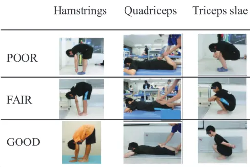
We graded tightness of the 3 muscle groups as good, fair, or poor at baseline and re - evaluated after 2 months of the stretching regimen (Figure 1). This grading method is arbitrary and impre-cise but can be performed rapidly. Specifically, we graded muscle tightness as follows.
Hamstrings : Patients were asked to bend the trunk forward from a standing position as far as possible. Muscle tightness was rated as good when patients could touch the floor with the palms, fair when they could touch the floor with the fingertips, and poor when they could not touch the floor at all.
Quadriceps : Patients were asked to bend each knee joint as far as possible while in the prone position, aiming to contact the but-tock with the heel. Muscle tightness was graded as good when the heel could make contact with the buttock, fair when the final dis-tance between the heel and buttock was equivalent to that of one fist, or poor when the final distance was more than one fist. Evalu-ation was performed for each side separately, and we took the worse
ORIGINAL
Active stretching for lower extremity muscle tightness in
pediatric patients with lumbar spondylolysis
Masahiro Sato(1)
, Yasuyoshi Mase(1), and Koichi Sairyo(2)
1)
Department of Orthopedic Surgery and Rehabilitation, Hachioji Sports Orthopedic Clinic, Hachioji, Japan,2)Department of Orthopedics,
Tokushima University Graduate School, Tokushima, Japan
Abstract : Background : It was reported that hamstring muscle tightness may increase mechanical loading on the lumbar spine. Therefore, we attempt to decrease tightness in the leg muscles in pediatric patients. Methods : Forty -six pediatric patients with spondylolysis underwent rehabilitation. We applied active stretching to the hamstrings, quadriceps, and triceps surae. Tightness in each muscle was graded as good, fair, or poor. We edu-cated each patient on how to perform active stretching at home. They were re-evaluated for muscle tightness 2 months later. Results : Tightness at baseline and after 2 months was as follows : for the hamstrings, good in 3 patients, fair in 9, and poor in 34 and significant improved after 2 months (p 0.05), with improvement by least 1 grade seen in 86%% of patients with fair or poor at baseline ; for the quadriceps, 7, 3, and 30 patients had good, fair and poor, with significant improvements in 72%% (p 0.05). For the triceps surae, 6, 3 and 10 patients had good, fair and poor, which improved significantly (p 0.05). Conclusion : Home-based active stretching was effective for relieving muscle tightness in the leg in a pediatric population. Adolescent athletes should perform such exercise to maintain flexibility and prevent lumbar disorders. J. Med. Invest. 64 : 136-139, February, 2017
Keywords : Active stretching, Reciprocal inhibition, low back pain, lumbar disorder, spondylolysis
Received for publication January 3, 2017 ; accepted January 25, 2017. Address correspondence and reprint requests to Koichi Sairyo, MD, PhD, Department of Orthopedics, Tokushima University, 3 - 18 - 15 Kuramoto, Tokushima 770 - 8503, Japan and Fax : +81 - 88 - 633 - 0178.
The Journal of Medical Investigation Vol. 64 2017
side as the patients’ tightness.
Triceps surae : Patients were asked to squat with their heels in contact with the floor. Muscle tightness was graded as good when they could squat completely while clasping both arms behind the back, fair when they could squat completely with the arms held in front, and poor when they could not squat completely even with the arms held in front.
Training regimen
On the first day of the study, one of our physical therapists
educated the patients on how to perform active stretching exercises for the hamstrings, quadriceps and triceps surae. Active stretching was performed for about 10 s for each muscle group at a time. The exercises were repeated 5 times daily. Reciprocal inhibition of ac-tive stretching was achieved by contraction of the hamstring, glu-teus maximus, and tibialis anterior muscles when stretching the quadriceps, hamstrings and triceps surae, respectively. The pa-tients attended rehabilitation once a week so that the therapists could confirm that the stretching exercises were being performed correctly. Figure 2 demonstrates the stretching method for each
POOR
FAIR
GOOD
Hamstrings
Quadriceps
Triceps slae
Figure 1 :Evaluation of tightness in the hamstring, quadriceps, and triceps surae muscle groups. Tightness was graded as good, fair, or poor.
Hamstrings Quadriceps Triceps
slae
Figure 2 :Stretching method for each muscle group. For the hamstrings (Figure 2 - left panel), Jackknife stretching was performed. With extending their knee joint, the hamstrings is stretched and simultaneously the quadriceps fomoris muscle contracts. For stretching the quadriceps (Figure 2 - central panel), the hip joint is extended with muscle contraction of the gluteus maximus. Figure 2 - right panel indicates the stretching method for the triceps slae muscle. The ankle joint gets dorsi - flexed with the muscle contraction of the tibialis anterior muscle.
0 50 100 (%) pre post hamstrings pre post triceps slae pre post quadriceps poor fair good muscle group. For the hamstrings in Figure 2 - left panel, Jackknife stretching was performed (11). With extending their knee joint, the hamstrings is stretched and simultaneously the quadriceps fomoris muscle contracts. For stretching the quadriceps (Figure 2 - central panel), the hip joint is extended with muscle contraction of the gluteus maximus. Figure 2 - right panel indicates the stretching method for the triceps slae muscle. The ankle joint gets dorsi -flexed with the muscle contraction of the tibialis anterior muscle. Statistical analysis
Data were collected at baseline and after performing the stretch-ing regimen for 2 months and compared usstretch-ing the chi - square test. P values less than 0.05 were taken as significant.
RESULTS
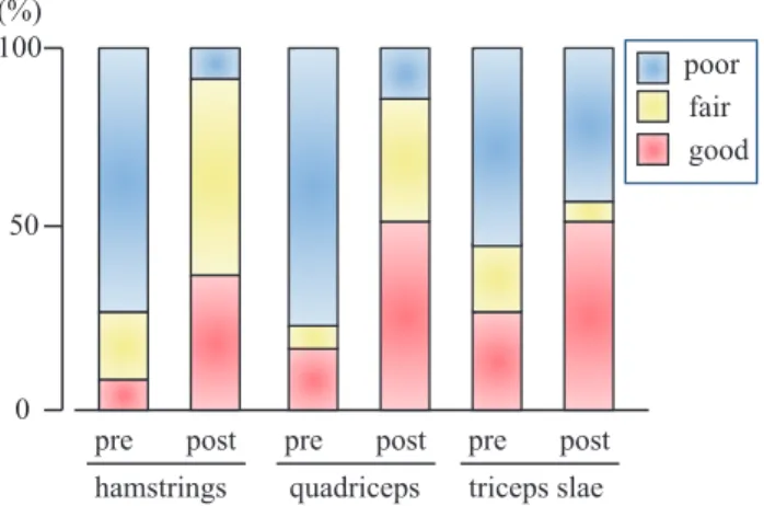
The results are shown in Figure 3. Tightness in the hamstring muscles was graded as good in 3 patients, fair in 9, and poor in 34 at baseline and improved significantly to good in 17, fair in 24, and poor in 8 at re - evaluation after 2 months of stretching exercises (p!0.05). Improvement by least 1 grade was seen in 86% of patients with fair or poor tightness at baseline. Tightness in the quadriceps muscles was graded as good in 7 patients, fair in 3, and poor in 30 at baseline and also improved significantly to good in 20, fair in 12, and poor in 8 after 2 months (p!0.05). Improvement by least 1 grade was seen in 72% of patients with fair or poor tightness at baseline. Tightness in the triceps surae muscles was graded as good in 6 patients, fair in 3, and poor in 10 at baseline and as good in 10, fair in 1, and poor in 8 after 2 months. Again, the improve-ment was significant (p!0.05).
DISCUSSION
In this paper, we describe the effects of active stretching on the hamstring, quadriceps, and triceps surae muscles in pediatric patients (aged!18 years) with lumbar spondylolysis. We found significant improvements after 2 months of a home - based active stretching regimen (p!0.05). It has been reported that low back pain may be related to muscle tightness in the lower extremities (8). Our active stretching regimen was very effective for improving the flexibility of these muscles, so it may be an effective strategy for
the prevention of low back pain in pediatric patients.
According to the joint- by - joint theory proposed by Cook (9, 10), the lumbar spine would be categorized as a stability joint. The tho-racic spine and hip and knee joints would be regarded as mobility joints. Therefore, stability of the lumbar spine and mobility of the hip and ankle joints would be important in the prevention of low back pain. Otaet al. (13) reported that exercise targeting the core abdominal muscles to stabilize the lumbar spine can ameliorate low back pain, which is consistent with the joint- by - joint theory (9, 10). Hasebeet al. (14) reported that flexibility of the hamstrings is closely related to lumbopelvic rhythm. They found that movement of the lumbar spine was dominant during lumbar flexion in subjects with tight hamstrings, which may cause low back pain related to mechanical stress. However, movement of the hip and pelvis was dominant in subjects with flexible hamstrings when bending the trunk forward. Therefore, they considered flexible hamstrings to be important for the prevention of low back pain. Their report is also somewhat consistent with the joint- by - joint theory (9, 10). With this in mind, we have been using a rehabilitation protocol of mobilization and stabilization for pediatric patients with low back pain.
The participants in this study were pediatric patients with lumbar spondylolysis, which in most cases would be attributable to me-chanical stress (6, 7). Therefore, our strategy of maintaining a good degree of flexibility of the mobile joints, such as the hip and ankle, could augment the core muscle strengthening exercises used to maintain stability of the lumbar spine (9, 10).
In a recent study by Sakaiet al. (15) reporting the results of con-servative treatment in 63 pediatric patients with lumbar spondyloly-sis, pars stress fractures were found to recur in 26.1% of pediatric patients in whom bony healing had been achieved with conserva-tive treatment, and the recurrence rate decreased when their pa-tients performed stretching exercises to reduce muscle tightness in the lower extremities. Taken together with our findings, active stretching of muscle groups designed to improve flexibility in the leg could be important for prevention of stress - induced lumbar disorders in children and adolescents. We believe that our home -based active stretching program is useful for improving the flexi-bility of joints responsible for moflexi-bility, in particular the hip and ankle. Active stretching of the hamstring, quadriceps, and triceps surae muscle groups may be beneficial for preventing low back pain in the pediatric population.
In this study, all cases had the lumbar spondylolysis at the ter-minal stage. Thus, it was not difficult to perform this type of stretch-ing at home. Clinically, we have been treatstretch-ing patients with any stage of the lumbar spondylolysis. According to the strategy for the conservative treatment (1 - 3, 15), patients having the early stage of the disorder would need to wear a hard brace to achieve a bony healing at least for three months. For such cases, first of all, the main purpose of the treatment would be pain control. After the pain disappears, they start to perform the stretching. The hard brace we use (1, 15) does not bother hip motion. Thus, with wearing the brace, they can perform the stretching. One of the limitations of this study would be the method to evaluate the rigidity of the mus-cles as shown in Figure 1. The method is not precise. It may be better and more precise, if we measure some distance indicating the rigidity. On the other hand, merit of this method is to be easy to evaluate even in a very busy out- patient clinic. To screen if the patients have the rigidity or not, this method would be very ef-fective. We would like to recommend this evaluating method in a very busy out- patient clinic as a screening.
We suggest that surgeons pay attention to tightness of muscles in the region of mobility joints, particularly the hip and ankle, in pedi-atric patients with low back pain in addition to treating the underly-ing lumbar pathology. Active stretchunderly-ing with reciprocal inhibition may be highly effective for improving the flexibility of these joints. Figure 3 :Results of measurement of tightness in the hamstring,
quad-riceps and tquad-riceps surae muscle groups. Tightness was significantly im-proved after 2 months of the stretching regimen in all muscle groups (p!0.05).
REFERENCES
1. Sairyo K, Sakai T, Yasui N, Dezawa A : Conservative treatment for pediatric lumbar spondylolysis to achieve bone healing using a hard brace : what type and how long? J Neurosurg Spine 16(6) : 610 - 4, 2012
2. Sairyo K, Sakai T, Yasui N : Conservative treatment of lumbar spondylolysis in childhood and adolescence : the radiological signs which predict healing. J Bone Joint Surg Br 91(2) : 206 -9, 2009
3. Nitta A, Sakai T, Goda Y, Takata Y, Higashino K, Sakamaki T, Sairyo K : Prevalence of Symptomatic Lumbar Spondylolysis in Pediatric Patients. Orthopedics 39(3) : e434 - 7, 2016 4. Sairyo K, Goel VK, Masuda A, Vishnubhotla S, Faizan A, Biyani
A, Ebraheim N, Yonekura D, Murakami R, Terai T : Three -dimensional finite element analysis of the pediatric lumbar spine. Part I : pathomechanism of apophyseal bony ring frac-ture. Eur Spine J 15(6) : 923 - 9, 2006
5. Faizan A, Sairyo K, Goel VK, Biyani A, Ebraheim N : Biome-chanical rationale of ossification of the secondary ossification center on apophyseal bony ring fracture : a biomechanical study. Clin Biomech (Bristol, Avon) 22(10) : 1063 - 7, 2007 6. Sairyo K, Katoh S, Sasa T, Yasui N, Goel VK, Vadapalli S,
Masuda A, Biyani A, Ebraheim N : Athletes with unilateral spondylolysis are at risk of stress fracture at the contralateral pedicle and pars interarticularis : a clinical and biomechanical study. Am J Sports Med 33(4) : 583 - 90, 2005
7. Terai T, Sairyo K, Goel VK, Ebraheim N, Biyani A, Faizan A, Sakai T, Yasui N : Spondylolysis originates in the ventral as-pect of the pars interarticularis : a clinical and biomechanical
study. J Bone Joint Surg Br 92(8) : 1123 - 7, 2010
8. Feldman DE, Shrier I, Rossignol M, Abenhaim L : Risk factors for the development of low back pain in adolescence. Am J Epidemiol 154(1) : 30 - 6, 2001
9. Cook G : The joint by joint theory (in Japanese). In Move-ment : functional moveMove-ment system, edited and translated by Nakamaruet al. Nap limited, Tokyo, Japan. pp 308-317, 2014 10. Motohashi E : The joint by joint theory (in Japanese). J Clin
Sports Med 33 (10) : 908 - 916, 2016
11. Sairyo K, Kawamura T, Mase Y, Hada Y, Sakai T, Hasebe K, Dezawa A : Jack - knife stretching promotes flexibility of tight hamstrings after 4 weeks : a pilot study. Eur J Orthop Surg Traumatol 23(6) : 657 - 63, 2013
12. White SG, Sahrmann SA : A movement system balance ap-proach to management of musculoskeletal pain. In : Grant R (ed). Physical therapy of the cervical and thoracic spine. Churchill Livingstone Inc, New York, pp 339 - 357, 1994 13. Ota M, Kaneoka K, Hangai M, Muramatsu T : Effectiveness
of Lumbar Stabilization Exercises Compared with Traditional Therapeutic Exercises for Chronic Low Back Pain. Journal of Spine Research 6(9) : 1385 - 1391, 2015
14. Hasebe K, Sairyo K, Hada Y, Dezawa A, Okubo Y, Kaneoka K, Nakamura Y : Spino - pelvic - rhythm with forward trunk bend-ing in normal subjects without low back pain. Eur J Orthop Surg Traumatol 24(Suppl 1) : 193 - 199, 2014
15. Sakai T, Tezuka F, Yamashita K, Takata Y, Higashino K, Nagamachi A, Sairyo K : Conservative Treatment for Bony Healing in Pediatric Lumbar Spondylolysis. Spine (Phila Pa 1976). 2016 Oct 17. [Epub ahead of print]