Muscle damage and inflammation in whole-body
strenuous exercises
著者
作山 悠子
学位授与機関
Tohoku University
修士論文
Muscle damage and inflammation in whole-body
strenuous exercises
(全身性激運動における筋損傷と炎症)
東北大学大学院医学系研究科
機能医科学講座障害科学専攻運動学分野
作山悠子
Abstract
Exertional or exercise-induced rhabdomyolysis can develop after strenuous physical activity. The purpose of this study was to elucidate whether whole-body strenuous exercises are associated with high serum creatine kinase (CK) values, exertional rhabdomyolysis, and/or renal failure after exercise. Moreover, the study also investigated which types of exercise cause elevated serum CK levels. Blood and urine samples were obtained from 37 subjects who performed whole-body strenuous exercises. The samples were collected before and 4 and 5 days after the exercises and analyzed. The analyzed parameters included an indicator of muscle damage (serum CK level), an indicator of inflammation (serum high-sensitivity C-reactive protein level), an indirect marker of skeletal muscle damage (delayed-onset muscle soreness), measures of renal function (serum sodium, potassium, chlorine, and creatinine levels; urinary protein and uric acid levels; and the presence/absence of occult blood in urine), and iron status indicators (serum iron levels;
unsaturated iron binding capacity, and total iron binding capacity). The mean serum CK levels at 4 and 5 days after the exercises were 4022.6 and 3214.6, respectively, which represented increases of 1500% and 1199% above the baseline level, respectively (P <0.01). None of the subjects’
creatinine levels were above the upper limit of normal at 4 or 5 days after the exercises. Only squat jump performance display a significantly strong correlation with the increase in the serum CK level (r = 0.721, P <0.001). The subjects’ serum iron levels were significantly decreased on post-exercise day 4 (P <0.001). In conclusion, whole-body strenuous exercises can produce marked elevations in serum CK levels without causing renal failure. The marked elevation of serum CK levels is associated with highly repetitive eccentric contraction-based exercises such as squat jumps. In addition, the subjects’ serum iron levels decreased after the exercises and were found to be associated with muscle damage and inflammation.
Table of contents
Introduction・・・・・・・・・・・・・・・・・・・・・・・・・・・・・・・・・・1
Materials and Methods・・・・・・・・・・・・・・・・・・・・・・・・・・・・・ 4
Subjects・・・・・・・・・・・・・・・・・・・・・・・・・・・・・・・・・・・4
Whole-body strenuous exercises・・・・・・・・・・・・・・・・・・・・・・・・・6
Blood sampling・・・・・・・・・・・・・・・・・・・・・・・・・・・・・・・・7
Urine sampling・・・・・・・・・・・・・・・・・・・・・・・・・・・・・・・・7
Delayed-onset muscle soreness (DOMS) ・・・・・・・・・・・・・・・・・・・・・7
Statistical analysis・・・・・・・・・・・・・・・・・・・・・・・・・・・・・・・ 7
Results・・・・・・・・・・・・・・・・・・・・・・・・・・・・・・・・・・・・ 8
Discussion・・・・・・・・・・・・・・・・・・・・・・・・・・・・・・・・・・ 10
Acknowledgements・・・・・・・・・・・・・・・・・・・・・・・・・・・・・・ 15
References・・・・・・・・・・・・・・・・・・・・・・・・・・・・・・・・・・ 16
1
Introduction
Exertional or exercise-induced rhabdomyolysis occurs when strenuous physical activity places stress on a muscle, resulting in damage to its fibers (Hamer, 1997; Knochel, 1990; Line et al., 1995; Walsworth et al., 2001). The primary pathophysiology of rhabdomyolysis typically involves strenuous eccentric exercises being performed at an intensity to which the participant is unaccustomed or by individuals that are unprepared or underprepared for physically demanding tasks (Knochel, 1982; Knochel, 1990). The more strenuous or prolonged an exercise is, the worse damage it causes (Knochel, 1982; Knochel, 1990). Such damage manifests as delayed-onset pain and soreness, weakness, and increases in the circulating concentrations of muscle proteins such as creatine kinase (CK) (Knochel, 1982; Knochel, 1990). Serum CK levels are used as a surrogate marker of muscle injury in the clinical setting to determine whether renal damage has occurred and whether treatment needs to be administered to prevent renal failure (Clarkson, 2006a). Rhabdomyolysis results from the disruption of muscle membrane integrity, which can arise from a variety of conditions; however, exercise-induced or exertional rhabdomyolysis can be diagnosed using the following criteria: a history of strenuous exercise, a serum CK level of >5 times normal (range, 58-280 U/L) or 290-1,400 U/L (Knochel, 1982; O’Connor et al., 2008), and the detection of blood during a urine dipstick test (i.e., a tea- or cola-colored dipstick) (O’Connor et al., 2008; Clarkson et al., 2006a). A serum CK level of >10 times the upper limit of normal, or approximately 2,000U/L, is frequently used as the criterion for statin myopathy and a reason for stopping statin therapy (Thompson et al., 2003), and a CK level of >10,000 U/L is accepted as being diagnostic of rhabdomyolysis (de Lemos et al., 2004). Although the clinical signs and symptoms of the condition vary widely, damaged muscles can swell excessively and lead to compartment syndrome, which often requires surgical intervention (Knochel, 1982; Moeckel-Cole et al., 2009; Rupert, 2002), or
2
acute renal failure due to the precipitation of myoglobin in the renal tubules (Clarkson et al., 2006a; Clarkson, 2007; Huerta-Alardin et al., 2005).
In rhabdomyolysis, the amount of myoglobin delivered to proximal renal tubule cells overwhelms their ability to convert iron to ferritin, resulting in intracellular ferrihemate accumulation. As a metal, iron has the ability to generate oxygen free radicals. This leads to oxidative stress and renal cell injury. However, there have not been any studies about the relationships between serum iron levels and indicators of renal function after strenuous exercise (Efstratisdis et al., 2007).
Although compartment syndrome and acute renal failure are rare in cases of exertional rhabdomyolysis, hospital admission is still reasonable and prudent (Sinert et al., 1994). However, there are no commonly accepted criteria for determining when individuals who present with elevated CK levels require hospitalization and/or treatment. The utility of serum CK cut-off values for making such decisions is disputed since such values do not take into consideration baseline differences, and normal CK values have not been clearly defined (O’Connor et al., 2008; Brewster et al., 2007). Moreover, the serum CK concentrations observed after physical exercise vary widely between individuals, and it has been suggested that they are affected by gender, race, and fitness (O’Connor et al., 2008; Brewster et al., 2007).
In the present study, the subjects performed whole-body strenuous exercises and then had their blood parameters analyzed. The analyzed parameters included an indicator of muscle damage (the serum CK level), an indicator of inflammation (the serum high-sensitivity C-reactive protein level, hs-CRP), an indirect marker of skeletal muscle damage (delayed-onset muscle soreness, DOMS), measures of renal function (serum sodium, potassium, chlorine, and creatinine levels; urinary protein and uric acid levels; and the presence/absence of occult blood in urine), and iron status
3
indicators (serum iron levels; unsaturated iron binding capacity, UIBC; and total iron binding capacity, TIBC). Factors such as heat stress, dehydration, drugs, alcohol abuse, and crush trauma are known to cause high serum CK levels, rhabdomyolysis, and renal failure (Singh et al., 2005). Clarkson described the relationships between exertional muscle damage, renal function, and profoundly elevated serum CK values in a large population of healthy subjects (Clarkson et al., 2006a). However, the subjects of the latter study only performed eccentric contractions of their elbow flexor muscles, and no studies examining the abovementioned relationships have involved the performance of whole-body strenuous exercises by healthy subjects.
The present study attempted to elucidate whether whole-body strenuous exercises are associated with high CK values, exertional rhabdomyolysis, and/or renal failure, and if so, which exercises are particularly associated with elevated serum CK levels. In addition, the relationships between serum iron levels and indicators of renal function after whole-body strenuous exercises were examined. It is hypothesized that whole-body strenuous exercise that results in markedly elevated serum CK levels is not sufficient to induce renal failure in healthy individuals. Improving our understanding of the relationships among markedly elevated serum CK levels, exertional rhabdomyolysis, and strenuous exercise would facilitate the appropriate treatment of individuals that suffer adverse effects of strenuous exercise.
4
Materials and Methods
Subjects
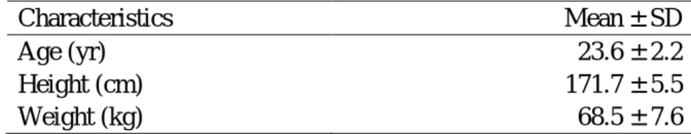
Thirty-seven healthy males (age: 23.6±2.2yr, height: 171.7±5.5cm, weight: 68.5±7.6kg; mean ± SD) volunteered for this study (Table 1). All of the subjects were freshmen at a protective service workers’ academy and lived in the same dormitory. The subjects were not participating in a supervised on-duty physical training program. The present study employed the physical fitness tests performed by freshman students at the academy each year. The subjects did not refrain from analgesic use, physical activity, or alcohol consumption during the study. In addition, all subjects were informed of the aims, benefits, and risks of the study; signed informed consent forms; and were told that they were free to withdraw from the study at any time without penalty. The study protocol was approved by the institutional review board of the Tohoku University Graduate School of Medicine.
Whole-body strenuous exercises
The subjects performed all of the whole-body strenuous exercises on the same day, and the exercises were separated by 10-min rest periods. The subjects were instructed to drink water during the exercises, and verbal and written reminders to maintain hydration were provided throughout the study.
1. Toe touch test. In the toe touch test, the subjects started by standing erect on a box with their feet hip-width apart. They were then asked to bend forward as far as possible, while ensuring that their knees, elbows, and fingers remained fully extended. The distance stretched by each subject was measured using lateral photographs of them standing in the erect and maximally flexed positions.
5
2. Standing triple jump test. The subjects performed three consecutive bounds, during which they were asked to jump as far as possible.
3. Back strength test. The subjects performed a back extension whilst pulling the handle of a portable dynamometer (Takei, Tokyo, Japan), during which they were asked extend their back as much as possible.
4. Seated overhead press test. The subjects performed an overhead press whilst sitting erect in a seat. First, the subjects grabbed a 30kg barbell and held it in front of their chest. Then, they raised the barbell to the overhead position. The subjects were asked to perform as many repetitions of the seated overhead press as possible until they dropped the barbell.
5. Three-stage shuttle run test. The subjects were asked to continuously run short distances; i.e., to run a short distance and back again, before immediately repeating the exercise (shuttle runs), at a constant speed. In the first stage of the exercise, each shuttle took about 10-25 seconds to complete, whereas in the second and third stages the shuttles took 50-65 seconds and 90-105 seconds to complete, respectively. The subjects performed each stage for 5 min and were asked to complete as many shuttles as possible within each set whilst running at a constant speed.
6. Pull-up test. The subjects hung on a high bar and pulled their bodies up with their arms without “kipping”. They were asked to perform as many pull-up repetitions as possible until they dropped off the bar.
6
7. Squat jump test. The subjects jumped from a starting position in which their knees were bent at a 90° angle. Their hands were placed on their hips during the jump, which prevented arm swinging. The subjects were asked to perform as many squat jumps as possible within 4 min.
8. Push-up test. The subjects started in the standard push-up position with their hands
shoulder-width apart and their elbows fully extended. Next, they lowered their bodies until their chests touched the ground whilst ensuring that the plane running from their toes through their hips to their shoulders remained approximately parallel to the ground. Finally, the subjects pushed themselves back up to the starting position. The subjects were asked to perform as many push-ups as possible within 3 min.
9. Sit-up test. The subjects lay in the supine position with their arms crossed over their chests, their hips flexed to approximately 45°, their knees flexed to approximately 90°, and their feet placed flat on the floor. Then, they were instructed to curl their head, shoulders, and trunk up until their shoulder blades were off the floor. The subjects were asked to perform as many sit-ups as possible within 3 min.
10. 275m sprint test. The subjects were asked to perform a 275m sprint as fast as possible.
Blood sampling
Blood samples were obtained from the subjects’ forearm veins at their dormitory 2 days before and 4 and 5 days after the exercises (Figure 1). The fourth post-exercise day was chosen as the first time point because this is when the peak serum CK response occurs. Blood was collected in 5-mL
7
siliconized vacuum glass tubes containing ethylenediaminetetraacetic acid (EDTA), and the samples were sent to Miyagi Prefectural Preventive Association of Lifestyle Diseases (Sendai, Japan) to be analyzed. To assess cartilage and muscle damage, the serum CK level was assayed on the same day, and aliquots of serum for use in the hs-CRP assay were subsequently frozen and stored at -20°C until use. The other examined markers were used to assess renal function (serum sodium, potassium, chlorine, and creatinine levels) or iron status (the serum iron concentration, UIBC, and TIBC).
Urine sampling
Urine samples were collected in the morning at the subjects’ dormitory 2 days before and 4 and 5 days after the exercises. The samples were used to analyze renal function (urinary protein and uric acid levels and the presence/absence of occult blood in urine).
Delayed-onset muscle soreness (DOMS)
The subjective presence and intensity of the pain experienced by the subjects were evaluated using a visual analog scale graded from 0 (no pain) to 10 (intolerable pain) arbitrary units (a.u.).
Statistical analysis
8
repeated-measures ANOVA was used to detect significant differences between different time points. When significant differences were detected, Bonferroni’s correction was used for pairwise comparisons. Differences between the pre- and post-exercise values of each parameter were evaluated using the paired Student’s t-test. Pearson’s correlation coefficient was used to analyze the relationships between the examined parameters (except for the electrolyte concentrations). Pearson’s correlation coefficient was used to analyze the associations between increases in the serum CK level and each exercise. A P value of <0.05 was used to indicate statistical significance.
9
Results
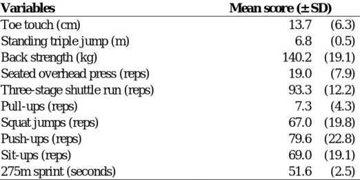
The whole-body strenuous exercise performance data are summarized in Table 2. None of the subjects experienced any adverse medical events during or after the exercises.
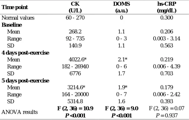
Indicators of muscle damage and inflammation (Table 3)
The mean CK levels on post-exercise days 4 and 5 were 4022.6 and 3214.6, respectively, which represented increases of 1500% and 1199% above the baseline level, respectively (P <0.01). Of the 37 participants, 10 had serum CK values of >2,000 U/L and 4 had serum CK values of >10,000 U/L; i.e., the levels used to diagnose myositis and rhabdomyolysis, respectively, on post-exercise day 4. There was a large degree of variability in the changes in the serum CK level induced by the exercises, but some subjects exhibited marked increases in their CK levels during the post-exercise period. Slight muscle soreness (0-3 a.u.) was reported prior to the exercises because of the daily training performed by the subjects. However, the subjects experienced significantly worse muscle pain on day 4 after the exercises (P <0.01). The nociceptive sensations perceived on post-exercise day 4 ranged from 0 to 6, with the mean value being 2.1 ± 1.7 a.u. The subjects were still experiencing significantly increased muscle pain compared with the baseline on post-exercise day 5 (P <0.01). Whilst the mean hs-CRP level was increased on post-exercise days 4 and 5, neither value was significantly higher than the baseline value (P = 1.00).
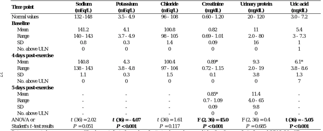
Serum and urinary indicators of renal function (Table 4)
Despite the fact that muscle injury results in the release of creatine, which would be expected to lead to increased serum creatinine levels, no impairment in renal function was detected after the exercises. In addition, none of the subjects had creatinine levels that were greater than the upper limit of normal at 4 or 5 days after the exercises. However, the mean creatinine level was significantly increased on post-exercise day 4 (P <0.001). The mean creatinine level was slightly
10
lower on post-exercise day 5, but remained significantly higher than the baseline value (P <0.001). The mean uric acid and serum potassium level were significantly increased on post-exercise day 4 (both P <0.001). Conversely, the mean serum levels of sodium and chloride and the mean urinary protein level did not change significantly after the exercises. All tests for occult urinary blood were negative.
Serum indicators of iron status (Table 5)
The serum iron level was significantly decreased at 4 days post-exercise (P <0.001). In fact, it fell below the lower reference limit of the assay. The serum iron level did not breach the upper limit of the normal range in any case. UIBC was significantly increased at 4 days post-exercise (P <0.001), whereas TIBC did not change significantly after the exercises (P = 0.077).
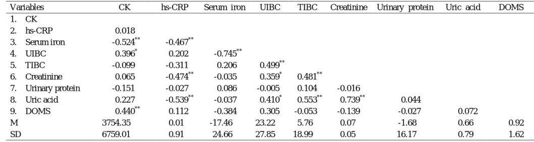
On post-exercise day 4, a strong correlation (r = 0.739) was detected between the serum creatinine level and the uric acid level (Table 6). In addition, the serum iron level was strongly correlated with UIBC (r = -0.745). A moderate correlation (r = -0.467) was detected between the serum hs-CRP level and the serum iron concentration. The subjects’ serum iron levels were also moderately correlated with their serum CK levels (r = -0.524). However, their serum iron levels were not significantly correlated with any of the other indicators of renal function.
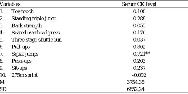
Only the squat jump test exhibited a significantly strong correlation with increased serum CK levels (r = 0.721, P <0.001) (Figure 2, Table 7)
11
Discussion
This study focused on the inter-subject variability in the effects of whole-body strenuous
exercises and examined whether such exercises are associated with high serum CK values, exertional rhabdomyolysis, and/or renal failure. This is the first study to suggest that a relationship exists between elevated serum CK levels caused by whole-body strenuous exercises and renal function. However, the results of the present study support the hypothesis that whole-body strenuous exercises that result in markedly elevated serum CK levels are not sufficient to induce renal failure in healthy individuals. In the present study, 4 subjects (11%) exhibited serum CK levels of >10,000 U/L, a concentration used to diagnose rhabdomyolysis, and of these 2 subjects (5%) displayed serum CK levels of >20,000 U/L, a concentration associated with renal failure in other studies (Sinert et al., 1994), but none of the subjects displayed impaired renal function after the whole-body strenuous exercises.
This study used a whole-body strenuous exercise protocol including both concentric and eccentric exercises to induce muscle damage. The above protocol was successful at inducing muscle damage, as evidenced by the significant differences between the pre- and post-exercise values of the examined parameters. As expected, a large degree of variability was detected in the subjects’ parameter values, particularly in their post-exercise serum CK levels. The latter result suggests that the whole-body strenuous exercises produced muscle fiber damage. Previous studies have reported that highly significant increases in the serum CK level were only observed after strenuous exercises, indicating that such exercises lead to exercise-induced muscle-fiber injury (Clarkson, 1997; Sorichter et al., 1999). The results of the present study support the findings of these studies.
12
muscle tissue damage (Rupert, 2002). In cases of rhabdomyolysis, the serum CK level is used as a surrogate measure of the serum myoglobin concentration because it CK released from muscle in proportion to myoglobin release, and the serum CK level can be assessed more quickly and cheaply than the serum myoglobin level (Clarkson, 2007). In the clinical setting, serum CK levels are commonly used to assess the severity of muscle damage and determine whether patients who present with symptoms of rhabdomyolysis should be hospitalized in order to prevent renal failure (Clarkson et al., 2006a; Rupert, 2002). However, no standard CK criteria for making such decisions exist because of the limited information available regarding the exercise-induced elevation of serum CK levels and the effects of these changes on renal function (Walsworth et al., 2001). Accordingly, there is currently no commonly accepted algorithm for determining whether individuals who present with elevated serum CK levels require treatment or hospitalization. During rhabdomyolysis, large quantities of CK and other muscle proteins are released into the bloodstream by the damaged muscles (Vanholder et al., 2000). In fact, the serum CK levels of rhabdomyolysis patients can reach approximately 100,000 U/L, whereas the normal range is less than 280 U/L (Knochel, 1982; O’Connor et al., 2008). O’Connor et al. (O’Connor et al., 2008) and others (Rupert, 2002; Harriston, 2004) have suggested that a serum CK level of 5 times greater than normal should lead to a suspicion of rhabdomyolysis. In the present study, no subjects developed renal failure, which suggests that the exercise-induced elevation of serum CK levels might not be a useful criterion for deciding whether hospitalization is necessary. Instead, such criteria should be based on other clinical features of exertional rhabdomyolysis.
The timing of CK release into and clearance from the blood after exercise primarily depends on the type, intensity, and duration of the exercise performed. In a previous study, a peak serum CK level of approximately two-fold higher than the baseline value was detected at 8 h after strength
13
training (Hurley et al., 1995). Furthermore, increased serum CK levels after eccentric exercise were found to be associated with muscle injury, and pronounced increases in the serum CK level were seen between 2 and 7 days after exercise (Serrão et al., 2003). Another study found that if an individual rests after prolonged exercise, they will only exhibit markedly increased serum CK levels for 24 h after the exercise. However, if they continue to train their serum CK levels will remain high for much longer (Stäubli et al., 1985). After eccentric exercise, CK release was found to peak at 96 h after the exercise, and participating in an additional exercise session only produced small increases in CK release, probably due to accelerated enzyme clearance (Hyatt et al., 1998). More intense activity, such as twice-daily football training, was found to lead to significant increases in the serum CK level on the 4th day of training. Subsequently, CK activity was found to decrease between post-exercise days 4 and 10, which probably represents adaptation to the training program (Ehlers et al., 2002). Circulating CK levels are also used to chart treatment progression because they decrease as rhabdomyolysis is resolved (Vanholder et al., 2000). In the present study, the subjects performed all of the strenuous exercises on a single day and did not engage in any exercise for the next two days. As reported, the subjects’ serum CK levels were markedly increased at 4 days after the exercises. However, their serum CK levels started to decrease on day 5 after the exercises. These observations indicate that rest days might be required after strenuous exercise in order to allow the participant’s serum CK levels to decrease, but it is unclear that how much rest is needed for recovery. Appropriate guidelines about when subjects should return to physical activity after exertional rhabdomyolysis are needed.
The present study detected a positive correlation between squat jump performance and the subsequent increase in the serum CK level. This indicates that performing a high number of squat jumps will result in a large increase in the serum CK concentration. Squat jumps are classified as a
14
high intensity eccentric exercise as the muscle groups of the lower extremities work eccentrically during the landing phase of squat jumps (Hoffman et al., 2005). It has been well documented that in individuals who are unaccustomed to high intensity exercise eccentric muscle activity causes greater muscle damage than concentric muscle activity (Dierking et al., 2000). Thus, it is speculated that the eccentric muscle activity employed during the landing phase of squat jumps causes considerable muscle damage in people who are unaccustomed to such exercises (Hoffman et al., 2005). Such high intensity eccentric exercise can lead to rhabdomyolysis. Clarkson reported the case of a 12-year-old boy who participated in an indoor physical education class during which he was required to perform an excessive number (>250) of repetitive squat jumps as punishment for talking in class. The boy reported intense muscle soreness in his thighs and produced dark urine at 2 days post-exercise; thus, his parents took him to the emergency department. His serum CK level rose to 244,006 U/L on post-exercise day 4, which resulted in him being hospitalized (Clarkson, 2006b). Muscle damage can also be induced by exercise involving repetitive eccentric contractions over a long period of time in untrained or undertrained individuals. Exercises that are particularly damaging usually involve eccentric loading, lengthening contractions, lowering weights against gravity, or negative repetitions (O’Connor et al., 2008). Taken together, these studies suggest that highly repetitive eccentric exercises, especially squat jumps, cause marked increases in the serum CK level and exertional rhabdomyolysis.
The reason why some individuals suffer renal failure after exertional exercise is not fully understood. To determine the factors correlated with indicators of renal function, the present study examined the relationships between serum iron levels and indicators of renal function. A negative correlation was detected between the serum iron level and the serum hs-CRP level. The subjects’ serum iron values decreased after the whole-body strenuous exercises. However, the opposite
15
result was obtained in a previous study, which detected an association between the serum iron level and the serum IL-6 concentration (Santos-Silva et al., 2002). IL-6 is a mediator of CRP production, which is produced by monocytes and macrophages in response to low-grade infection or other inflammatory stimuli. It has been suggested that elevated non-immediate production of hs-CRP is mediated by a regulatory pathway involving IL-6 that is similar to the pathway involved in immediate hs-CRP production (Fried et al., 1998). Peeling et al. showed that the hemolytic nature of running creates post-exercise increases in the serum levels of iron and IL-6. Post-exercise increases in serum iron levels have often been reported (Buchman et al., 1998; Davidson et al., 1987; Pattini et al., 1990) and might be the direct result of red blood cell destruction (hemolysis) caused by exercise-induced load-bearing and circulatory stress (Peeling et al., 2009; Telford et al., 2003). As a result of hemolysis, senescent cells release their hemoglobin and associated iron content into the circulation, which results in increased serum iron levels (Giblett, 1963). Left unattended, elevated levels of free iron can lead to oxidative tissue damage due to iron’s reactive oxygen potential (Giblett, 1963). There is accumulating evidence that myoglobin-mediated oxidative renal injury plays a causative role in the development of rhabdomyolysis-induced renal failure, and a hypothesis has been proposed to explain the mechanism by which myoglobin causes oxidative injury to the kidneys; i.e., it is suggested that the release of free iron from myoglobin catalyzes Fenton reactions (Boutaud et al., 2011). However, the results of the present study do not support this hypothesis, indicating that another mechanism is responsible for the development of rhabdomyolysis-induced renal failure. Kelvenen et al. reported that infections and inflammation are associated with low serum iron levels (Kervinen et al., 2004). Under physiological conditions, iron is tightly bound to carrier proteins in its oxidized ferric form. When it is released and reduced to its ferrous form, iron catalyzes the formation of highly reactive hydroxyl radicals. The
16
formation of these radicals in phagocytic vesicles is one of the basic defense mechanisms of the body against invasive organisms (Kervinen et al., 2004). Inflammation caused by strenuous exercise might lead to a reduction in serum iron levels. One limitation of the present study is that some useful indicators of iron status, e.g., myoglobin, hepcidin, and ferritin levels, were not measured. Therefore, the mechanism responsible for the relationship between serum iron levels and serum hs-CRP levels remains unclear.
In conclusion, whole-body strenuous exercises can result in markedly elevated serum CK levels without causing renal failure. More interestingly, such increases in serum CK levels are associated with exercises involving highly repetitive eccentric contractions such as squat jumps. After the whole-body strenuous exercises, the subjects’ serum iron levels decreased and were found to be correlated with markers of muscle damage and inflammation.
17
Acknowledgements
I would like to express my deep gratitude to my master thesis advisor, Prof. Ryoichi Nagatomi. I have learned many things since I became Prof. Nagatomi’s student. I also thank Miyagi Prefectural Preventive Association of Lifestyle Diseases (Sendai, Japan) for helping me to collect and analyze the blood and urine samples. In addition, I am grateful to the study participants for their cooperation with this study.
18
References
Boutaud, O., Roberts, LJ., 2nd. (2011) Mechanism-based therapeutic approaches to rhabdomyolysis-induced renal failure. Free Radic Biol Med., 51, 1062-1067.
Brewster, LM., Mairuhu, G., Sturk, A., van Montfrans, GA. (2007) Distribution of creatine kinase in the general population: Implications for statin therapy. Am Heart J., 154, 655-661. Buchman, AL., Keen, C., Commisso, J., et al. (1998) The effect of a marathon run on plasma and
urine mineral and metal concentrations. J Am Coll Nutr., 17, 124-127.
Clarkson, PM. (1997) Eccentric exercise and muscle damage. International J Sports Med., 18, S314-S317.
Clarkson, PM., Kearns, AK., Rouzier, P., Rubin, R., Thompson, PD. (2006) Serum creatine kinase levels and renal function measures in exertional muscle damage. Med Sci Sports Exerc., 38, 623-627.
Clarkson, PM. (2006) Case report of exertional rhabdomyolysis in a 12-year-old boy. Med Sci
Sports Exerc., 38, 197-200.
Clarkson, PM. (2007) Exertional rhabdomyolysis and acute renal failure in marathon runners.
Sports Med., 37, 361-363.
Davidson, RJ., Robertson, JD., Galea, G., Maughan, RJ. (1987) Hematological changes associated with marathon running. Int J Sports Med., 8, 19-25.
de Lemos, JA., Blazing, MA., Wiviott, SD., Lewis, EF., Fox, KA., White, HD., Rouleau, JL., Pedersen, TR., Gardner, LH., Mukherjee, R., Ramsey, KE., Palmisano, J., Bilheimer, DW., Pfeffer, MA., Califf, RM., Braunwal,d E. (2004) Early intensive vs a delayed conservative simvastatin strategy in patients with acute coronary syndromes - Phase Z of the A to Z trial.
19
Dierking, JK., Bemben, MG., Bemben, DA., Anderson, MA. (2000) Validity of diagnostic ultrasound as a measure of delayed onset muscle soreness. J Orthop Sports Phys Ther., 30, 123-125.
Efstratisdis, G., Voulgaridou, A., Nikiforou, D., Kyventidis, A., Kourkouni, E., Vergoulas, G. (2007) Rhabdomyolysis updated. Hippokratia., 11, 129-137.
Ehlers, GG., Ball, TE., Liston, L. (2002) Creatine kinase levels are elevated during 2-a-day practices in collegiate football players. J Athl Train., 37, 151-156.
Fried, SK., Bunkin, DA., Greenberg, AS. (1998) Omental and subcutaneous adipose tissues of obese subjects release interleukin-6: depot difference and regulation by glucocorticoid. J
Clin Endocrinol Metab., 83, 847-850.
Giblett, ER. (1963) The haptoglobins of human serum. J Forensic Sci., 8, 446-452.
Hamer, R. (1997) When exercise goes awry: Exertional rhabdomyolysis. Southern Med J., 90, 548-551.
Harriston, S. (2004) A review of rhabdomyolysis. Dimens Crit Care Nurs., 23, 155-161.
Hoffman, JR., Ratamess, NA., Cooper, JJ., Kang, J., Chilakos, A., Faigenbaum, AD. (2005) Comparison of loaded and unloaded jump squat training on strength/power performance in college football players. J Strength Cond Res., 19, 810-815.
Huerta-Alardin, AL., Varon, J., Marik, PE. (2005) Bench-to-bedside review: Rhabdomyolysis - an overview for clinicians. Critical Care., 9, 158-169.
Hurley, BF., Redmond, RA., Pratley, RE., Treuth, MS., Rogers, MA., Goldberg, AP. (1995) Effects of strength training on muscle hypertrophy and muscle cell disruption in older men. Int J
Sports Med., 16, 378-384.
20
following repeated bouts of eccentric exercise. Med Sci Sports Exerc., 30, 1059-1065. Kervinen, H., Tenkanen, L., Palosuo, T., Roivainen, M., Manninen, V., Mänttäri, M. (2004) Serum
iron, infection and inflammation; effects on coronary risk. Scand Cardiovasc J., 38, 345-348.
Knochel, JP. (1982) Rhabdomyolysis and myoglobinuria. Annual review MED., 33, 435-443. Knochel, JP. (1990) Catastrophic medical events with exhaustive exercise - white-collar
rhabdomyolysis. Kidney Intl., 38, 709-719.
Line, RL., Rust, GS. (1995) Acute exertional rhabdomyolysis. American Family Physician., 52, 502-506.
Moeckel-Cole, SA., Clarkson, PM. (2009) Rhabdomyolysis in a collegiate football player. J
Strength Cond Research., 23, 1055-1059.
O'Connor, FG., Brennan, FH., Campbell, W., Heled, Y., Deuster, P. (2008) Return to Physical Activity After Exertional Rhabdomyolysis. Current Sports Med Reports., 7, 328-331. Pattini, A., Schena, F., Guidi, GC. Serum ferritin and serum iron changes after cross-country and
roller ski endurance races. (1990) Eur J Appl Physiol Occup Physiol., 61, 55-60.
Peeling, P., Dawson, B., Goodman, C., Landers, G., Wiegerinck, ET., Swinkels, DW., Trinder, D. (2009) Training surface and intensity: inflammation, hemolysis, and hepcidin expression.
Med Sci Sports Exerc., 41, 1138-1145.
Rupert SA. (2002) Pathogenesis and treatment of rhabdomyolysis. J Am Academy of Nurse
Practitioners., 14, 82-87.
Santos-Silva, A., Rebelo, I., Castro, E., Belo, L., Catarino, C., Monteiro, I., Almeida, MD., Quintanilha, A. (2002) Erythrocyte damage and leukocyte activation in ischemic stroke.
21
Serrão, FV., Foerster, B., Spada S,. et al. (2003) Functional changes of human quadriceps muscle injured by eccentric exercise. Braz J Med Biol Res., 36, 781-786.
Sinert, R., KOHL, L., Rainone, T., Scalea, T. (1994) Exercise-induced rhabdomyolysis. Annals of
Emergency Med., 23, 1301-1306.
Singh, D., Chander, V., Chopra, K. (2005) Rhabdomyolysis. Methods and Findings in Experimental and Clinical Pharmacology., 27, 39-48.
Sorichter, S., Puschendorf, B., Mair, J. (1999) Skeletal muscle injury induced by eccentric muscle action: Muscle proteins as markers of muscle fiber injury. Exerc Immunology Review., 5, 5-21.
Stäubli, M., Roessler, B., Köchli, HP., Morales, MM., Monteiro-Pedro, V., Tannús, A., Salvini, TF. (1985) Creatine kinase and creatine kinase MB in endurance runners and in patients with myocardial infarction. Eur J Appl Physiol Occup Physiol., 54, 40-45.
Telford, RD., Sly, GJ., Hahn, AG., Cunningham, RB., Bryant, C., Smith, JA. (2003) Footstrike is the major cause of hemolysis during running. J Appl Physiol (1985)., 94, 38-42.
Thompson, PD., Clarkson, P., Karas, RH. (2003) Statin-associated myopathy. JAMA., 289, 1681-1690.
Vanholder R, Sever MS, Erek E, Lameire N. (2000) Rhabdomyolysis. J Am Soc Nephrol., 11, 1553-1561.
Walsworth, M., Kessler, T. (2001) Diagnosing exertional rhabdomyolysis: A brief review and report of two cases. Military Med., 166, 275-277.
22
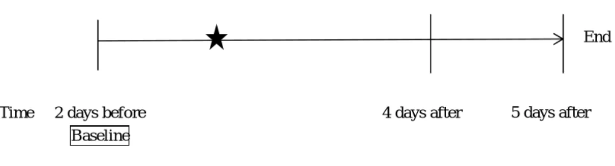
Figure 1. Schematic of the exercise and blood and urine sample collection timeline
Blood samples were obtained each evening. Urine samples were collected each
morning. Star: whole-body strenuous exercises
Time 2 days before Baseline
4 days after 5 days after End
23
Figure 2. Relationship between serum creatine kinase levels and squat jump
performance
The graph shows the values obtained for each subject at 4 days post-exercise (r =
0.721) (N = 37).
24
Table 1. General characteristics of the subjects (N = 37)
Characteristics
Mean ± SD
Age (yr)
23.6 ± 2.2
Height (cm)
171.7 ± 5.5
25
Table 2. Descriptive data for the whole-body strenuous
exercises
Variables
Mean score (± SD)
Toe touch (cm)
13.7 (6.3)
Standing triple jump (m)
6.8 (0.5)
Back strength (kg)
140.2 (19.1)
Seated overhead press (reps)
19.0 (7.9)
Three-stage shuttle run (reps)
93.3 (12.2)
Pull-ups (reps)
7.3 (4.3)
Squat jumps (reps)
67.0 (19.8)
Push-ups (reps)
79.6 (22.8)
Sit-ups (reps)
69.0 (19.1)
26
Table 3. Indicators of muscle damage and inflammation
Time point
CK
(U/L)
DOMS
(a.u.)
hs-CRP
(mg/dL)
Normal values
60 - 270
0
0.300
Baseline
Mean
268.2
1.1
0.206
Range
92 - 735
0 - 3
0.003 - 3.14
SD
140.9
1.1
0.563
4 days post-exercise
Mean
4022.6*
2.1*
0.219
Range
182 - 26940
0 - 6
0.006 - 4.39
SD
6776
1.7
0.703
5 days post-exercise
Mean
3214.6*
1.9*
0.179
Range
164 - 20000
0 - 7
0.006 - 2.42
SD
5314.8
1.6
0.393
ANOVA results
F (2, 36) = 10.9
F (2, 36) = 9.0
F (2, 36) = 0.07
P <0.001
P <0.001
P = 0.937
Data are presented as mean, range, or SD values (N = 37).
CK, creatine kinase; DOMS, delayed-onset muscle soreness; hs-CRP,
high-sensitivity C-reactive protein. * P <0.05 versus the baseline value; the
significance levels accompanied by asterisks refer to P values obtained with
ANOVA followed by Bonferroni’s correction. Statistically significant ANOVA
results are shown in bold type.
27
Table 4. Serum and urinary indicators of renal function
Time point Sodium
(mEq/L) Potassium (mEq/L) Chloride (mEq/L) Creatinine (mg/dL) Urinary protein (mg/dL) Uric acid (mg/dL) Normal values 132 -148 3.5 - 4.9 96 - 108 0.60 - 1.20 20 - 120 3.0 - 7.2 Baseline Mean 141.2 4.1 100.8 0.82 11 5.4 Range 140 - 143 3.7 - 4.9 98 - 105 0.69 - 1.01 2.0 - 80 3 - 7.3 SD 0.8 0.3 1.4 0.09 16 1
No. above ULN 0 0 0 0 0 1
4 days post-exercise
Mean 140.8 4.3 100.4 0.89* 9.3 6.1*
Range 138 - 143 3.8 - 4.8 97 - 104 0.72 - 1.15 2.0 - 19 3.8 - 8.6
SD 1.1 0.3 1.5 0.1 3.8 1.3
No. above ULN 0 0 0 0 0 7
5 days post-exercise
Mean - - - 0.85* 11.4
-Range - - - 0.7 - 1.09 4.0 - 65
-SD - - - 0.09 9.8
-No. above ULN - - - 0 0
-t (36) = 2.02 t (36) = -4.07 t (36) = 1.61 F (2, 36) = 45.0 F (2, 36) = 0.4 t (36) = -5.05
P = 0.051 P < 0.001 P = 0.117 P < 0.001 P = 0.685 P < 0.001
Data are presented as mean, range, or SD values or indicate the number of patients whose values were higher than the upper limit of normal (ULN) (N = 37).
* P < 0.05 versus the baseline value; the significance levels accompanied by asterisks refer to P values obtained with ANOVA followed by Bonferroni's correction or the paired Student's t-test.
Statistically significant ANOVA or Student's t -test results are shown in bold type. Table 4. Serum and urinary indicators of renal function.
ANOVA or
28
Table 5. Serum indicators of iron status
Time point
Serum iron
(μg/dL)
UIBC
(μg/dL)
TIBC
(μg/dL)
Normal values
50 - 200
140 - 330
270 - 425
Baseline
Mean
66.1
262.8
328.9
Range
15 - 129
167 - 356
251 - 388
SD
21.6
33.1
31.3
4 days post-exercise
Mean
48.7*
286*
334.6
Range
23 - 77
190 - 381
239 - 415
SD
14.4
38.3
33.6
Student's t-test results
t (36) = 4.25
t (36) = -5.00
t (36) = -1.82
P <0.001
P <0.001
P = 0.077
Data are presented as mean, range, or SD values (N = 37).
* P <0.05 versus the baseline value; the significance levels accompanied by
asterisks refer to P values obtained with the paired Student's t-test. Statistically
significant Student's t-test results are shown in bold type.
29
Table 6. Indicators of muscle damage, inflammation, renal function, or iron status: Correlations and descriptive statistics (N = 36)
Variables CK hs-CRP Serum iron UIBC TIBC Creatinine Urinary protein Uric acid DOMS
1. CK ― 2. hs-CRP 0.018 ― 3. Serum iron -0.524** -0.467** ― 4. UIBC 0.396* 0.202 -0.745** ― 5. TIBC -0.099 -0.311 0.206 0.499** ― 6. Creatinine 0.065 -0.474** -0.035 0.359* 0.481** ― 7. Urinary protein -0.151 -0.027 0.086 -0.005 0.104 -0.016 ― 8. Uric acid 0.227 -0.539** -0.037 0.410* 0.553** 0.739** 0.044 ― 9. DOMS 0.440** 0.112 -0.384 0.305 -0.053 -0.139 -0.027 0.072 ― M 3754.35 0.01 -17.46 23.22 5.76 0.07 -1.68 0.66 0.92 SD 6759.01 0.91 24.66 27.85 18.99 0.05 16.17 0.79 1.62 * P <0.05, ** P <0.01
30
Table 7. Correlations between the serum CK level and whole-body strenuous exercises (N = 36)
Variables Serum CK level
1. Toe touch 0.108
2. Standing triple jump 0.288
3. Back strength 0.055
4. Seated overhead press 0.176
5. Three-stage shuttle run 0.037
6. Pull-ups 0.302 7. Squat jumps 0.721** 8. Push-ups 0.263 9. Sit-ups 0.237 10. 275m sprint -0.092 M 3754.35 SD 6852.24 ** P <0.01