Arch. histol. jap., Vol. 50, No. 5 (1987) p. 485-493
Lysine-Mediated
Tissue
Osmication
in Combination
with
a Tannin-Osmium
Conductive
Staining
Method
for
Non-Coated
Scanning
Electron
Microscopy
of
Biological
Specimens
Takuro
MURAKAMI1, Zhen-lan SONG1*, Hitoshi HINENOYAI, Aiji OHTSUKA1,
Takehito TAGUCHI1, Jing-jie LIU1* and Tadashi SANO2
Department of Anatomy (Prof. T.MURAKAMI)1, Okayama University Medical School, and Okayama Kosei Hospital (Dr. T.SANO)2, Okayama, Japan
Received July 25, 1987
Summary. Glutaraldehyde fixed rat kidney blocks showed no charging effect when treated with lysine and osmic acid and viewed in a scanning electron microscope using an accelera-tion voltage of 5-30 kV and a specimen current of 1 •~ 10-10A. The podocyte processes and endothelial micropores of the glomerulus were visible without metal coating. Glutar-aldehyde fixed, tannin-osmium impregnated and lysine-osmium treated specimens also showed no charging effect in the scanning electron microscope, yielding instead much clearer scanning images which were comparable to those obtained from gold-coated specimens or from tannin-osmium-thiocarbohydrazide-osmium impregnated specimens (MURAKAMI and JONES, 1980).
Biological specimens exposed in the scanning electron microscope have the potential for charging effects that must be rendered electron-conductive prior to viewing. This is usually accomplished by evaporating or sputtering a coating of carbon, gold, plati-num or other metals in a vacuum chamber (INGRAM et al., 1976; MUNGER,1977; ECHLIN and KAYE,1979). In the secondary electron mode for three-dimensional visualization of morphological details, only the coated metal is observed; resolution depends upon the thickness of the coated metal (MUNGER, 1977; MURPHY, 1978, 1980). To avoid or reduce this and other limitations such as the heating and etching effects of evaporation or sputtering, ligand-mediated osmium impregnation methods exemplified by the tannin-osmium and tannin-osmium-thiocarbohydrazide-tannin-osmium methods have been used (MURAKAMI, 1973; KELLEY et al., 1973; MALICK and WILSON, 1975; MURPHY, 1978, 1980; TAKAHASHI, 1979, 1986). This paper describes a new lysine-mediated osmium impregnation method and its combination with the tannin-osmium method (MURAKAMI, 1973). This combina-tion (tannin-osmium-lysine-osmium impregnation), like the tannin-osmium-thiocarbo-hydrazide-osmium method (MURAKAMI and JONES, 1980), produces well contrasted scanning images even at high magnifications.
*visiting research fellows from China (Department of Internal Medicine
, Dalian University Medical School)
buffer containing 2-4% glutaraldehyde and 2% tannic acid (pH 7.0-7.4), washed for 3 hrs or longer in distilled water, stained for 3 hrs with 2% osmic acid in distilled water, washed for 3 hrs or longer in distilled water, rinsed for 3 hrs in the 2% L-lysine solution (see above), washed for 3 hrs or longer in distilled water, stained again for 3 hrs with 2% osmic acid in distilled water, and washed for 3 hrs or longer in distilled water (tannin-osmium-lysine-osmium method).
The lysine-osmium or tannin-osmium-lysine-osmium treated blocks were de-hydrated through a graded series of ethanol, freeze-cracked in liquid nitrogen, dried in a critical point drier, and observed free of any metal coating, in a scanning electron microscope (HHS-2R, Hitachi, Ibaragi, Japan) using an acceleration voltage of 5-30 kV and a specimen current of 1 x 10⁻¹⁰A. In some cases, the lysine-osmium or tannin-osmium-lysine-osmium impregnated blocks were partially sputter-coated with gold (about 100 A in thickness). They were also observed in the scanning electron micro-scope.
For supplementary experimentation, the non-perfused kidney of an adult rat was cut into small pieces (about 0.5>< 1.0 x 1.0 mm), fixed in the phosphate-buffered 2-4% glutaraldehyde solution and treated or impregnated with lysine and osmic acid or tannic acid, osmic acid, lysine and osmic acid in the same manner as above. These
pieces were embedded in an epoxy resin, cut into ultra-thin sections, and observed, without any additional metal staining, in a transmission electron microscope (H-700 or
H-500, Hitachi, Ibaragi, Japan) using an acceleration voltage of 75-100 kV.
RESULTS
Rat kidney blocks of 1.0-1.5 mm thickness were completely stained or blackened by 3 hr-treatments with lysine and osmic acid solutions or with tannic acid, osmic acid, lysine and osmic acid solutions.
The lysine-osmium and tannin-osmium-lysine-osmium methods here imparted suf-ficient electron-conductivity to allow scanning electron microscopic study of uncoated specimens. In both methods, no charging was noted at an acceleration voltage of 5-30 kV and a specimen current of 1 x 10⁻¹⁰A (Fig. l-4). Neither was charging noted in the surfaces exposed by freeze-cracking (Fig. 2, 4).
The lysine-osmium impregnated specimens, including their cracked surfaces, showed adequate contrast to allow operation at magnifications up to 10,000-15,000 x
Lysine-Mediated Osmication in Combination with Tannin-Osmium Method 487
fenestrations of the renal glomerulus, were visible without metal coating at these magnifications (Fig. 2, Insets A, B).
The tannin-osmium-lysine-osmium impregnated specimens, including their crack-ed surfaces, produccrack-ed more contrasting or more highlighted scanning images than those obtained from the lysine-osmium impregnated specimens (Fig. 3). This was especially apparent at high magnifications. The microvilli of the urinary tubules were clearly visible without metal coating when the tannin-osmium-lysine-osmium method was used (Fig. 3, Inset A).
The scanning images from the tannin-osmium-lysine-osmium impregnated speci-mens were comparable to those from the gold-coated areas. As shown in Inset B of Figure 3, the podocyte foot processes of the renal glomerulus stained by the tannin-osmium-lysine-osmium method yield excellent contrast at high magnifications. Similar findings were obtained even for the freeze-cracked surfaces. As shown in the Figure 4A, the endothelial fenestrations and other structures of the glomerular capillary wall stained by the tannin-osmium-lysine-osmium method and exposed by freeze-cracking showed as good a contrast as when coated with gold (Fig. 4B).
The lysine-osmium impregnated specimens produced adequate images in the transmission electron microscope, though additional metal stainings such as lead staining were omitted (Fig. 5). The endothelium, basement membrane and podocyte foot process of the glomerular capillary were well preserved. Neither marked damage of these delicate structures nor a marked thickening of the cell surface or plasma membrane was noted. The tannin-osmium-lysine-osmium impregnated specimens gave much darker images in the transmission electron microscope (Fig. 5, Inset A). Careful examination revealed that the tannin-osmium-lysine-osmium method, like the lysine-osmium method, caused no marked damage of tissue structures.
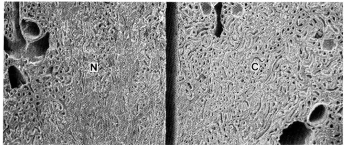
Fig. 1. A scanning electron micrograph of a razor blade-cut surface of a rat kidney block fixed with glutaraldehyde, treated with tannic acid, osmic acid, lysine and osmic acid, and partially coated with gold. Note that the non-coated area (N) shows no charging and its brightness is comparable to that of the gold-coated area (C). 25 kV accelera-tion voltage. x50
DISCUSSION
The present tannin-osmium-lysine-osmium method is advantageous in making possible more contrasting or more highly magnified scanning observations of uncoated speci-mens than did earlier methods. Only the tannin- osmium-thiocarbohydrazide-osmium method (MURAKAMI and JONES, 1980; MURAKAMI et al., 1983) may be comparable to the tannin-osmium-lysine-osmium method. Preliminary experiments in this study have shown that repeated staining with lysine and osmium (lysine-osmium-lysine-osmium impregnation) is less effective in highlighting the scanning images of uncoated speci-mens than the tannin-osmium-lysine-osmium method. These preliminary experiments
also have shown that in highlighting the scanning images of uncoated specimens the osmium-thiocarbohydrazide-osmium-thiocarbohydrazide-osmium (MALICK and WILSON, 1975),osmium-tannin-ferrocyanide-osmium-lead (TAKAHASHI, 1986) and tannin-osmium-cationic iron colloid (MURAKAMI et al., 1986) methods are inferior to the tannin-osmium-lysine-osmium method.
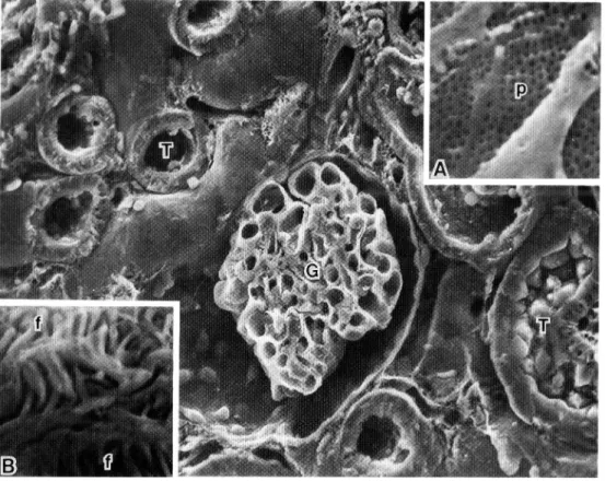
Uncoated specimens prepared by the original tannin-osmium method could be Fig. 2. A scanning electron micrograph of a rat kidney block fixed i~:i glutaraldehyde,
treated with lysine and osmic acid, and freeze-cracked in liquid nitrogen. Note that no charging is observed in any area of the cracked surface. Also note in Insets A and B (closer views of the glomerulus, G) that the micropores (p) of the glomerular endothelium and the fine processes (f) of the glomerular podocyte are visible, though not so sharply contrasted. T urinary tubules. 20kV acceleration voltage. X 550, Inset A: x18,000, B : x15,000
Lysine-Mediated Osmication in Combination with Tannin-Osmium Method 489
studied at useful magnifications of up to 10,000 -15,000 X (MURAKAMI, 1973). Immersion in the mixed amino acid solution slightly enhances the tannin-osmium impregnation (MURAKAMI, 1974). Similar enhancement was achieved by the use of guanidine hydro-chloride, arginine hydrochloride and glycine with tannic acid (MURAKAMI et al., 1977; MURAKAMI, 1978). Non-coated observation here attained by the lysine-osmium method may be comparable to that by the original tannin-osmium (MURAKAMI, 1973), osmium-thiocarbohydrazide-osmium (KELLEY et al., 1973), ruthenium red-osmium (FERIA-VELASCO and ARAUZ-CONTRERAS, 1981) or osmium-hydrazine-osmium (MURAKAMI et al., 1982) method.
Transmission electron microscopy of ultrathin sections reveals that the lysine-osmium or tannin-lysine-osmium-lysine-lysine-osmium methods cause neither marked damage nor marked contamination of tissues. This proves that the present methods are reliable and useful for the study of tissue structures under the scanning electron microscope. Tannic acid and lysine sometimes contain granular or muddy substances which contaminate the tissues. There can be eliminated by filtering tannic acid and lysine through a cellophane sheet prior to their use (MURAKAMI and JONES, 1980).
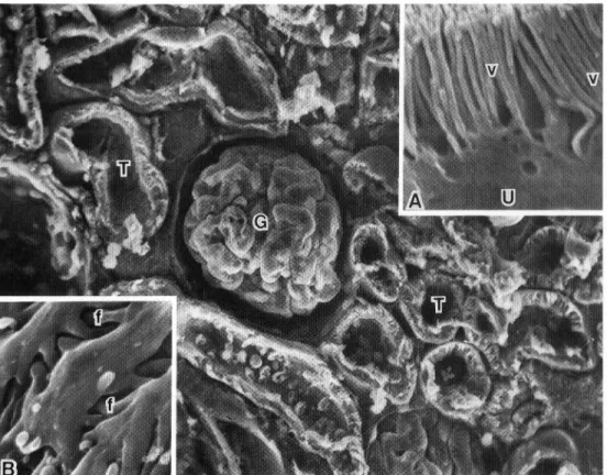
Fig. 3. A scanning electron micrograph of a razor blade-cut surface of a rat kidney block fixed with glutaraldehyde, and treated with tannic acid, osmic acid, lysine and osmic acid. Note that no charging is observed in any area of the cut surface. Also note in Insets A and B (closer views of the glomerulus, G, and urinary tubule, T) that the microvilli (v) of the urinary epithelium (U) and the fine processes (j) of the glomeru-lar podocyte are clearly visible, though not coated with any metal. 20 kV accelera-tion voltage. X 450, Inset A : x 11,200, B : x 17,000
It is well known that glutaraldehyde or osmic acid combines with tissues as a fixative. The present methods allow for intense osmification in the following manner.
In the lysine-osmium method, lysine may combine with tissue-fixed glutaraldehyde to form tissue-glutaraldehyde-lysine-osmium complexes. It is also possible that lysine acts
as directly a cationic agent to combine with tissue anionic sites, and forms tissue-lysine-osmium complexes. In the tannin-osmium-lysine-osmium method, tannic acid may combine with tissue proteins and saccharides to form tissue-tannin-osmium complexes. It should further be mentioned that, in this method, lysine may combine with tissue-fixed osmic acid and tannic acid to form tissue-osmium-lysine-osmium and tissue-tannin-lysine-osmium complexes. In fact, tissue-lysine-osmium complexes may be formed even in the tannin-osmium-lysine-osmium method. This intense osmication with lysine or tannic acid and lysine may enhance electron-conductivity and also secondary electron emission of the tissues. Preliminary experiments have shown that L-arginine (free base), paraphenylenediamine and hydrazine hydrate with rather small molecular sizes can be used as a substitute for lysine in the tannin-osmium-lysine-osmium method. These agents are also useful as a substitute for lysine in the lysine-osmium method.
Fig. 4. Non-coated (A) and gold-coated (B) scanning images of the freeze-cracked surfaces of the rat kidney glomeruli fixed with glutaraldehyde and treated with tannic acid, osmic acid, lysine and osmic acid. Note that the non-coated or tannin-osmium-lysine-osmium impregnated specimen yields adequate contrast for clear observation of the endothelial micropores (p) and foot processes (f) of the glomerulus (A), and that these non-coated images of the micropores and foot processes (A) are compatible with the gold-coated ones (B, p and f). L glomerular capillary lumen. A and B: x7,500
Lysine-Mediated Osmication in Combination with Tannin-Osmium Method 491
Specimens stained by the original or modified tannin-osmium methods were occa-sionally further treated by metal evaporation or sputtering in a vacuum chamber to enhance secondary electron emission or to obtain more highlighted images (FuJITA,
1974). In the lysine-osmium method, such metal coating is recommended to obtain good scanning images at high magnifications. In the tannin-osmium-lysine-osmium method,
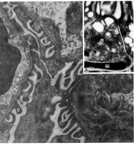
Fig. 5. A transmission electron micrograph of a rat kidney glomerulus fixed with glutar-aldehyde and treated with lysine and osmic acid. Inset A shows a transmission electron micrograph of a urinary tuble fixed with glutaraldehyde and treated with tannic acid, osmic acid, lysine and osmic acid. Note that the tannin-osmium-lysine-osmium method stains the tissues much darker than the lysine-tannin-osmium-lysine-osmium method. Also note that no marked damage of tissue structures is observed in either method. L glomerular capillary lumen, U urinary epithelial cell, b glomerular basement membrane, e glomerular endothelium, f podocyte foot processes, m mitochondria, p endothelial micropores, v microvilli of the urinary epithelial cell. x20,000, Inset A:
is strongly basic and damages the glutaraldehyde-fixed tissues, the pH values of the lysine solution in the lysine-osmium method should be adjusted to 7.0-7.4. In contrast, such pH adjustment of the lysine solution is not always needed in the tannin-osmium-lysine-osmium method since the specimens are firmly fixed with glutaraldehyde, tannic acid and osmic acid prior to the lysine treatment. TAKAHASHI (1979) repeatedly stained the osmium-fixed specimens with glutaraldehyde-tannic acid mixture and osmic acid. Our preliminary experiments have shown that in highlighting the scanning images this repeated tannin-osmium impregnation is inferior to the tannin-osmium-lysine-osmium or tannin-osmium-thiocarbohydrazide-osmium methods (MURAKAMI and JONES, 1980; MURAKAMI et al., 1983). The primary reason for this may be that tannic acid is larger in molecular size than lysine or thiocarbohydrazide and therefore is less able to pene-trate into tannin-osmium impregnated specimens. It has recently been shown that the glutaraldehyde-lysine complex is a useful fixative for the transmission electron microscopy of some tissue elements such as actin filament and glycocalyx (BOYES, 1984). This fixative is suitable for the lysine-osmium and tannin-osmium-lysine-osmium methods, particularly for the latter because tannic acid reacts with lysine (see above). To stain the inner structures of large blocks, freeze-fracture prior to the lysine or tannin treatment (or after the glutaraldehyde or osmic acid fixation) is recommended (IIDA, 1984).
REFERENCES
Boyes, J.: The use of primary amines to improve glutaraldehyde fixation. In: (ed. by) J.-P. Revel, T.Barnard and G.H.Haggis: The science of biological specimen preparation
for microscopy and microanalysis. Scanning Electron Microscopy, Inc., AMF O'Hare
(Chicago), 1984 (p. 7-21).
Echlin, P. and G.Kaye: Thin films for high resolution conventional scanning electron microscopy. Scanning Electron Microscopy/1979/II: 21-30 (1979).
Feria-Velasco, A. and J.Arauz-Contreras: Ruthenium red-mediated osmium binding for examining uncoated biological material under the scanning electron microscope.
Stain Technol. 56: 71-78 (1981).
Fujita, T.: A scanning electron microscope study of the human spleen. Arch.histol.jap. 37: 187-216 (1974).
Iida, N.: Freeze-fracture of biological specimens prior to conductive staining.Arch.histol.
Lysine-Mediated Osmication in Combination with Tannin-Osmium Method 493
Ingram, P., N.Morosoff, L.Pope, F.Allen and C.Tisher : Some comparisons of the techniques of sputter (coating) and evaporative coating for scanning electron
scopy. Scanning Electron Microscopy/1976/I: 75-81 (1976).
Kelley, R.O., R.A.F.Dekker and J.G.Bluemink: Ligand-mediated osmium binding: its application in coating biological specimens for scanning electron microscopy. J.
Ultrastr.Res. 45: 254-258 (1973).
Malick, L.E. and R.B.Wilson: Modified thiocarbohydrazide procedure for scanning tron microscopy: routine use for normal, pathological, experimental tissues. Stain
Technol. 50: 265-269 (1975).
Munger, B.L.: The problem of specimen conductivity in electron microscopy. Scanning Electron Microscopy/1977/I: 481-490 (1977).
Murakami, T.: A metal impregnation method of biological specimens for scanning electron microscopy.Arch.histol.jap. 35: 323-326 (1973).
A- : revised tannin-osmium method for non-coated scanning electron microscope specimens.Arch.histol,jap. 36: 189-193 (1974).
- : T
annin-osmium conductive staining of biological specimens for non-coated ning electron microscopy. Scanning 1: 127-129 (1978).
Murakami, T. and A.L.Jones: Conductive staining of biological specimens for non-coated scanning electron microscopy: double staining by tannin-osmium and
thiocarbohydrazide-osmium methods. Scanning Electron Microscopy/1980/I: 221-226
(1980).
Murakami, T., K.Yamamoto, T.Itoshima and S.Irino: Modified tannin-osmium conductive staining method for non-coated scanning electron microscope specimens. Its
tion to microdissection scanning electron microscopy of the spleen.Arch.histol. jap.
40: 35-40 (1977).
Murakami, T., A.Kubotsu, A.Ohtsuka, S.Akita, K.Yamamoto and A.L.Jones: Double staining by vaporized osmium tetroxide-hydrazine hydrate of biological specimens
for non-coated scanning electron microscopy. Scanning Electron Microscopy/1982/I:
459-464 (1982).
Murakami, T., N.Iida, T.Taguchi, O.Ohtani, A.Kikuta, A.Ohtsuka and T.Itoshima:
Conductive staining of biological specimens for scanning electron microscopy with pecial reference to ligand-mediated osmium impregnation. Scanning Electron
scopy/1983/I: 235-246 (1983).
Murakami, T., T.Taguchi, A.Ohtsuka, K.Sano, T.Kaneshige, R.L.Owen and A.L.Jones:
A modified method of fine-granular cationic iron colloid preparation: its use in light and electron microscopic detection of anionic sites in the rat kidney glomerulus and
certain other tissues.Arch.histol.jap. 49: 13-23 (1986).
Murphy, J.A.: Non-coating techniques to render biological specimens conductive. Scanning Electron Microscopy/1978/II: 175-193 (1978).
- : N
on-coating techniques to render biological specimens conductive/1980 update. Scanning Electron Microscopy/1980/I: 209-220 (1980).
Takahashi, G.: Conductive staining method (In Japanese). The Cell (Tokyo) 11: 114-123 (1979).
- : The tannin-ferrocyanide-OSO4 method for TEM and SEM (In Japanese). The Cell (Tokyo) 18: 176-180 (1986). 村 上 宅 郎 〒700岡 山 市 鹿 田 町2-5-1 岡 山 大 学 医 学 部 解 剖 学 教 室
Prof. Takuro MURAKAMI Department of Anatomy
Okayama University Medical School 2-5-1 Shikata-cho