− 1 −
1 Annual Report of Iwate Medical University
Center for Liberal Arts and Sciences No. 55(2020), 1–4
1. Introduction
To perform quasi-monochromatic X-ray imaging, we developed several photon-counting energy-dispersive X-ray computed tomography (PC-CT) scanners [1-4], and enhanced K-edge CT was carried out using iodine and gadolinium contrast media. When performing PC-CT, we usually measure the X-ray dose rate using an ionization chamber [5,6] to calculate the incident dose for objects.
Ideally, the X-ray dose rate is proportional to the second power of the tube voltage when ignoring the production of characteristic X rays. However, the dose rate is not in proportion to the second power owing to the energy dependence of the ionization chamber; the chamber sensitivity increases with decreasing photon energy
Abstract
We measured the X-ray dose rate with copper (Cu) filtration. The X-ray spectra and dose rate were measured using a cadmium telluride detector and an ionization chamber, respectively. Without fi ltration, the maximum-photon and bremsstrahlung-peak energies increased with increasing tube voltage, and the dose rate was proportional to the 1.7th power of the tube voltage. Using a 0.3-mm-thickness Cu fi lter, the bremsstrahlung peak energy substantially increased when the tube voltage was increased, and the dose rate was in proportion to the 3.3rd power of the tube voltage. Using a glass-window X-ray tube, the dose rate was proportional to about the second power of the tube voltage, and the ionization chamber was highly sensitive to low-energy photons.
(Accepted December 4, 2020)
Keywords:X-ray dose rate, ionization chamber, copper fi ltration, tube voltage dependence, X-ray spectra
a
Department of Physics, Iwate Medical University, 1-1-1 Idaidori, Yahaba, Iwate 028-3694, Japan
b
Department of Radiology, School of Medicine, Iwate Medical University, 2-1-1 Idaidori, Yahaba, Iwate 028-3694, Japan
c
Department of Surgery, Toho University Ohashi Medical Center, 2-22-36 Ohashi, Meguro, Tokyo 153-8515, Japan
Eiichi SATO
a, Yasuyuki ODA
a, Sohei YOSHIDA
b, Satoshi YAMAGUCHI
b, Kunihiro YOSHIOKA
b, Hodaka MORIYAMA
c, Osahiko HAGIWARA
c,
Toshiyuki ENOMOTO
c, Manabu WATANABE
cVariations in X-ray dose rate with copper fi ltration
− 2 − Eiichi SATO et al.
2
at tube voltages below 150 keV.
In the present research, major objectives are as follows: to measure variations in X-ray dose rate with copper (Cu) fi ltration, to measure X-ray spectra with the fi ltration, and to observe the energy dependence of an ionization chamber. Therefore, we constructed the experimental setups for measuring X-ray spectra and dose rates and confi rmed the energy dependence of the chamber.
2. Methods
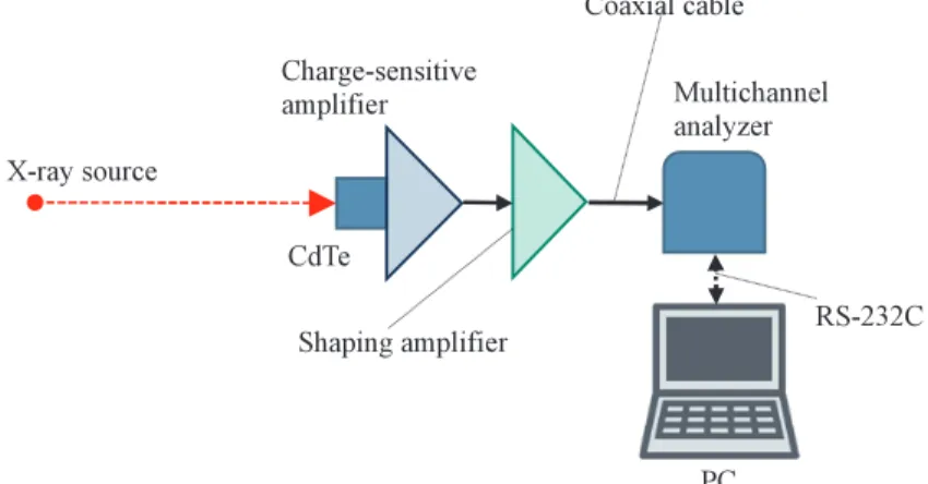
Figure 1 shows the experimental setup for measuring X-ray spectra, and we used a CdTe detector (Amptek, XR-100T) with charge-sensitive and shaping amplifi ers. When an X-ray photon is absorbed by the CdTe crystal, the electric charges are produced in the CdTe diode and are converted into a pulse voltage using the charge- sensitive amplifier. The pulse voltage is amplified to an event pulse using the shaping amplifier. The event pulses produced from the shaping amplifi er are sent to a multichannel analyzer to perform pulse-height analysis.
The X-ray spectra were observed on the PC monitor, and the tube current was 8 μA without fi ltration. Next, the current was increased to 15 μA to increase the photon count rate when using a 0.3-mm-thickness Cu fi lter.
The method for measuring X-ray dose rate is shown in Fig. 2. The dose rate was measured using a dosimeter (Toyo Medic, RAMTEC 1000 plus) and an ionization chamber (Scanditronix, DC300) placed 1.0 m from the X-ray source. When using the Cu fi lter, the fi lter was attached to the X-ray source (R-tec, RXG-0152).
Fig. 1. Experimental setup for measuring X-ray spectra using the CdTe detector.
Fig. 2. Method for measuring X-ray dose rate at 1.0 m from the source.
− 3 −
Variations in X-ray dose rate with copper fi ltration 3
3. Results
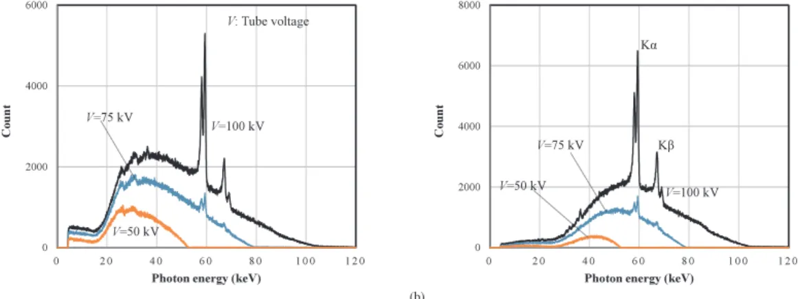
Figure 3 shows the X-ray spectra with changes in the tube voltage. Without filtration at a tube current of 8 μA [Fig. 3 (a)], the maximum-photon and bremsstrahlung-peak energies increased with increasing tube voltage. At a tube voltage of 100 kV, sharp tungsten Kα and Kβ lines were observed. Compared with the tube voltages, the maximum energies were slightly high owing to the ripple of the tube voltage. Subsequently, the bremsstrahlung peak energy shifted to high energy using the Cu filter, since the low-energy photons were absorbed effectively [Fig. 3 (b)].
Variations of the X-ray dose rate with the tube voltage are shown in Fig. 4. Without the fi lter, the dose rate increased with increasing tube voltage, and the rate was proportional to the tube current [Fig. 4 (a)]. Using the Cu fi lter, although the dose rate substantially increased with increases in the tube voltage, the dose rate was in proportion to the tube current [Fig. 4 (b)].
4. Discussion
We measured the X-ray dose rate with changes in the tube voltage, and the dose rate was not proportional to the second power of the tube voltage. Using the dose rates at tube voltages of 50 and 100 kV, the rate increased in proportional to the 1.7th power of the tube voltage.
Assuming that the total photon number including characteristic photons is proportional to the dose rate,
KĮ
Kȕ
Fig. 3. Tube voltage dependence of the X-ray spectra. (a) Without fi ltration at a tube current of 8 μA and (b) using a 0.3-mm-thickness Cu fi lter at a tube current of 15 μA.
Fig. 4. Tube voltage dependence of X-ray dose rate measured at 1.0 m from the X-ray source at tube currents of 0.3 and 0.6 mA. (a) Without fi ltration and (b) using a 0.3-mm-thickness Cu fi lter.
− 4 − Eiichi SATO et al.
4
the rate increased in proportion to the 2.3rd power. Therefore, the rate was roughly proportional to the second power, and the chamber sensitivity increased slightly with decreasing photon energy.
Using the Cu fi lter, low-energy photons were absorbed effectively, and the photon number substantially increased with increasing tube voltage. Therefore, the rate increased in proportion to the 3.4th power of the tube voltage.
If we assume that the dose rate is proportional to the x-power of the tube voltage, the x was below 1 when using an X-ray tube with a 0.5-mm-thickness beryllium window in our former research.
5. Conclusion
We measured the X-ray spectra and dose rate with changes in the tube voltage. Without fi ltration, the dose rate from the glass-window X-ray tube was roughly proportional to the second power of the tube voltage. Using the 0.3-mm-thickness Cu fi lter, the dose rate was proportional to the 3.4th power of the tube voltage. Therefore, the dosimeter was highly sensitive to low-energy photons, and the dose rate was not just proportional to the second power of the tube voltage.
Acknowledgments
This work was supported by Grants from JSPS (17K10371, 17K09068, 17K01424, and 17H00607). This was also supported by a Grant-in-Aid for Strategic Medical Science Research (S1491001 and 2014-2018) from the Ministry of Education, Culture, Sports, Science and Technology of Japan.
References
[1] Matsukiyo, H., Sato, E., Oda, Y., Ishii, T., Yamaguchi, S., Sato, Y., Hagiwara, O., Enomoto, T., Watanabe, M., Kusachi, S., “Investigation of quad-energy photon counting for X-ray computed tomography using a cadmium telluride detector,” Appl. Radiat. Isot. 130, 54–59 (2017).
[2] Sato, E., Sato, T., Oda, Y., Sato, Y., Yoshida, S., Yamaguchi, S., Hagiwara, O., Matsukiyo, H., Enomoto, T., Watanabe, M., Kusachi, S., “Triple-energy high-count-rate X-ray computed tomography scanner using a cadmium telluride detector,” Health Technol. 8, 197–203 (2018).
[3] Sato, T., Sato, E., Oda, Y., Sato, Y., Yoshida, S., Yamaguchi, S., Hagiwara, O., Matsukiyo, H., Enomoto, T., Watanabe, M., Kusachi, S., “Dual-energy high-count-rate X-ray computed tomography scanner using a cerium-doped yttrium aluminum perovskite crystal and a small-photomultiplier tube,” Health Technol. 8, 179–187 (2018).
[4] Moriyama, H., Watanabe, M., Kusachi, S., Oda, Y., Sato, E., “Low-dose low-scattering X-ray computed tomography with high-spatial-energy resolutions using a cooled cadmium telluride detector,”
Ultramicroscopy 199, 62–69 (2019).
[5] AlMasri, H., Funyu, A., Kakinohana, Y., Murayama, S., “Investigation of thermal and temporal responses of ionization chambers in radiation dosimetry,” Rad. Phys. Tech. 5, 172–177 (2012).
[6] Kato, T., Arai, K., Sagara, T., Kato, R., Yamazaki, Y., Oyama, S., “Patient-specific quality assurance for proton depth dose distribution using a multi-layer ionization chamber in a single-ring wobbling method,”
Rad. Phys. Tech. 12, 305–311 (2019).