Acta med. Nagasaki. 13: 211-227
The N-Terminal Structure of Adult Hemoglobin from Japanese Monkey (Macaca fuscata fuscata) and the N-Terminal Amino Acids of All the Tryptic Peptides
from its α andβ Polypeptide Chains*
Yoshiro TANAKA**
Department of Biochemistry, Nagasaki University School of Medicine, Nagasaki, Japan
Received for publication, March 5, 1969
The N-terminal structure of adult Japanese monkey (macaca fuscata fuscata) hemoglobin and the N-terminal amino acid analyses of the tryptic
peptides from this hemoglobin were performed.
Hemoglobin, after removal of heme, was dinitrophenylated. The DNP- globin was hydrolyzed with HCl, and from the hydrolysate DNP-derivatives were extracted with ether or ethyl acetate. They were isolated and iden- tified by silica gel-Celite column chromatography, and quantitative ana- lyses were performed by spectrophotometry. Consequently, it was known that one mole of hemoglobin of the Japanese monkey consisted of two
polypeptide chains whose N‑terminal structure was Val‑Leu(α. chains)and two polypeptide chains whose N‑terminal structure was Val‑His‑Leu(β chains). On the other hand, globin was separated into the subunits, α and,β polypeptide chains by countercurrent distribution. Each polypeptide
chain was hydrolyzed with trypsin. The hydrolyzed peptides were isolated and purified by column chromatography and paper chromatography. The N-terminal amino acids of the peptides thus isolated and purified were dis- cussed by comparing with human hemoglobin. It was known that the N-
terminal sturcture of,βT2 peptide consisted of serine in the case of human
hemoglobin, but asparagine in the case of Japanese monkey hemoglobin and that other 21 peptides has same N-terminal amino acids as those from human hemoglobin do, respectively.
INTRODUCTION
In this laboratory, hemoglobins from various animals have comp- aratively been studied. Evolutional problems of living organisms have generally been investigated with comparative studies of their physical shapes and habits, however, in the light of the fact that all the living organisms are of chemical constituents it is surely another good way
`This work was presented at the 41st meeting of the Japanese Biochemical society , October 1968.
**田 中 義 郎
for elucidation of the problems to compare those chemical constituents at molecular level among different species of living organisms. The recent information that the amino acid sequences of proteins are under the control of basic sequence of gene, DNA proves that it is one of the most available ways to investigate on the species' specificity in the primary structure of proteins whose physiological functions are common. For this investigation hemoglobin is a convenient experimental material since it widely distributes among the animal kingdom and because it can be isolated and purified more easily than other proteins.
The structure of the heme part of hemoglobin, which was deter- mined by 11. FISHER so early as 1928, is known to be common to all the hemoglobins from various kinds of animals. With respect of the protein moiety, globin, hemoglobins from various kinds of animals are being studied since BRAUNITZER et al .43 and KONIGSBERG et al.93 worked out the primary structure of adult human hemoglobin.
Primate hemoglobins are generally studied with interest in how different they are from human hemoglobin. ZUCKERKANDL et a122) 23). pres- umed that chimpanzee hemoglobin was similar to human hemoglobin, and that the a and j3 polypeptide chains of gorilla hemoglobin were diffe- rent from those of human hemoglobin in one amino acid residue, res- pectively. BUETTNER-JANUSCH et al." analyzed the amino acid compositions of the tryptic peptides from hemoglobins of such monkeys as the hylo- bates, the papio, the perodicticus, the galogo, and the lumur, and then
they compared the results with human hemoglobin. According to their
report, considerably many differences are found among them. Conce- rning hemoglobins of rvracaca genus, the whole primary structure of rhesus monkey hemoglobin was determined in this laboratory. 13' Rhe- sus monkey hemoglobin proved to be different from human hemoglobin at four positions in the a polypeptide chain and at eight positions in
the ~ polypeptide chain. The Japanese monkey (macaca fuscata fuscata) which inhabits only on the Japanese Islands belongs to the same genus with the rhesus monkey. In an attempt to investigate on differences
in the amino acid sequences of hemoglobins between these two species, the primary structure of Japanese monkey hemoglobin is studied in this laboratory.
The present author determined the N-terminal structure of adult Japanese monkey hemoglobin by the DNP method and furthermore determined the N-terminal amino acids of the tryptic peptides from the
a and 8 polypeptide chains.
MATERIALS AND METHODS
1) Preparation of Globin
Hemoglobin was obtained by the method of DRABKIN. 53 The blood
was added 3.8% citric acid solution as an anticoagulant and then was
subjected to centrifugation at 3,000 r . p . m . for 10 minutes in order to
remove plasma. The erythrocytes obtained were washed three times with 0.9% NaCl solution and hemolyzed by the addition of an equal volume of deionized water and half a volume of toluene.
The hemolysate was stirred in the cold room overnight and centri- fuged at 12,000 r. p . m . for 60 minutes. From the middle layer between the stroma and toluene, hemoglobin solution was obtained. For removal of heme from hemoglobin, 50m1 of hemoglobin solution was dropped with stirring into HCl-acetone mixture (15ml of concentrated HCl, 500m1 of acetone) which was cooled at -20°C.2' Globin precipitated in the
acetone solution was isolated by centrifugation and then washed three times with a mixture of HC1 and acetone. The globin thus obtained was dissolved in deionized water. It was dialyzed against deionized water and finally lyophilized.
2) Separation of a and ~ Polypeptide Chains of the Globin by Countercurrent Distribution
The globin was separated into a and j3 polypeptide chains with a mixture of 1650 ml of sec-butanol containing 1.56 g of trichloroacetic acid, 225 ml of propionic acid, and 1305 ml of water on an automatic countercurrent distribution machine with 100 tubes of 20 ml capacity, Shibata CDA -100; 500 mg of the globin was dissolved in 80 ml of the above described solvent, separated into the upper and lower phases, and then placed in the four tubes of the machine, Tube No. 4 to No.
7. 150 upper phase transfers were carried out by setting the machine for a 30-second shifting time and a 20-minute settling time. The solvent in the tubes was made transparent by the addition of 0.5 ml of 50%
ethanol, and then the absorbance at 280 m,u of each tube was measured.
The solvents at the peaks were combined and dialyzed against deion- ized water before lyophilization.
3) Preparation of the Tryptic Peptides from the Polypeptide Chains
In 50 ml of 8 M urea solution, 1g of the a or S polypeptide chain was dissolved and denatured at 60°C for 45 minutes. After removal of urea by dialysis, the polypeptide chain was suspended in 80 ml of deionized water and brought to pH 8.0 by the addition of 0.1 N NaOH.
To this solution was added 15 mg of trypsin (Worthington Biochemical Corp. Twice crystallized) which was dissolved in 1/16N HCl and remo- ved its chymotryptic activity15' . Hydrolysis was carried out at 37°C for 4 hours, the pH being maintained at 8.0. The hydrolysate was adjusted
to pH 6.4 with 1 N acetic acid and then the insoluble peptide was preci- pitated. The soluble peptides and insoluble peptides were separated from each other by centrifugation. Isolation and purification of the soluble peptides were performed as follows by column and paper chro- matographies.
a) Column Chromatography of the Soluble Tryptic Peptides on Dowex
1x2
The resin (Dowex 1x2, 200-400 mesh) was washed with 1 N NH4OH, water, acetic acid, and water successively, and finally sus- pended in the starting buffer. It was loaded in a column (2.0 x 150 Cm).
The column was further equilibrated with the starting buffer. The sample, the soluble peptide after adjusted to pH 10.0 with NaOH, was placed on the column. As a developer was used the acetate buffer containing organic bases such as pyridine, collidine, picoline, and lutidine. In addition, gradient elution with acetic acid was employed.
Elution was carried out at a flow rate of 180-200m1 per hour, the column being kept at 37°C. The eluate was collected in 18 ml-fractions.
According to the method of YEMM and COCKING,") a 0.2 ml portion from each fraction subsequent to alkali hydrolysis was subjected ` to the ninhydrin color reaction. The eluate at each peak in the ninhydrin color reaction was dried under reduced pressure below 30°C and disso- lved in 7 ml of deionized water.
b) Paper Chromatography of the Soluble Tryptic Peptides
Toyo filter paper No. 50 was used. Development was performed by descending method. The developer used was the upper phase of a mixture of n-butanol, acetic acid, and water (4:1:5). Peptides were detected by spraying 0.2% ninhydrin-n-butanol solution and by heating to color with an iron. Besides, PAULY'S reaction,"' SAKAGUCHI'S reac- tion"' , EHRLICH' S17) reaction,,,) a-nitrosonaphtol reactions' were carried out on the paper chromatogram for identification of peptides. The peptides were eluted with 5% acetic acid and the eluates were dried under reduced pressure.
4) Analysis of the N-terminal structure of the globin by the DNPmethod a) Dinitrophenylation of the globin
In 75ml of deionized water. was suspended 500mg of the globin and to this was added 0.1N NaOH to be pH 9.0. This suspension was further added 100 mg of Na2 CO3 and 0.5 ml of dinitrof.luorobenzene (DNFB) and stirred for 3 hours at 40°C. At the termination of the reaction, the solution was brought to pH 2.0 by the addition of 1N HC1. The DNP- globin, which was recognized as a yellow precipitate, was isolatad by centrifugation and washed 3 times with 0.1 N HC1. It was thorou- ghly washed on the glass filter with acetone and ether, and then dried by allowing to stand overnight at 50°C in an incubator.
b) Hydrolysis of the DNP-globin and extraction of DNP-derivatives
from the hydrolysate
In 15m1 of constant boiling point HC1 (twice distilled) was susp-
ended 100mg of the dried DNP-globin. This suspension was hydrolyzed
under refluxing for an appropriate time. DNP-derivatives were extra-
cted from the hydrolysate with ether and ethyl acetate by the proce- dure illustrated in Fig. 1. The ether or ethyl acetate extracts were dried under reduced pressure.
Fig. 1. Procedure of extraction of the DNP-derivatives from the hydrolysate of DNP-globin
c) Sillica gel-Celite column chromatography for the DNP-derivatives
Column Chromatography for the DNP-derivatives was performed by the modification of the method of GREEN et al.6', in which silica gel- Celite was used as an adsorbent."' Silica gel (100 mesh, Mallinckrodt Chemical Work, U. S. A .) and Celite 545 (Wako Chemical Industry, Ltd.) was mixed at the rate of 2 to 1, and the mixture was loaded in a column (0.9x20 cm) at a height of 15cm. The column was washed with 5 ml of ether, 10 ml of ether-acetone mixture (1:1), 2ml of ether, and 7ml of ligroin, successively and was finally equilibrated with 7ml of the starting developer.
The DNP-derivative dissolved in 3 ml of the appropriate solvent was applied to the a-foreprepared column. The developer used was the mixture containing ligroin, acetic acid, acetone, formic acid, and ethyl
acetate. Movement of the yellow band of the DNP-derivatives in the column was recorded every 7ml of the developer. The isolated yellow band was taken out and eluated on a glass filter with ether-alcohol mixture (4:1). The eluate was dried under reduced pressure.
d) Estimation of the DNP-derivatives
The DNP-derivatives isolated by column chromatography were dissolved in an aliquot of glacial acetic acid and their absorbance at 340 mg were measured. Regarding the molecular extinction coefficient of the DNP-derivatives at 340 m,a as 16,100, their quantities were cal- culated according to the following formula.
DNP-derivative (amole) = A x 6.2 x a/100
In the formula, a denotes the quantity (ml) of the acetic acid in which a DNP- derivative was dissolved, and A, its absorbance at 340 ma.
e) Identification of the DNP-derivatives
Each DNP-derivative after dissolved in 4ml of constant boiling point HC1 was hydrolyzed at 105°C for 24 hours in a sealed tube. The hydrolysate was extracted with three 10 ml-portions of ether, and then the ether extract after dried under reduced pressure was subjetced to the paper chromatography in order to identify and estimate its DNP- amino acids. The aqueous phase was also subjected to an amino acid analyzer, Hitachi KLA-2 in order to identify and estimate its amino acids.
5) N-terminal amino acid analysis of the tryptic peptides by the DNPmethod
In 10 ml of 1% NaHCO3 solution was dissolved 0.2-0.5 /,mole of tryptic peptides, and to this was added 0.05 ml of dinitrofluorobenzen (DNFB). The mixture was stirred at 40°C for 2 hours. The excess of DNFB was removed by extraction with three 10 ml-portions of ether.
The aqueous phase was acidified with HC1 and extracted with three 10 ml-portions of ether. By this treatment, dinitrophenol (DNP) which was a byproduct in dinitrophenylation was extracted in the ether phase.
The DNP-tryptic peptides in the aqueous phase, after dried under reduced pressure, were dissolved in constant boiling HC1 and hydr- olyzed at 105°C for 20 hours in a sealed tube. From this hydrolysate, DNP-amino acids were extracted with three 10 ml-portions of ether, and dried under reduced pressure in a heart-shaped flask, a modifica- tion of MILLS' apparatus. The flask was connected to a cold finger and completely aspirated by a vacuum pump in order to remove as much dinitrophenol (DNP) as possible." Identification of DNP-amino acids was performed as follows by two dimensional paper chromatography.
The sample dissolved in a small portion of acetone was applied on a
sheet of Toyo filter paper No. 50 (40 x 40 cm). Development was per-
formed by the ascending method with the upper phase of n-butanol
and 1N NH4OH (1 : 1) for the first dimension and with 1.5 M phosphate
buffer for the second dimension. The DNP-amino acids were estimated
by measuring the absorbance at 360 ma of the eluates which were eluted
with 5 ml of warm water from the yellow spots on the paper chroma-
togram. 19)
RESULTS AND DISCUSSION
1) 'The N-terminal structure of the hemoglobin from the Japanese monkey
Globin was prepared by removal of heme from hemoglobin of Ja- panese monkeys (macaca fuscata fuscata). It was dinitrophenylated in aforementioned method; 510 mg of dry DNP-globin was obtained from 500 mg of globin. Three 100 mg-portions of the DNP-globin were hydro- lyzed with constant boiling point HC1 under refluxing for 1 hour, 6 hours, and 20 hours, respectively. From the hydrolysates, DNP-deriv-
atives were extracted by ether or ethylacetate and then they were isolated by silica gel-Celite column chromatography. The ether extracts were dissolved in 3 ml of Solvent I, a mixture of 0.06 ml of acetic acid, 0.3 ml of acetone and 2.6 ml of ligroin, and applied on the column.
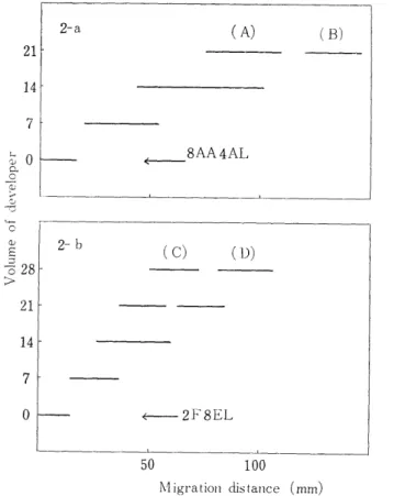
Development carried out by using 8AA4AL. The movement of the yellow. band was given in Fig. 2-a. As seen in this figure, two yellow bands, A and B, were separated at the time when 21m1 of the
Fig. 2. Silica gel-Celite chromatography of ether-extracted DNP-derivatives from the hydrolysate of the DNP-globin.
8AA4AL: The mixture of acetic acid (8m1) and acetone (4m1) was added ligroin so that the whole volume was 100ml.
2F1EL; The mixture of formic acid (2m1) and ethyl acetate (8ml) was added ligroin so that the whole volume was 100ml.
developer had been flowed. The mobility of B-band was similar to that of DNP-amino acids (DNP-Val, DNP-Leu, DNP-Ileu) of IV group which were isolated by the chromatography using the system of GREEN et al. 11 The yellow substance taken out from the column was eluted with ether-alcohol mixture and subjected to the two dimensional paper chromatography. As a result, only a spot which was identical with the standard DNP-Val was recognized. Therefore, this substance was identified as DNP-Val. In the similar way, A-band was subjected to
the two dimensional paper chromatography. Two spots were found.
One accorded with dinitroaniline (DNA) and the other accorded with none of the DNP-amino acids. Therefore, DNP-derivatives contained in A-band were dissolved in 3 ml of Solvent I and subjected to column chromatography with 2F8EL developer. The result was given in Fig.
2-b. It was separated into two yellow bands, C and D. D-band corres- ponded to DNA both in the mobility in the chromatography with this same system and in the location in the two dimensional paper chr-
omatography. C-band corresponded to none of the DNP-amino acids on the paper chromatogram. Therefore, an aliquot of C-band was hydrolyzed with constant boiling HCl at 105°C for 24 hours in a sea- led tube. Yellow substance was extracted with ether. The ether phase was subjected to paper chromatography, and the aqueous phase after dried was subjected to an amino acid analyzer. Consequently,
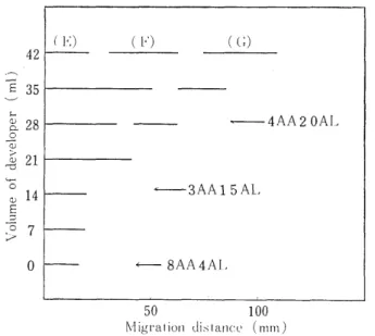
Fig. 3. Silica gel-Celite chromatography of ethylacetate-extracted DNP-derivatives
from the hydrolysate of the DNP-globin 8AA4AL; See Fig. 2.
3AA15AL; The mixture of acetic acid (3m1)and acetone (15m1) was added ligroin so that the whole volume was 100ml.
4AA20AL; The mixture of acetic acid (4m1) and acetone (20 ml) was added ligroin so that the whole volume was 100ml.
DNP-Val was only recognized in the ether phase. Its quantity was 0.130 amole. In the aqueous phase, leucine was only recognized. Its quantity was 0.163 amole. These facts suggest that C-band could be identified as DNP-Vai-Leu.
The ethylacetate extract was dissolved in 3m1 of Solvent II, a mixture of 0.6m1 of acetic acid and 2.4ml of benzene, and then isolated by column chromatography with 8AA4AL, the starting deve- loper. As seen in Fig. 3, the yellow band did not move in the develo- pment with 21m1 of 8AA4AL. Therefore, development with 14 ml of 3AA15AL and 14 ml of 4AA20AL followed it. As a result of it, three yellow bands were separated. E-band gave many spots on the two- dimensional paper chromatography. However, from the ether extract from the HCl hydrolysate of E-band, nothing like the yellow substance was extracted. On the contrary, from the aqueous phase, s-mono-DNP- Lys and many free amino acids were found, suggesting that E-band
was a mixture of s-mono-DNP-Lys and peptides containing a-mono-DNP- Lys and that it had nothing to do with the N-terminal structure of the globin. As for F-band, only one spot was recognized on the paper chromatogram. The ether extract of F-band subsequent to HCl hydrol-
ysis contained only DNP-Val, and the aqueous phase, only imid-mono- DNP-His which was eluted with about 50 ml of buffer, pH 5.28 on a 15 cm column of the automatic amino acid alalyzer.111 Consequently, the DNP-derivative of F-band was identified as a dipeptide of DNP- Val-His. G-band also gave one spot on the paper chromatogram. After HCl hydrolysis of it, DNP-Val, imid-mono-DNP-His, and Leu were recognized in the same mole number. Therefore, in due consideration of the results on F-band, the DNP-derivative of G-band was identified as DNP-Val-His-Leu.
DNP-Val, DNP-Val-Leu, DNP-Val-His, and DNP-Val-His-Leu thus obtained were analyzed quantitatively by spectrophotometry. Their values were calculated per 1 mole of the DNP-globin, regarding 100 mg
Table I
Number of N-terminal residues of adult hemoglobin from the Japanese monkey
Hydrolysis time (hr) DNP-Val DNP-Val-Leu DNP-Val-His DNP-Va1-His-Leu Total
1 0.481 1.382 0.842 0.312 I 3.017
6 2.121 0.535 0.364 0.108 3.128
20 3.647 0.229 0.108 - 3.984
These values are expressed as molar ratios
DNP-globin, 100mg, is assumed to be 1.14 iimole.
of the DNP-globin as 1.14 amole.l13' Table I gives the results. The percentage recoveries of the DNP-derivatives were not corrected except that that of DNP-Val obtained by 20 hour hydrolysis was corrected to be 90%. As seen in the table, Japanese monkey hemoglobin turned out, just as in the case of human hemoglobin, to be composed of two polypeptide chains whose N-terminal structure was Val-Leu, and two polypeptide chains whose N-terminal Structure was Val-His-Leu.
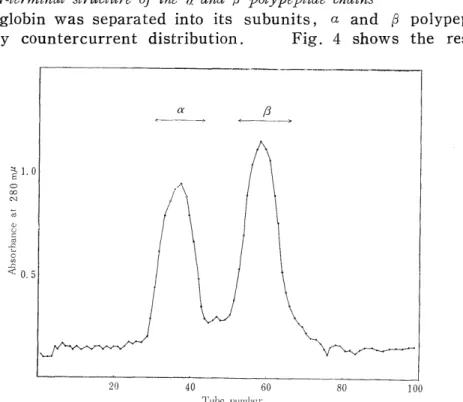
2) The N-terminal structure of the (,i and a polypeptide chains
The globin was separated into its subunits, a and ~ polypeptide chains by countercurrent distribution. Fig. 4 shows the result.
Fig. 4 Countercurrent distribution of the globin from Japanese monkey hemoglobin
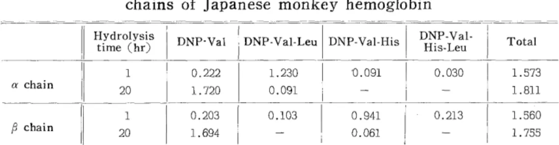
Analyses of the N-terminal structures of the polypeptide chains were per- formed in quite the same ways as in the case of the globin. The analysis values were converted into those per 3,300 g (2 moles) of a polypeptide chain and given in Table II. As seen in this table, the a polypeptide chains have the N-terminal structure of Val-Leu and the j3 polypeptide chains, that of Val-His-Leu. The values of DNP-Val, DNP-Val-His, and DNP-Val-FIis-Leu obtained after one hour hydrolysis suggest that the purity of each chain separated by countercurrent distribution. of
this system was over 93%.
3) The N-terminal amino acids of the tryptic peptides from a and i polypeptide
chains
The a and S polypeptide chains were digested with trypsin subse-
Table II
Number of N-terminal residues in the a and ~polypeptide
chains of Japanese monkey hemoglobin
Hydrolysis ti DNP -Val DNP -Val-Leu DNP-Val-His DNP-Val- Total
me (hr) His-Leu
1 0.222 1.230 '0.091 0.030 1.573
a chain 20 1.720 0.091 - - 1.811
1 0.203 0.103 0.941 0.213 1.560
chain 20 1.694 - 0.061 - 1.755
These values are expressed as molor ratios
The molecular weight of the polypeptide chain is assumed to be 33,000.
quently to denaturation in 8 M urea. Peptides in the digest were first fractionated by the precipitating method at pH 6.4 into the soluble tryptic peptides and the insoluble one called "core". The soluble tryptic peptides were isolated and purified both by column chromatography of Dowex 1 x 2 with acetate buffer containing such organic bases as pyri- dine, collidine, and picoline and by paper chromatography developed with the mixture of n-butanol, acetic acid and water. The results are shown in Figs. 5, 6, 7, and 8.
A portion of each peptide thus isolated was hydrolyzed with HCl and then subjected to amino acid analysis. As described above, iden-
Fig. 5. Column chromatogram of the soluble tryptic peptides from the a chain of Japanese monkey hemoglobin. Column: 2.'0 x 150 cm, Starting developer:
1% pyridine,l% collidine acetate buffer, pH 9.0. Step 2: 1% pyridine 1%
collidine acetate buffer. pH 8.5. Gradient 1: Mixing chamber, 1500m1 of
1% pyridine 1% lutidine 1% picoline acetate buffer, pH 7.5, Upper cham-
ber, 0.08 N acetic acid. Gradient 2: Upper chamber 1.0 N acetic acid.
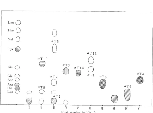
Fig..6.'^Paper- chromatogram of the soluble tryptic peptides from the a chain of Japanese monkey hemoglobin
Fig. 7. Column chromatogram of the soluble tryptic peptides from the a chain of Japanese monkey hemoglobin. Column: 2.0x150 cm, Starting developer; 1%
pyridine 1% collidine acetate buffer,pH 9.0. Step 2: 1% pyridine 1% colli-
dine acetate buffer, pH8.5. Gradient 1: Mixing chamber, 1500 ml of 1%
pyridine 1% lutidine 1% picoline acetate buffer, pH 7.5 Upper chamber,
0.08 N acetic a'id. Gradient 2: Upper chamber, 1.0 N acetic acid
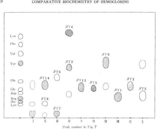
Fig. 8. Paper chromatogram of the soluble tryptic peptides from the 0 chain of Japanese monkey hemoglobin
tification, estimation of the purity, and estimation of quantity were per- formed. Concerning amino acid compositions of these peptides, TERAO20>
and TACHIKAWA19 have already reported. The peptides on the paper chromatogram in Figs. 6 and 8 are numberd according to their reports, in which they were designated in the light of those of human hemoglobin.
The insoluble tryptic peptide was throughly washed with sodium acetate buffer, pH 6.4, suspended in deionized water, and lyophilized.
A portion of the insoluble tryptic peptide thus prepared was hydrolyzed with HC1 and submitted to amino acid analysis, and then the N-ter- minal structure was analyzed.
Dinitrophenylation of the insoluble tryptic peptides was carried out in NaHCO3 solution and then the excess DNFB was removed by ether extraction. The aqueous phase acidified with HCl was again subjected to ether extraction. By this treatment, all the DNP-tryptic peptides containing lysine and arginine remained in the aqueous phase, and dinitrophenol (DNP), the byproduct during the reaction, could be removed in the ether phase. This treatment was convenient because dinitrophenol was obstructive to paper chromatography for identification of DNP-amino acids. DNP-peptides were hydrolyzed with HCl and then DNP-amino acids due to their N-terminus were extracted with ether.
They were isolated and identified by the two dimensional paper chr-
omatography and estimated by spectrophotometry. Fig. 9 shows the
Fib'. 9 Paper chromatogram of the DNP-amino acids and DNP-peptide;
Table III
N-terminal amino acids and amino acid compositions of all the tryptic peptides from the a-polypeptide chain of Japanese Money hemoglobin
Peptide No DNP-amino acids Amino acid composition
. found 1amole
aTl DNP-Val 0.72 Val-(Asp 1, Ser 1, Pro 1, Ala 1, Leu 1)-Lys aT2 DNP-Ser 0.71 Ser-(Asp 1, Val 1)-Lys
aT3 DNP-Ala 0.56 Ala-(Gly 1, Ala 1, Try 1)-Lys
aT4 DNP-Val 0.63 Val-(His 1, Glu 3, Gly 4, Ala 3, Leu 1, Tyr 1)-Arg aT5 DNP-Met 0.34 Met-(Thr 2, Ser 1, Pro 1, Leu 1, Phe 2)-Lys
DNP-MetO 0.12
aT6 DNP-Thr 0.68 Thr-(His 2, Asp 1, Ser 2, Glu 1, Pro 1, Gly 1, Ala 1,
Val 1, Leu 1, Tyr 1, Phe 2)-Lys
aT7 DNP-Gly 0'.43 Gly-(His 1, Gly 1)-Lys
aT8 Lys
aT9 DNP-Val 0.71 Val-(His 3, Asp 5, Thr 1, Ser 2, Pro 1, Gly 1, Ala 6,
Val 2, Met 1, Leu 5)-Lys
aT10 DNP-Leu 0.83 Leu-Arg
aTll DNP-Val 0.65 Val-(Asp 2, Pro 1, Val 1, Phe 1)-Lys
aT12,13 DNP-Leu 0.71 Leu-(Lys 1, His 3, Asp 1, Thr 4, Ser 5, Glu 1, Pr-) 2,
Ala 6, Cys 1, Val 4, Leu 9, Phe 2)-Lys
aTl4 di DNP-Tyr 0.34 Tyr-Arg
Table IV
N-terminal amino acids and amino acid compositions of all the tryptic peptides from the j3-polypeptide chain of Japanese monkey hemoglobin
Peptide No. DNP-amino acids found moles Amino acid composition
j T1 DNP-Val 0.72 Val-(His 1, Thr 1, Glu 2, Pro 1, Leu 1)-Lys (9T2 DNP-Asp 0.63 Asp-(Ser 2, Gly 1, Ala 1, Val 1, Leu 1, Try 1)-Lys
~,T3 DNP-Val 0.68 VaJ-(Asp 2, Glu 2, Gly 3, Ala 1, Val 2, Leu 1)-Arg P3T4 DNP-Leu 0.80 Leu-(Thr 1, Glu 1, Pro 1, Val 2, Leu 1, Tyr 1, Try 1)
-Arg
~T5 DNP-Phe 0.b6 Phe-(Asp 3, Ser 3, Glu 1, Pro 2, Gly 2, Ala 1, Val 1,
Met 1, Leu 1, Phe 2)-Lys
8T6 DNP-Val 0.80 Val-Lys
(3T7 DNP-Ala 0.63 Ala-(His 1, Gly 1)-Lys
J3T8 Lys
,GT9 DNP-Val 0.64 LVal-(His 1, Asp 4, Ser 1, Gly 2, Ala 1, Leu 4, Phe 1)- ys
l3Tl'0,11 DNP-Gly 0.39 Gly-(Lys 1, His 2, Asp 3, Thr 1, Ser 1, Glu 3, Pro 1,
Ala 1, Cys 1, Val 1, Leu 3, Phe 2)-Lys
2T12 DNP-Leu 0.51 Leu-(His 2, Asp 1, Gly 2, Ala 1, Cys 1, Val 3, Leu 3,
Phe 1)-Lys
j3T13 DNP-Glu 0.58 Glu-(Thr 1, Glu 3, Pro 1, Ala 2, Val 1, Tyr 1, Phe 1)- L ys
(3T14 DNP-Val 0.55 Val-(His 1, Asp 1, Gly 1, Ala 4, Val 2, Leu 1)-Lys (3T15 di DNP-Tyr 0.33 Tyr-His
location of the DNP-amino acids on the paper chromatogram together with the location of the DNP-peptides obtained by the hydrolysis of the DNP-globin. The percentage recoveries of the DNP-amino acids from the tryptic peptides were not corrected and the values were converted into those per one mole of a peptide. Table III and IVshow the results
together with amino acid composition.
From aT12,13-.and fT10,11,12, the so-called insoluble tryptic peptides, many DNP-amino acids were detected. In the case of aT12,13, however, the N-terminal residue is considered to be leucine since DNP-Leu was overwhelmingly more than others. j3T10,11,12 contained plenty of DNP- Leu and DNP-Gly. Since it is said that DNP-Gly shows weak resistance against HCI hydrolysis and that its recovery is smaller than others'4' ,
this result indicates that QT10,11,12 contains the peptide whose N-ter-
minus is glycine as much as or more than the peptide whose N-terminus
is leucine. It turns out that 3T10,11,12 consists of two kinds of pep-
tides whose N-termina are composed of glycine and leucine, respe-
ctively. TACHIKAWA, in his study of the (S polypeptide chain by amino-
ethylation and tryptic hydrolyses, found three peptides, (T10,11, ~T12a
and 13T12b instead of an insoluble tryptic peptide19~. It is considered
that the peptide whose N-terminal amino acid was glycine is identical
with j3T10,11 and that the peptide whose N-terminal amino acid was leucine is identical with j3T12.
The N-terminal amino acids of the tryptic peptides thus investigated were compared with those from human hemoglobin. They are quite the same with each other except fT2. The N-terminal amino acid of jjT2 from Japanese monkey hemoglobin was identified as DNP-Asp, which originated from asparagine, since j3T2 peptide ,.was basic from the result of paper electrophoresis. On the other hand, the N-terminal
amino acid residue of j3T2 of human hemoglobin is serine.
CONCLUSION
The N-terminal structure of Japanese monkey (macaca fuscata fuscata) was analyzed by the DNP method. It was known that Japanese monkey hemoglobin was composed of four polypeptide chains i.e. two a polype- ptide chains (a chains) whose N-terminal structure was Val-Leu and two (3 polypeptide chains ((3 chains) whose N-terminal structure was Val-His-Leu. Next, these polypeptide chains were separated by coun- tercurrent distribution and tryptic peptides were obtained. The N-ter- minal structures of all the tryptic peptides were determined. The results were compared with those from human hemoglobin. One diffe- rence was found in j3T2 peptide; The N-terminal amino acid of fT2 is serine in the case of human hemaglobin but asparagine in the case of Japanese monkey hemoglobin.
ACKNOWLEDGEMENTS
The author wishes to express his cordial gratitude to Prof. Dr. G.
MATSUDA who gave him valuable advice and constant encouragement, and
also to Dr . T . MALTA and Dr. H . OTA for their helpful advice. His thanks
are due to Miss S. HAZAMA for amino acid analyses and to Miss S. ARA-
KAWA for her help in preparing this manuscript.
REFERENCES
1) ACHER, R. and CROCKER, C.: Biochim. Biophys. Acta. 9: 704 (1952) 2) ANSON, M.L. and MIRSKY, A.E.: J. Gen. Phisiol. 13: 469 (1930) 3) BUETTNER-JANUSH, J. and HILL, R.L.: Science 147: 836 (1965)
4) BRAUNITZER, G., GEHRING-MIJLLER, R., HILSCHMAN, H. HILSE, K., HOBOM, G., RUDLOFF, V., and WITTMAN-LIEBOLD, B.: I-Ioppe-Seyler's 7, . physiol. Chem.
325: 283 (1961)
5) DRABKIN, D.L.: J. Biol. Chem. 164: 703 (1946)
6) GREEN, F.C. and KAY, L.M.: Analytical Chem. 24: 726 (1952)
7) IWAI, K. and FuJIOKA, H.: Protein, .Nucleic aci1, Enzyme 8: No.5, 5 (1963) 8) JEPSON, J.B. and SMITH, I.: Nature 172: 1100 (1953)
9) KONIGSBERG, W., GUIDOTTI, G. and HILL, R.: J. Biol. Chem. 236: PC 55 (1961) 10) LEVEY, A.L.: Nature 174: 126 (1954)
11) MAEDA, K.: Acta Medica Nagasaki. 10: 106 (1965)
12) MATSUDA, G., MAITA, T., TAKEI, H., MAEKAWA, T., SHIKAYA, T., MAEDA, K., and FEJIWARA, M.: Acta Melica Nagasaki. 8: 28 (1963)
13) MATSUDA, G., MAITA, T., TAKEI, H., OTA, H., YAMAGUCHI, M., MIYAUCHI,
T., and MIGITA, M.: J. Riochem. 64: 279 (1968)
14) PORTER, R.R. and SANGER, F.: Biochem. J. 42: 287 (1948)
15) REDFIELD, R.R. and ANFINSEN, C.B.: J. Biol. Chem. 221: 385 (1956)
16) RHINESMITH, H.S., SCHROEDER, W.A. and PAULING, L.: J. Am. Chem. Soc.
76: 609 (1957)
17) SMITH, I.: Nature 171: 43 (1953)
18) SMITH, I.: Chromatographic Techniques p,60, William Heineman Medical Book, Ltd., London (1958)
19) TACHIKAWA I.: Acts Medica Nagasaki. 13:157 (1969) 20) TERAO, Y.: Acta Me,lica Nagasaki. 13:170 (1969)
21) YEMM, E.M. and COCKING, E.C.: Analyst 80: 209 (1955)
22) ZUCKERKANDL, E., JONES, R.T., and PAULING, L.: Proc. Nat'. Acad. Sci. 46:
1349 (1960)
23) ZUCKERKANDL, E., and SCHROEDER, W.A.: Nature, 192: 984 (1961)