Fukushima Medical University
福島県立医科大学 学術機関リポジトリ
This document is downloaded at: 2021-11-08T00:02:57Z
Title
Lipopolysaccharide inhibits myogenic differentiation of C2C12 myoblasts through the Toll-like receptor 4-nuclear factor-κB signaling pathway and myoblast-derived autocrine/paracrine tumor necrosis factor-α( 本文 )
Author(s) 大野, 雄康
Citation
Issue Date 2018-03-21
URL http://ir.fmu.ac.jp/dspace/handle/123456789/736
Rights
© The Author(s). This thesis/dissertation is slightly modified from "PLoS One. 2017 Jul 24;12(7):e0182040. doi:
10.1371/journal.pone.0182040", used under Creative Commons Attribution License.
DOI
Text Version ETD
学 位 論 文
学位論文名
Lipopolysaccharide inhibits myogenic differentiation of C2C12 myoblasts through the Toll–like receptor 4–nuclear factor–κB signaling pathway and
myoblast–derived autocrine/paracrine tumor necrosis factor–α
(Lipopolysaccharide は Toll–like receptor 4–nuclear factor–κB および autocrine/paracrine tumor necrosis factor–α 経路を介してマウス筋芽細
胞の筋形成を抑制する)
福島県立医科大学大学院医学研究科 分子薬理学分野
申請者氏名: 大野雄康
To advance our knowledge of muscle wasting associated with sepsis or metabolic endotoxemia
Table of contents
Abstract (Japanese) ... 1
Abstract ... 2
Abbreviations ... 4
Introduction ... 5
Materials and Methods ... 8
Myogenic cell culture ... 8
Total RNA isolation and reverse-transcription polymerase chain reaction ... 9
Histological assessment ... 11
Western blot analysis ... 12
NF-κB assay ... 13
Statistical analysis ... 14
Results ... 15
TLR4 mRNA is expressed constitutively in differentiating myoblasts ... 15
LPS inhibits myogenesis in a dose-dependent manner ... 17
Effect of LPS on muscle regulatory factors and NF-κB DNA-binding activity ... 20
The inhibitory effect of LPS on myogenesis is reversible ... 23
Effect of TAK-242 on LPS-induced inhibition of myogenesis ... 25
TLR4–NF-κB signaling mediates LPS-induced dysregulation of muscle regulatory factors . 28 LPS signals through both TLR2 and TLR4 to activate NF-κB ... 31
Myoblast-derived autocrine/paracrine TNF-α is involved in LPS-induced inhibition of myogenesis ... 33
Proposed mechanism of LPS inhibition of myogenesis in differentiating myoblasts ... 36
Discussion ... 37
Conclusions ... 44
Acknowledgements ... 45
Competing interests ... 45 Funding ... 46 References ... 47
1
論 文 内 容 要 旨(和文)
学位論文題名
Lipopolysaccharide inhibits myogenic differentiation of C2C12 myoblasts through the Toll–like receptor 4–nuclear factor–κB signaling pathway and myoblast–derived autocrine/paracrine tumor necrosis factor–α
(LipopolysaccharideはToll–like receptor 4–nuclear factor–κBおよび
autocrine/paracrine tumor necrosis factor–α経路を介してマウス筋芽細胞の筋形 成を抑制する)
背景:
Lipopolysaccharide (LPS) はグラム陰性菌細胞壁の主な構成成分である。循環血液中のLPS
濃度上昇は、敗血症だけでなく糖尿病、末期腎不全、肝硬変等の様々な内因性疾患でも認め られる。(後者はmetabolic endotoxemiaとして知られている)。骨格筋の病的萎縮はこれら全て の病態に共通する徴候である。筋力低下は日常生活動作を著しく制限し、患者の生命予後を 悪化させ、大きな健康被害を引き起こす。骨格筋は、筋芽細胞から筋管細胞を経て再生修復 される。この骨格筋形成プロセスの障害は、病的筋萎縮の主要な原因となり得る。しかしな がら、LPSが骨格形成過程にどのような機序で、どのように影響を与えるか不明であった。
方法:
確立した骨格筋分化モデル細胞株である、マウス筋芽細胞 C2C12 を分化誘導培地で LPS (0.1 または 1 μg/mL)、Toll like receptor (TLR) 4 signalの特異的阻害剤であるTAK–242(1 μM)、 およびtumor necrosis factor (TNF) –α中和抗体 (5 μg/mL) 存在下/非存在下で培養した。骨格筋 特異的タンパク質であるミオシン重鎖Ⅱ、正の骨格筋分化誘導因子であるmyogenin、MyoD、 および負の分子シグナルである myostatin の発現量をウエスタンブロット法で定量した。
Nuclear factor–κB (NFκB)の活性はenzyme-linked immunosorbent assayを用いて測定した。
結果:
LPSは筋管細胞の形成、およびミオシン重鎖Ⅱの発現を用量依存的に抑制した。LPSは用量 依存的に筋芽細胞の NFκB を活性化し、myogenin および MyoD の発現量を低下させ、更に
myostatinの発現量を上昇させた。LPSの筋形成抑制作用は可逆的であり、細胞死誘導などの
変化ではない事が示唆された。TAK-242 および TNF–α 中和抗体は、それぞれ LPS投与によ って誘導されるNFκBの活性を抑制し、myogenin、MyoDの発現量を回復させ、さらにmyostatin の発現量を低下させた。その結果、筋形成能は部分的に回復した。
結語:
これらのデータは、LPSが筋芽細胞のTLR4–NFκBおよび autocrine/paracrine TNF–α誘発経 路を通して骨格筋形成を抑制することを示唆している。これらの経路が、敗血症やmetabolic
endotoxemiaにおける病的骨格筋萎縮の一因になっている可能性がある。本研究によりTLR4–
NFκBおよびTNF–αの薬理学的な制御が、LPS誘発性骨格筋萎縮の新規の治療ターゲットに
なる可能性が示された。
2
Abstract
Background: Circulating lipopolysaccharide (LPS) concentrations are often elevated in patients
with sepsis or with various endogenous diseases that are associated with metabolic endotoxemia.
Involuntary loss of skeletal muscle, termed muscle wasting, is commonly observed in these
conditions, suggesting that circulating LPS might play an essential role in its development.
Although impairment of muscle regeneration is an important determinant of skeletal muscle
wasting, it is unclear whether LPS affects this process and, if so, by what mechanism. Here, we
used the C2C12 myoblast cell line to investigate the effects of LPS on myogenesis.
Methods: C2C12 myoblasts were grown to 80% confluence and induced to differentiate in the
absence or presence of LPS (0.1 or 1 μg/mL); TAK-242 (1 μM), a specific inhibitor of Toll-like
receptor 4 (TLR4) signaling; and a tumor necrosis factor (TNF)-α neutralizing antibody (5 μg/mL).
Expression of a skeletal muscle differentiation marker (myosin heavy chain II), two essential
myogenic regulatory factors (myogenin and MyoD), and a muscle negative regulatory factor
(myostatin) was analyzed by western blotting. Nuclear factor-κB (NF-κB) DNA-binding activity
was measured using an enzyme-linked immunosorbent assay.
Results: LPS dose-dependently and significantly decreased the formation of multinucleated
myotubes and the expression of myosin heavy chain II, myogenin, and MyoD, and increased NF-
3
κB DNA-binding activity and myostatin expression. The inhibitory effect of LPS on myogenic
differentiation was reversible, suggesting that it was not caused by nonspecific toxicity. Both
TAK-242 and anti-TNF-α reduced the LPS-induced increase in NF-κB DNA-binding activity,
downregulation of myogenic regulatory factors, and upregulation of myostatin, thereby partially
rescuing the impairment of myogenesis.
Conclusions: Our data suggest that LPS inhibits myogenic differentiation via a TLR4–NF-κB-
dependent pathwayand an autocrine/paracrine TNF-α-induced pathway. These pathways may be
involved in the development of muscle wasting caused by sepsis or metabolic endotoxemia.
4
Abbreviations
ANOVA: Analysis of variance
CK2: casein kinase 2a2
DM: differentiation medium
ELISA: enzyme-linked immunosorbent assay
GAPDH: glyceraldehyde 3-phosphate dehydrogenase
LPS: lipopolysaccharide
MyHC: Myosin heavy chain
NF-κB: nuclear factor-κB
PBS: phosphate-buffered saline
SEM: standard error of the mean
TLR: Toll-like receptor
TNF: tumor necrosis factor
RT-PCR: reverse-transcription polymerase chain reaction
5
Introduction
Lipopolysaccharide (LPS), the major molecular component of the outer membrane of
gram-negative bacteria, binds to Toll-like receptor 4 (TLR4) and induces formation of a TLR4–
CD14 complex that increases nuclear factor-κB (NF-κB) activity [1,2]. LPS can cause a
dysregulated inflammatory response leading to life-threatening organ dysfunction; a syndrome
termed sepsis [3]. Increased levels of circulatory LPS are observed in patients with sepsis [4],
elderly subjects [5,6] and individuals with diabetes mellitus [7], obesity [7], human
immunodeficiency virus infection [8,9], cancer [10,11], liver cirrhosis [12], and end-stage kidney
disease [13,14]. In the latter cases, increased LPS levels are caused by bacterial translocation from
the intestinal tract to the circulation [15], a phenomenon known as metabolic endotoxemia [15].
Severe involuntary loss of skeletal muscle, termed muscle wasting, can be observed in all of these
conditions [16], suggesting a potential role for circulating LPS in its development. Muscle wasting
contributes to generalized weakness and debilitation, worsens quality of life, and increases
mortality and economic burden [17]. Thus, there is an urgent need to advance our knowledge of
its molecular pathogenesis.
One important cause of muscle wasting is breakdown of muscle protein through the
ubiquitin–proteasome-dependent pathway [18]. Previous studies have shown that LPS activates
6
the ubiquitin–proteasome pathway through TLR4 and induces catabolism both in cultured C2C12
muscle cells [19] and in rat muscle in vivo [20]. In agreement with these findings, increased
ubiquitin–proteasome activity has been reported in elderly subjects [21] and in patients with
metabolic endotoxemia due to diabetes mellitus [22], obesity [23], liver cirrhosis [24], and chronic
kidney disease [25,26]. Damaged or degenerated myofibers are repaired or replaced through
myogenesis, the process by which myoblasts fuse to form multinucleated myotubes. Although
reduced myogenic capacity is another important determinant of skeletal muscle wasting [27–31],
it is not known whether LPS affects this process.
Vertebrate skeletal muscle myogenesis is under the strict control of muscle-specific
transcription factors such as MyoD and myogenin [32,33] and negative regulatory factors such as
myostatin [34–36]. Previous work with cultured C2C12 myoblasts suggests that exogenous tumor
necrosis factor α (TNF-α) inhibits myoblast differentiation by downregulating myogenin and
MyoD via NF-κB activation [27–31]. Hyperammonemia [37] and reactive oxygen species [38]
also act through NF-κB to induce myostatin expression in mouse myoblasts. Whether and how
LPS affects myogenesis regulatory factors is unknown. Since TLR4 is expressed in skeletal
muscle [39–41] and circulating LPS can reach peripheral tissues [42], we hypothesized that LPS
might perturb both positive and negative regulatory factors via TLR4–NF-κB signaling in
differentiating myoblasts, thereby suppressing muscle regeneration.
7
LPS stimulates expression of proinflammatory cytokines, including TNF-α, not only in
classical immune tissues but also in skeletal muscle [40,41,43]. Since TNF-α contributes to many
pathogenic processes, including insulin resistance [44,45] and carcinogenesis [46], through both
autocrine and paracrine mechanisms, it is possible that LPS-induced TNF-α secretion by
myoblasts might also play a role in muscle wasting.
Here, we aimed to evaluate the effect of LPS on myogenesis, including the possible
roles of TLR4–NF-κB signaling and autocrine/paracrine TNF-α on both positive and negative
muscle regulatory factors. We found that selective inhibition of TLR4 signaling or neutralization
of TNF-α activity had a beneficial effect on LPS-treated C2C12 myoblasts. Thus, TLR4–NF-κB
signaling and myoblast-derived TNF-α play key roles in the impairment of muscle regeneration.
8
Materials and Methods
Myogenic cell culture
The murine C2C12 myoblast cell line was obtained from the RIKEN Cell Bank (Cell
No. RCB0987, Tsukuba, Japan). Myoblasts were cultured in growth medium consisting of high-
glucose Dulbecco’s modified Eagle’s medium (DMEM; Wako, Osaka, Japan), 10% (vol/vol) fetal
bovine serum (Equitech Bio, Kerrville, TX), 100 U/mL penicillin, and 100 μg/mL streptomycin
(Wako) at 37°C in a humidified atmosphere of 5% CO2. When the cells reached 80% confluence,
the culture medium was changed to differentiation medium (DM), consisting of high-glucose
DMEM, 2% heat-inactivated horse serum (Thermo Fisher Scientific, Waltham, MA), 100 U/mL
penicillin, and 100 μg/mL streptomycin, to induce myogenesis.
C2C12 myoblasts were treated with LPS from Escherichia coli 026:B6 (Sigma Aldrich,
St. Louis, MO) dissolved in phosphate-buffered saline (PBS) at a concentration of 0.1 or 1 μg/mL
for various times between 2 h and 144 h, as indicated. MyoD and myogenin expression and NF-
κB (p65) DNA-binding activity were analyzed at 48 h, while expression of myosin heavy chain
(MyHC) II, a myogenic marker, and myostatin was measured at 144 h. The TLR4 signaling
inhibitor TAK-242 (Merck Millipore, Darmstadt, Germany) was added to cells at a final
concentration of 1 μg/mL in dimethyl sulfoxide (DMSO, 0.1% vol/vol) immediately after the
9
addition of LPS. The anti-TNF-α-neutralizing antibody (goat polyclonal, Cat. No. AB-410-NA;
R&D Systems, Minneapolis, MN) was added at 5 μg/mL in PBS at 1 h prior to the addition of
LPS. The anti-TLR2 neutralizing antibody T2.5 (mouse monoclonal, Cat. No. mab-mtlr2;
InvivoGen, San Diego, CA) was added at 10 μg/mL in PBS at 1 h prior to the addition of LPS.
The cell stimulators and inhibitors were present throughout the incubation. DM containing the
appropriate concentrations of vehicle, LPS, TAK-242, and antibodies was exchanged every other
day.
Total RNA isolation and reverse-transcription polymerase chain reaction (RT-PCR)
Total RNA was extracted from C2C12 cells using ISOGEN (Wako) according to the
manufacturer’s protocol. Precipitated total RNA was dissolved in diethylpyrocarbonate-treated
water. To remove contaminating genomic DNA, the samples were treated with recombinant
DNaseI (Takara Bio, Kusatsu, Japan) for 15 min at 37°C and re-precipitated. First-strand cDNA
was prepared from total RNA (1 μg) using random hexamer priming and Moloney murine
leukemia virus reverse transcriptase (Thermo Fisher Scientific) in a final reaction volume of 20
μL. The cDNA was diluted 5-fold with water and used as a template for PCR analysis. Primer
sequences for murine TLR4, glyceraldehyde 3-phosphate dehydrogenase (GAPDH), and casein
10
kinase 2a2 (CK2) were: TLR4 Fw: 5′-CAAGAACATAGATCTGAGCTTCAACCC-3′ and Rv:
5′-GCTGTCCAATAGGGAAGCTTTCTAGAG-3′; GAPDH Fw: 5′-
ACCACAGTCCATGCCATCAC-3′ and Rv: 5′-CACCACCCTGTTGCTGTAGCC-3′; and CK2
Fw: 5′-GGAGGCCCTAGATCTTCTTG-3′ and Rv: 5′-CGCGTTAAGACGTTTTGATT-3′. PCR
was carried out with 35 amplification cycles and an annealing temperature of 58°C using a
MyCycler Thermal Cycler (Bio-Rad Laboratories, Hercules, CA) and Taq DNA polymerase
(Takara Bio). The PCR products were separated by 1.5% agarose gel electrophoresis and
visualized by ethidium bromide staining.
Quantitative analysis of TLR4 transcripts was performed in duplicate by real-time RT-
PCR using SYBR Premix Ex Taq (Takara Bio), reaction capillaries (Roche Diagnostics,
Mannheim, Germany), and a Light Cycler 1.5 (Roche Diagnostics). CK2 transcripts were
quantified as an internal control. Real-time RT-PCR was performed as described previously
[47,48], and included an initial denaturing step at 95°C for 30 s; 40 cycles of denaturing at 95°C
for 5 s, annealing at 58°C for 10 s, and extension at 72°C for 15 s; and a final heating step for
dissociation analysis. Crossing points were determined by the second derivative maximum
method [49], and expression levels were calculated by a modified version of the standard curve
method [50] using Light Cycler software version 3.5 (Roche Diagnostics).
11
Histological assessment
Cells were subjected to May–Grünwald and Giemsa staining to allow clear visualization
of nuclei and myotube structures for quantitative measurements. After incubation in DM for 144
h, C2C12 cells were washed in cold PBS, fixed in 100% methanol, and stained as previously
described [51] with minor modifications. Briefly, May–Grünwald staining solution (Wako) was
diluted 1:3 in sodium phosphate buffer (1 mM NaH2PO4•H2O and 1 mM Na2HPO4, pH 6.0) and
added to the cells for 5 min. Cells were then washed in distilled water and incubated in Giemsa
staining solution (Wako) diluted 1:10 in distilled water for 10 min. Finally, cells were washed
twice with distilled water and visualized with an inverted microscope (Olympus CK40, Tokyo,
Japan) equipped with a camera (Olympus DP21). The myogenic index [52,53] was used as a
morphological parameter of muscle differentiation. The number of nuclei in each myotube
containing ≥3 nuclei and the total number of nuclei were counted in 5 randomly selected fields
per well. The myogenic index (in %) was then calculated as: ([number of nuclei in myotubes in 5
fields] / [total number of nuclei in 5 fields] × 100) using ImageJ software version 1.39 (National
Institutes of Health, Bethesda, MD). A total of 50–60 fields from 10–12 independent experiments
was evaluated for each treatment group. Myotube widths were measured with ImageJ software
using a modification of a published method [54–56]. In brief, cells were evaluated in 5 randomly
selected fields per well. The width of each myotube containing ≥3 nuclei was measured at 3
12
different points on the cell and the average width per myotube was calculated. A total of 175–296
myotubes (50–60 fields, 10–12 independent experiments) was evaluated for each treatment group.
Western blot analysis
Cells were washed twice in cold PBS and lysed in ice-cold radioimmunoprecipitation
assay buffer consisting of 50 mM Tris-HCl (pH 8.0), 150 mM NaCl, 1% Nonidet P-40, 0.1%
sodium dodecyl sulfate, and 0.5% sodium deoxycholate supplemented with 1% protease inhibitor
cocktail (Thermo Fisher Scientific). Cell lysates were incubated on ice for 10 min, sonicated twice
for 5 s each, and centrifuged at 4°C for 10 min at 15,000 g. The supernatants were collected and
protein concentrations were determined using a protein assay kit (Bio-Rad Laboratories) with
bovine serum albumin (Wako) as a standard. Equal amounts of protein (10 μg) per lane were
resolved by 10% polyacrylamide gel electrophoresis, and proteins were transferred to a
polyvinylidene difluoride membrane (Merck Millipore) using a wet transfer method and an XCell
SureLock System (Thermo Fisher Scientific). The membrane was blocked in 5% (wt/vol) non-fat
dried milk for 1 h at room temperature and washed in PBS-Tween 20 (0.1% vol/vol). Membranes
were then incubated for 1 h at room temperature with antibodies specific for myosin heavy chain
(MyHC) II (mouse monoclonal, Cat. No. 14-6503; Affymetrix, San Diego, CA. 1:200 dilution),
MyoD (mouse monoclonal, sc-32758; Santa Cruz Biotechnology, Santa Cruz, CA. 1:100 dilution),
13
myogenin (mouse monoclonal, sc-12732; Santa Cruz Biotechnology. 1:100 dilution), myostatin
(rabbit polyclonal, ab71808; Abcam, Cambridge, UK. 1:500 dilution), or β-tubulin (rabbit
polyclonal, ab6046; Abcam. 1:1000 dilution). After 3 washes of 10 min each, the blots were
probed with a horseradish peroxidase-conjugated secondary antibody (goat anti-rabbit IgG, sc-
2004 [Santa Cruz Biotechnology], 1:5000 dilution; or goat anti-mouse IgG, 62-6520 [Thermo
Fisher Scientific],1:3000 dilution) as appropriate.Chemiluminescent signals from protein bands
was quantified using ChemiDoc XRS Plus image analysis software (Bio-Rad Laboratories).
NF-κB assay
NF-κB (p65) DNA-binding activity was determined using a TransAM enzyme-linked
immunosorbent assay (ELISA) kit (Active Motif, Carlsbad, CA) according to the manufacturer’s
protocol. In brief, nuclear extracts of C2C12 cells were prepared using the Nuclear Extract Kit
(Active Motif), added to the oligonucleotide-coated plate, and incubated with the NF-κB p65-
specific primary antibody (1:1000 dilution) and horseradish peroxidase-conjugated secondary
antibody (1:1000 dilution) contained in the ELISA kit. After washing, the colorimetric reaction
reagents were added, and sample absorbance at 450 nm was measured in a spectrophotometer
(Multiskan GO, Thermo Fisher Scientific). The assay was performed in duplicate.
14
Statistical analysis
The data are presented as the means ± standard error of the mean (SEM) unless
otherwise indicated. Data were analyzed by one-way analysis of variance (ANOVA), and the
treatment groups were compared with Tukey’s post hoc test for honest significant difference. All
statistical analyses were performed using IBM SPSS Statistics for Windows, version 21.0 (IBM
Corp., Armonk, NY). A p value <0.05 was considered statistically significant.
15
Results
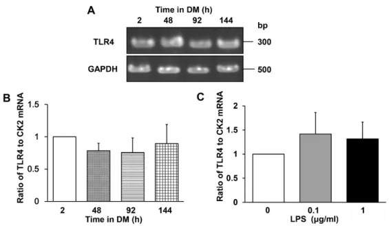
TLR4 mRNA is expressed constitutively in differentiating myoblasts
Although TLR4 is abundantly expressed in skeletal muscle of various vertebrates,
including human and mouse [39–41], its expression pattern in differentiating myoblasts is unclear.
We examined TLR4 mRNA expression at 2, 48, 92, and 144 h after induction of C2C12
differentiation and found that it is expressed constitutively (Fig 1A). Quantitation of TLR4 mRNA
by real-time RT-PCR confirmed that similar levels were expressed throughout the differentiation
period (Fig 1B). Similarly, TLR4 mRNA levels did not change after culture of C2C12 cells in
DM with LPS (0.1 or 1 μg/mL) for 144 h (Fig 1C), in agreement with previous observations by
Frost et al. [43,57] and Lang et al. [41]. These results indicate that TLR4 would be expected to be
available for LPS binding throughout C2C12 myogenesis and was unaffected by the presence of
LPS.
16
Fig 1. TLR4 mRNA expression in differentiating C2C12 myoblasts. (A–C) TLR4 mRNA
levels in C2C12 myoblasts cultured in DM for 2, 48, 96, or 144 h with or without LPS. (A)
Agarose gel of TLR4 and GAPDH (internal standard) mRNA levels during differentiation. (B)
Real-time RT-PCR quantification of TLR4 mRNA levels. Data were normalized to CK2 mRNA
levels and the ratio in cells cultured in DM for 2 h was set at 1.0. Data are the mean ± SEM of 3–
4 independent experiments, each performed in duplicate. (C) Quantification of TLR4 mRNA by
real-time RT-PCR. C2C12 cells were cultured with the indicated concentrations of LPS for 144 h.
Data were normalized to CK2 mRNA levels and the ratio in untreated cells was set at 1.0. Data
are the mean ± SEM of 3 independent experiments performed in duplicate. In (B) and (C),
P > 0.05 for all comparisons.
17
LPS inhibits myogenesis in a dose-dependent manner
To examine the effect of LPS on myogenesis, C2C12 cells were cultured in DM for 144
h in the presence or absence of 0.1 or 1 μg/mL LPS. These concentrations were employed for
consistency with a previous study [19] that showed that LPSinduces protein breakdown via the
ubiquitin–proteasome pathway in differentiated C2C12 cells. LPS was found to inhibit the
formation of multinucleated myotubes (Fig 2A). To quantify the morphological changes, the
proportion of total nuclei that were present in myotubes was calculated (the myogenic index; see
Materials and Methods). As shown in Fig 2B, the myogenic index was significantly lower in LPS-
treated than untreated myoblasts (control, 21 ± 3%; LPS 0.1 μg/mL, 10 ± 2%; LPS 1 μg/mL, 8 ±
2%). LPS also dose-dependently decreased the average myotube width (control, 20 ± 0.4 μm;
LPS 0.1 μg/mL, 15 ± 0.5 μm; LPS 1 μg/mL, 11 ± 0.3 μm; Fig 2C and 2D), in agreement with
earlier observations that LPS induces muscle proteolysis [19]. We also confirmed by western
blotting that LPS dose-dependently suppressed expression of the differentiation marker MyHC II
(LPS 0.1 μg/mL vs control, 44% decrease [p < 0.01]; LPS 1 μg/mL vs control, 85% decrease [p
< 0.001]; Fig 2E and 2F). Thus, LPS directly inhibits C2C12 myogenesis in a dose-dependent
manner in the absence of immune cells, which are the major sources of inflammatory cytokines.
18
Fig 2. Effect of LPS on C2C12 myogenesis. (A) LPS inhibits the formation of myotubes.
Myoblasts were cultured for 144 h in DM alone (control), DM plus LPS 0.1 μg/mL, or DM plus
LPS 1 μg/mL, then fixed and subjected to May–Grünwald–Giemsa staining. Representative
images are shown. Scale bar = 100 μm. Arrows indicate differentiated myotubes. (B) LPS
decreases the myogenic index. Cells were treated as described in (A). Data are the mean ± SEM
of 10–12 independent experiments, each examining 5 randomly selected fields (total 50–60 fields
per treatment group). (C) Frequency distribution of myotube widths. LPS-treated myotube
diameters show a left shift compared to the control group. (D) LPS decreases myotube width in a
dose-dependent manner. Data are the mean ± SEM of 10–12 independent experiments, each
examining 5 randomly selected fields (total 175–296 myotubes from 50–60 fields per treatment
19
group). Cells were treated as described in (A). (E) Representative western blot probed with
antibodies to MyHC II or β-tubulin (internal standard). Cells were treated as described in (A). (F)
Quantification of the data presented in (E). Data are the mean ± SEM of 13–14 independent
experiments. ***p < 0.001, **p < 0.01, ###p < 0.001, ##p < 0.01 by one-way ANOVA followed
by Tukey’s honest significant difference test.
20
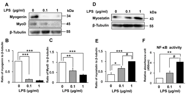
Effect of LPS on muscle regulatory factors and NF-κB DNA- binding activity
The myogenic regulatory factors myogenin and MyoD are responsible for the induction
and maintenance of early-phase muscle differentiation [32,33]. To understand the mechanism of
LPS-induced myogenic inhibition, we examined the expression of these regulatory factors in LPS-
treated C2C12 myoblasts. As shown in Fig 3A–3C, LPS treatment for 48 h dose-dependently
downregulated the expression of both myogenin (LPS 0.1 μg/mL vs control, 86% decrease [p <
0.001]; LPS 1 μg/mL vs control, 92% decrease [p < 0.001]) and MyoD (LPS 0.1 μg/mL vs control,
42% decrease [p < 0.01]; LPS 1 μg/mL vs control, 62% decrease [p < 0.001]).
We next examined whether LPS-induced inhibition of myogenesis is associated with
increased expression of myostatin, which is a critical autocrine and paracrine inhibitor of skeletal
muscle growth and differentiation [34–36]. Indeed, myostatin expression was increased dose
dependently by incubation with LPS for 144 h (LPS 0.1 μg/mL vs control, 185% increase [p <
0.05]; LPS 1 μg/mL vs control, 283% increase [p < 0.001]; Fig 3D and 3E).
Previous studies have suggested that NF-κB mediates both the downregulation of MyoD
and myogenin and the upregulation of myostatin [27–31,37,38]. Thus, we next evaluated NF-κB
(p65) DNA-binding activity in LPS-treated myoblasts. C2C12 nuclear extracts were prepared
after 48 h of LPS treatment and analyzed by ELISA. This time point was chosen for convenience
21
because the LPS-containing medium was refreshed every other day. As shown in Fig 3F, LPS
treatment dose-dependently increased NF-κB DNA-binding activity (LPS 0.1 μg/mL vs control,
201% increase [p = 0.37]; LPS 1 μg/mL vs control, 417% increase [p < 0.01]). Collectively, these
data suggest that LPS-induced MyoD and myogenin downregulation and myostatin upregulation
is associated with increased NF-κB activity.
Fig 3. Effect of LPS on muscle regulatory factors and NF-κB activity in differentiating
myoblasts. (A) LPS downregulates expression of the positive myogenic regulatory factors
myogenin and MyoD in a dose-dependent manner. C2C12 myoblasts were incubated in DM with
or without LPS (0.1 or 1 μg/mL) for 48 h. A representative western blot probed with antibodies
to myogenin, MyoD, or β-tubulin (internal standard) is shown. (B and C) Quantification of the
22
data presented in (A). Data are the mean ± SEM of 6–7 independent experiments. (D) LPS
upregulates expression of the negative myogenic regulatory factor myostatin in a dose-dependent
manner. C2C12 myoblasts were treated for 144 h as described in (A). A representative western
blot probed with antibodies to myostatin or β-tubulin (internal standard) is shown. (E)
Quantification of the data presented in (D). Data are the mean ± SEM of 14 independent
experiments. (F) LPS increases NF-κB DNA-binding activity in a dose-dependent manner.
C2C12 myoblasts were incubated in DM with or without LPS (0.1 or 1 μg/mL) for 48 h, and NF-
κB activity was analyzed using a TransAM ELISA kit. Data are the mean ± SEM of 5 independent
experiments performed in duplicate. ***p < 0.001, **p < 0.01, *p < 0.05, #p < 0.05 by one-way
ANOVA followed by Tukey’s honest significant difference test.
23
The inhibitory effect of LPS on myogenesis is reversible
Next, we investigated whether the inhibitory effect of LPS on myogenesis was
reversible. To address this question, we adapted the methodology of Langen et al. [28], who
showed that the toxic effect of TNF-α on C2C12 myogenesis was reversible. The cells were
incubated with LPS for 48 h, washed, and cultured in fresh LPS-free DM. Myogenin expression
was examined at 24 and 48 h after LPS washout. Notably, while myogenin expression was initially
reduced by LPS, it was restored after washing and removal of LPS (Fig 4A and 4B). These results
demonstrate that the inhibitory effect of LPS on myogenic differentiation is reversible, suggesting
that it is not due to a nonspecific toxic effect such as induction of cell death.
24
Fig 4. LPS-induced inhibition of myogenin expression is reversible. C2C12 cells were cultured
in DM with or without LPS (1 μg/mL) for 48 h, washed free of LPS, and then cultured for an
additional 24 h (72 h total) or 48 h (96 h total) in fresh DM. (A) Representative western blot
probed with antibodies to myogenin or β-tubulin (internal standard). (B) Quantification of the
data presented in (A). Data are the mean ± SEM of 5 independent experiments.
***p < 0.001,*p < 0.05, #p < 0.05, ##p < 0.01 by one-way ANOVA followed by Tukey’s honest
significant difference test.
25
Effect of TAK-242 on LPS-induced inhibition of myogenesis
We next assessed whether pharmacological inhibition of TLR4 signaling can ameliorate
the harmful effect of LPS on C2C12 myogenesis. Myoblasts were incubated for 144 h in the
presence or absence of LPS (1 μg/mL) and/or TAK-242 (1 μM), a small molecule specific
inhibitor of TLR4 signaling [58,59]. This concentration of TAK-242 was previously found to have
a beneficial effect on LPS-induced insulin resistance in L6 myoblast cells [60] and primary human
skeletal muscle cells [61]. We found that TAK-242 increased the abundance of multinuclear
myotubes in LPS-treated C2C12 cells (myogenic index: LPS 1 μg/mL, 7 ± 2% vs LPS 1 μg/mL
+ TAK-242 1 μM, 13 ± 2%, p < 0.05; Fig 5A and 5B) as well as myotube width (LPS 1 μg/mL,
11 ± 0.3 μm vs LPS 1 μg/mL + TAK-242 1 μM, 16 ± 0.4 μm, p < 0.001; Fig 5C and 5D). Moreover,
TAK-242 partially restored MyHC II expression (LPS 1 μg/mL + TAK-242 1 μM vs LPS 1 μg/mL,
251% increase, p < 0.05; Fig 5E and 5F), confirming its beneficial effect. Higher TAK242
concentrations (10 μM and 100 μM) did not further improve myogenesis and, in fact, were toxic
to the cells (data not shown). Collectively, these results indicate that TLR4 signaling is responsible,
at least in part, for the inhibition of myogenesis by LPS, since TAK-242 treatment attenuated the
effect.
26
Fig 5. Effect of TAK-242 on LPS-induced inhibition of myogenesis. (A) May–Grünwald–
Giemsa staining of C2C12 cells cultured for 144 h in DM alone, DM plus LPS (1 μg/mL), or DM
plus LPS (1 μg/mL) and TAK-242 (1 μM). Representative images are shown. Scale bar = 100 μm.
Arrows indicate differentiated myotubes. (B) TAK-242 partially reversed the LPS-induced
decrease in myogenic index. Cells were treated as described in (A). Data are the mean ± SEM of
10 independent experiments per treatment group, each examining 5 randomly selected fields (total
50 fields). (C and D) TAK-242 partially ameliorated the LPS-induced myotube atrophy. Cells
were treated as described in (A). Frequency distribution of myotube widths for each treatment
group are shown in (C), and comparisons of myotube widths for each treatment group are shown
in (D). Data are the mean ± SEM of 10–12 independent experiments, each examining 5 randomly
27
selected fields (total 194–296 myotubes from 50–60 fields per treatment group). (E)
Representative western blot probed with antibodies to MyHC II or β-tubulin (internal standard).
Cells were treated as described in (A). (F) Quantification of the data presented in (E). Data are
the mean ± SEM of 12–13 independent experiments. ***p < 0.001, ###p < 0.001, #p < 0.05 by
one-way ANOVA followed by Tukey’s honest significant difference test.
28
TLR4–NF-κB signaling mediates LPS-induced dysregulation of muscle regulatory factors
Next, we evaluated whether TLR4 signaling mediates the LPS-induced perturbation of
muscle regulatory factors. As shown in Figs 6A–6C, co-administration of TAK-242 (1 μM)
partially reversed the LPS-induced decrease in expression of myogenin (LPS 1 μg/mL + TAK-
242 1 μM vs LPS 1 μg/mL, 251% increase; p < 0.05) but not of MyoD (101% increase, p = 1.0).
Similarly, LPS-induced upregulation of myostatin was partially blocked by TAK-242 (30%
decrease vs LPS 1 μg/mL, p < 0.05; Fig 6D and 6E). However, at 0.1 μg/mL LPS, TAK-242 failed
to rescue the upregulation of myostatin expression (5% decrease, p = 0.7). TAK-242 also
decreased NF-κB DNA-binding activity in LPS-treated myoblasts (48% decrease vs LPS 1 μg/mL,
p < 0.05; Fig 6F). These data therefore suggest that TLR4 acts as an upstream regulator of NF-
κB-mediated dysregulation of myogenic regulatory factors.
29
Fig 6. Effect of TAK-242 on LPS-induced perturbation of muscle regulatory factors and NF-
κB activity. (A) TAK-242 partially reverses the LPS-induced downregulation of myogenin, but
not of MyoD. C2C12 myoblasts were cultured for 48 h in DM alone, with LPS (1 μg/mL or 0.1
μg/mL), or with a combination of LPS (1 μg/mL) and TAK-242 (1 μM). A representative western
blot probed with antibodies to myogenin, MyoD, or β-tubulin (internal standard) is shown. (B and
C) Quantification of the data presented in (A). Data are the mean ± SEM of 5–7 independent
experiments. (D) TAK-242 partially reverses the LPS-induced upregulation of myostatin
expression. C2C12 myoblasts were cultured for 144 h as described in (A). A representative
western blot probed with antibodies to myostatin or β-tubulin (internal standard) is shown. (E)
Quantification of the data presented in (D). Data are the mean ± SEM of 14–16 independent
30
experiments. (F) TAK-242 inhibits NF-κB activity in LPS-treated differentiating myoblasts. Cells
were treated as described in (A), and NF-κB activity was analyzed using a TransAMELISA kit.
Data are the mean ± SEM of 5 independent experiments performed in duplicate. ***p < 0.001,
*p < 0.05, #p < 0.05 by one-way ANOVA followed by Tukey’s honest significant difference test.
31
LPS signals through both TLR2 and TLR4 to activate NF-κB
As demonstrated in Fig 5 and Fig 6, selective inhibition of TLR4 signaling did not fully
suppress LPS-induced NF-κB activation and dysregulation of muscle regulatory factors. Since
LPS is also a ligand for TLR2 [62–64], and TLR2 mRNA is expressed in C2C12 myoblasts
[41,43], we considered that LPS might be signaling through both TLR2 and TLR4. To examine
this, myoblasts were incubated with 1 μg/mL LPS in the presence or absence of a TLR2-
neutralizing antibody (T2.5, 10 μg/mL), and NF-κB activity was measured after 48 h. This
concentration of anti-TLR2 antibody was previously found to prevent serum amyloid A-induced
TLR2-dependent signaling in C2C12 myotubes [56]. As shown in Fig 7A, addition of anti-TLR2
antibody decreased LPS-induced NF-κB activation by about 30%, although the difference did not
reach the level of statistical significance (p = 0.62). The selective TLR4 inhibitor had a greater
effect on NF-κB activity (Fig 7B), suggesting that LPS signals predominantly through TLR4 but
at least partially through TLR2 in these cells. This could be one possible explanation for the failure
of TAK-242 to completely suppress the LPS-induced effects on NF-κB and myogenic regulatory
factors.
32
Fig 7. Effect of a TLR2-neutralizing antibody on NF-κB activity in LPS-treated myoblasts. (A)
C2C12 myoblasts were cultured for 48 h in DM alone, LPS (1 μg/mL), or LPS (1 μg/mL) plus
TLR2-neutralizing antibody (10 μg/mL). Data are the mean ± SEM of 7 independent experiments
performed in duplicate. (B) C2C12 myoblasts were cultured for 48 h in DM with LPS (1 μg/mL),
LPS (1 μg/mL) plus TAK-242 (1 μM), or LPS (1 μg/mL) plus TLR2-neutralizing antibody
(10 μg/mL). NF-κB activity was measured by ELISA. Data are the mean ± SEM of 7–10
independent experiments performed in duplicate. **p < 0.01, *p < 0.05 by one-way ANOVA
followed by Tukey’s honest significant difference test.
33
Myoblast-derived autocrine/paracrine TNF-α is involved in LPS-induced inhibition of myogenesis
There is considerable evidence that LPS regulates the expression of proinflammatory
cytokines, such as TNF-α and interleukin 6, in mouse myoblasts and skeletal muscle [40,41,43],
and TNF-α has previously been shown to inhibit myogenic differentiation [27–31]. Therefore, we
hypothesized that myoblast-derived TNF-α may be involved in LPS-induced inhibition of
myogenesis. To test this hypothesis, we examined C2C12 cells after treatment with LPS (1
μg/mL) in the absence or presence of a TNF-α-neutralizing antibody (5 μg/mL). This
concentration of antibody was previously shown to inhibit TNF-α secretion and subsequent NF-
κB activation induced by serum restriction of C2C12 myoblasts [65]. We found that the TNF-α-
neutralizing antibody partially but significantly reversed the LPS-induced downregulation of
myogenin (LPS + anti-TNF-α vs LPS, 255% increase, p < 0.05; Fig 8A and 8B), MyoD (179%
increase, p < 0.001; Fig 8A and 8C), and MyHC II (262% increase, p < 0.01; Fig 8D and 8E)
expression, and additionally suppressed the LPS-induced upregulation of myostatin expression
(40% decrease, p < 0.01; Fig 8D and 8F) and NF-κB activation (40% decrease, p < 0.05; Fig 8G).
These results suggest that NF-κB activation and subsequent effects on myogenic regulatory
factors induced by LPS are partially mediated by myoblast-derived TNF-α. Thus, it is possible
that such an autocrine/paracrine TNF-α loop is involved in the impairment of muscle regeneration.
34
35
Fig 8. Effect of a TNF-α-neutralizing antibody on LPS-induced perturbation of muscle
differentiation. (A) C2C12 myoblasts were cultured for 48 h in DM alone, LPS (1 μg/mL), or
LPS (1 μg/mL) plus TNF-α-neutralizing antibody (5 μg/mL). A representative western blot
probed with antibodies to myogenin, MyoD, or β-tubulin (internal standard) is shown. (B and C)
Quantification of the data presented in (A). Data are the mean ± SEM of 7–15 independent
experiments. (D) C2C12 myoblasts were cultured for 144 h as described in (A). A representative
western blot probed with antibodies to MyHC II, myostatin, or β-tubulin (internal standard) is
shown. (E and F) Quantification of the data presented in (D). Data are the mean ± SEM of 5–21
independent experiments. (G) Cells were treated as described in (A), and NF-κB activity was
analyzed by ELISA. Data are the mean ± SEM of 7 independent experiments performed in
duplicate. ***p < 0.001, **p < 0.01, *p < 0.05, ###p < 0.001, ##p < 0.01, #p < 0.05 by one-way
ANOVA followed by Tukey’s honest significant difference test.
36
Proposed mechanism of LPS inhibition of myogenesis in differentiating myoblasts
Based on these collective data, we propose a mechanistic model for the inhibition myogenesis by
LPS in differentiating myoblasts (Fig 9).
Fig 9. Proposed mechanism of LPS inhibition of myogenesis in differentiating myoblasts.
37
Discussion
In the present study, we found that LPS inhibits C2C12 myogenesis through the TLR4–
NF-κB and autocrine/paracrine TNF-α-mediated pathways. We found that LPS downregulated
MyoD and myogenin expression and upregulated myostatin expression in a dose-dependent
manner. Both pharmacological inhibition of TLR4 signaling and antibody-mediated
neutralization of TNF-α reduced NF-κB activity and attenuated the LPS-induced dysregulation of
muscle regulatory factors.
For our in vitro experiments, we employed two concentrations of LPS, 0.1 and 1 μg/mL,
and observed a dose-dependent inhibitory effect on murine myoblast differentiation. A previous
study suggested that humans were more than 10,000-fold more sensitive than mice to LPS [66],
raising the possibility that human muscle regeneration could be much more vulnerable to the
effects of LPS. Circulating LPS levels are commonly elevated in conditions such as sepsis [4] and
endogenous diseases [5-15], and muscle atrophy can also be observed in these conditions [16].
Therefore, LPS-induced derangement of myogenesis might be a cause of muscle wasting in
patients with sepsis or metabolic endotoxemia.
In this study, we demonstrated that the TLR4 signaling pathway mediated LPS-induced
activation of NF-κB, downregulation of MyoD and myogenin expression, and upregulation of
38
myostatin expression. Our findings are in agreement with several earlier observations that
exogenous TNF-α-induced activation of NF-κB inhibited myogenesis in C2C12 cells by
suppressing MyoD and myogenin expression [27–31]. NF-κB-mediated upregulation of
myostatin has also been observed in other model systems, such as H2O2-treated cultured
myoblasts [38] and mouse models of liver cirrhosis and hyperammonemia [37]. In contrast,
several studies have suggested that myostatin expression in skeletal muscle is not increased in
sepsis models. For example, Smith et al. observed that myostatin mRNA levels were reduced and
myostatin protein levels were unchanged in rat skeletal muscle 16 h after cecal ligation and
puncture [67]. Lang et al. reported that myostatin mRNA was not increased 24 h after LPS
administration to rats [68]. One possible explanation for this discrepancy is the shorter endotoxin
exposure times, since we observed increased myostatin expression in C2C12 cells after 144 h of
LPS treatment. Martin et al. noted a time-dependent increase in myostatin mRNA expression after
administration of LPS to mice; they found that the levels remained unchanged at 24 h after LPS
injection but increased significantly after 76 h [69]. In the clinical setting, sepsis survivors often
display systemic inflammation for protracted periods and also develop muscle wasting.
Circulating myostatin is commonly elevated in patients with conditions such as chronic liver and
kidney disease [25,37,70], diabetes mellitus [71], and human immunodeficiency virus infection
[72], and in the elderly [73,74]. All of these populations are likely to be chronically exposed to
39
LPS due to bacterial translocation [15]. Taken together, these findings suggest that persistent
exposure to LPS or inflammation may be required to induce myostatin.
We found that myogenin expression was rapidly restored after switching from LPS (1
μg/mL)-containing medium to fresh medium, suggesting that the inhibition of myogenesis was
reversible and not simply a toxic effect of LPS, such as induction of apoptosis. Shang et al.
examined C2C12 cell viability after exposure to various concentrations of LPS [75]. They found
that LPS at 1–10 μg/mL had no effect on C1C12 apoptosis, whereas higher concentrations (100–
150 μg/mL) induced apoptosis through caspase-3 activation. Our findings are thus consistent with
their data. Reversible inhibition of myogenesis has also been observed upon treatment of C2C12
myoblasts with TNF-α [28]. Taken together, these data suggest that persistent exposure to LPS or
inflammatory cytokines, and subsequent NF-κB activation may be required to block myogenesis
and promote muscle wasting.
While the role of TLR4 in innate immunity is well characterized, its role in skeletal
muscle development has been unclear. To address this knowledge gap, we examined the effect of
a selective TLR4 signaling pathway inhibitor on the LPS-induced events. We observed that TAK-
242 partially rescued the LPS-induced inhibition of myogenesis, activation of NF-κB,
downregulation of myogenin, and upregulation of myostatin, suggesting that TLR4 is an upstream
regulator of skeletal muscle myogenesis. Previous studies have suggested that TLR4 plays an
40
important role in muscle protein breakdown. Doyle et al. [19] found that TLR4-mediated LPS
signaling induced muscle catabolism via coordinate activation of the ubiquitin–proteasome and
autophagy pathways. According to Dehoux et al. [20] and Martin et al. [69], ubiquitin ligase
mRNA expression was induced in both rat and mouse skeletal muscle after LPS injection. In
addition to these previous findings, we observed that LPS dose-dependently decreased the
myogenic capacity. Collectively, these findings indicate that LPS may induce muscle wasting via
synergistic effects on myogenesis and muscle proteolysis through TLR4. Skeletal muscle TLR4
is upregulated in diabetic and obese subjects [76] as well as in the elderly [5,77], suggesting that
the TLR4–NF-κB pathway may be elevated in these populations. Thus, inhibition of the TLR4
signaling axis might be a useful method for preventing or reversing LPS-induced muscle wasting
in patients with sepsis or metabolic endotoxemia. Future studies should address the effects of
TLR4 antagonists on LPS-induced muscle wasting.
We found that antibody-mediated neutralization of TNF-α reduced the LPS-induced
increase in NF-κB binding activity, downregulation of myogenin and MyoD, and upregulation of
myostatin in C2C12 cells, suggesting that LPS-induced autocrine/paracrine TNF-α might be
involved in the impairment of muscle regeneration. Autocrine/paracrine regulation of TNF-α has
also been observed in C2C12 myoblasts following serum restriction [65] and in various other cell
lines and tissues, including cancer cells [46,78], immune cells [79,80], and microglia [81]. In
41
skeletal muscle myoblasts, TNF-α is a strong activator of NF-κB [65] and of its own synthesis
[82]. Therefore, as is the case in cancer cells [46,78], a positive TNF-α autocrine/paracrine loop
in response to LPS may lead to persistent NF-κB activation in myoblasts, further inhibiting
myogenesis and thus inducing muscle wasting.
Interestingly, we found that TNF-α neutralization, but not TLR4 inhibition by TAK-242,
reversed the LPS-induced inhibition of MyoD expression. We speculate that MyoD expression
might be more sensitive to regulation by TNF-α than by TLR4 signaling. In support of this
possibility, the pattern of NF-κB activation by LPS and TNF-α has been shown to differ. In C2C12
myotubes, TNF-α was found to persistently activate NF-κB in a biphasic manner, while LPS did
not [31]. Moreover, in human epithelial cells, LPS from Haemophilus influenzae (exogenous
activation) and TNF-α (endogenous activation) synergistically induced NF-κB activation via two
distinct signaling pathways [83]. This could be one explanation for the failure of TAK-242 to
fully ameliorate the harmful effect of LPS on differentiating myoblasts.
Another possible explanation for the inhibition of TLR4 signaling did not fully suppress
LPS-induced NF-κB activation was the involvement of TLR2 mediated signaling. Although the
difference did not reach the level of statistical significance, anti-TLR2 antibody decreased LPS-
induced NF-κB activation by about 30%. Therefore, as in immune cells, microglia and astrocytes
[62–64], LPS-induced NF-κB activation in C2C12 myoblasts may be partially mediated through
42 TLR2 signaling pathway.
In vivo, LPS induces circulating immune cells to produce copious amounts of
inflammatory cytokines, which have been implicated as potential mediators of muscle wasting
via inhibition of myogenesis [27–31] and acceleration of muscle proteolysis [18,84,85]. In fact,
inflammatory cytokine concentrations are elevated in the circulation of patients with sepsis [86]
and metabolic endotoxemia [87–89]. Our study extends these observations by demonstrating that
LPS itself can directly inhibit myogenesis through TLR4–NF-κB signaling and myoblast-derived
TNF-α. Systemic and local inflammatory reactions may synergize to induce muscular wasting.
In this study, cells were co-incubated with TAK-242 dissolved in DMSO (final
concentration 0.1% [vol/vol]) and LPS dissolved in PBS. We acknowledge that a DMSO vehicle
control was not included in two experiments shown in Figs 5 and 6. High concentrations of DMSO
(1–2% [vol/vol]) have been shown to augment the LPS effect on immune cells (i.e., increase
inflammatory cytokine secretion) [90]; therefore, in our study, the relative effect of TAK-242 on
the LPS response may have been weakened. Nevertheless, previous studies have shown that, even
at concentrations as high as 1–2% (vol/vol), DMSO has no effect on NF-κB activity in various
cell lines, reducing this concern [90–92].
We observed that TAK-242 significantly decreased myostatin expression when cells
were stimulated with LPS at 1 μg/mL (30% decrease, p < 0.05) but not at 0.1 μg/mL (5% decrease,
43
p = 0.7). At present, we do not have a plausible explanation for this discrepancy.
To date, no drugs have been approved for the treatment of skeletal muscle wasting. Our
finding that blockade of the TLR4–NF-κB pathway or TNF-α can reverse impaired myogenesis
suggests a new set of drug targets for clinical intervention in sepsis- or metabolic endotoxemia-
induced muscle debilitation. Clinical trials with a TLR4 antagonist [93–95] and TNF-α inhibitor
[96] have shown no improvement of the mortality rate of severe sepsis patients; however, those
trials did not examine long-term muscle function [93–96]. The results presented here provide a
rationale to test the effects of TLR4 and TNF-α antagonists on LPS-induced muscle wasting in
sepsis or metabolic endotoxemia patients. Our data should also stimulate further studies to clarify
the role of TLR4–NF-κB and TNF-α signaling in muscle wasting.
44
Conclusions
LPS inhibits C2C12 myogenesis by dose-dependently downregulating myogenin and
MyoD expression and upregulating myostatin expression. TAK-242, a selective inhibitor of
TLR4-mediated signaling, and a TNF-α-neutralizing antibody both reduced NF-κB activity and
attenuated the downregulation of myogenic regulatory factors and upregulation of myostatin in
LPS-treated C2C12 myoblasts. Consequently, myogenesis was partially restored. These data
suggest that LPS inhibits myogenic differentiation of skeletal muscle myoblasts through TLR4–
NF-κB signaling and myoblast-derived autocrine/paracrine TNF-α, raising the possibility that
these pathways also contribute to the development of muscle wasting in patients with sepsis or
metabolic endotoxemia.
45
Acknowledgements
First, the author thank Kazuho Sakamoto, Ph.D. (Department of Pharmacology, School
of Medicine, Fukushima Medical University, Fukushima, Japan) for his excellent advice and daily
mentation. The author also thank Tomoyuki Ono, Ph.D. (Department of Pharmacology, School of
Medicine, Fukushima Medical University) for technical support; Yayoi Shikama, M.D., Ph.D.
(Center for Medical Education and Career Development, Fukushima Medical University) for help
in reviewing the manuscript; and Mr. Kuniyuki Hara and Ms. Nao Katayose (Department of
Pharmacology, School of Medicine, Fukushima Medical University) for their assistance in
generating Fig 7 and Fig 8. Finally, the author thank Nozomi Ono, M.D. (Department of
Psychiatry, Hoshigaoka Hospital, Koriyama, Japan), for her consistent assistance in this study.
Competing interests
The authors declare that they have no competing interests.