The relationship between uterine, milk, and environmental microbiota in postpartum
dairy cows
2020, March
NGUYEN THI THUONG
Graduate School of
Environmental and Life Science (Doctor’s Course)
OKAYAMA UNIVERSITY
I
TABLE OF CONTENTS
Table of contents ... I Declaration... III Acknowledgments ... IV Publications arising from this thesis ... V Conference proceedings ... VI List of figures ... VII List of tables ... VIII
Chapter 1. General Introduction... 1
Chapter 2. Literature Reviews ... 2
The postpartum uterine microbiota... 2
Physiology of uterus after calving ... 2
The microbiota in uterus after calving ... 5
The uterine diseases after parturition... 6
Therapy of the uterine diseases ... 8
The milk microbiota ... 9
Physiology of lactation ... 9
The microbiota in milk ... 11
Mastitis ... 12
Treatment and prevention of mastitis ... 13
The environment microbiota ... 13
The relationship between uterine and environmental microbiota ... 13
The relationship between milk and environmental microbiota ... 14
Chapter 3. The relationship between uterine, fecal, bedding, and airborne dust microbiota from dairy cows and their environment ... 15
Abstract ... 15
Introduction ... 15
Materials and methods ... 16
Results ... 19
Discussion ... 21
Chapter 4. An investigation of seasonal variations in the microbiota of milk, feces, bedding, and airborne dust ... 32
Abstract ... 32
II
Introduction ... 32
Materials and methods ... 34
Results ... 36
Discussion ... 38
Chapter 5. General Conclusion ... 46
References ... 47
III
DECLARATION
I hereby declare that my thesis/dissertation entitled:
“The relationship between uterine, milk, and environmental microbiota in postpartum dairy cows”
has been composed by myself, that the work contained here in is my own except where explicitly stated otherwise in the text, and that this work has not been submitted for any other degree or processional qualification except as specified.
Parts of this work have been published in Animals (2019).
Date
Signature
IV
ACKNOWLEGMENTS
Firstly, I would like to express my sincere gratitude to my supervisor Prof. Nishino Naoki for the continuous support of my Ph.D study and research, for his patience, motivation, and immense knowledge. His guidance helped me in all the time of research and writing of this thesis. I could not have imagined having a better advisor and mentor for my Ph.D study.
I am very grateful to Prof. Saito Noboru, Prof. Funahashi Hiroaki and Prof. Morita Hidetoshi for their support as co-supervisor. Their lectures and extensive knowledge helped me a lot during my Ph.D study. And I would like to appreciate Assoc. Prof. Tsuruta Takeshi for his kind support and encouragement in my Ph.D time.
I would like to thank Ms. Ayumi Miyake and Mr. Tanabe Yuji from Okayama Livestock Research Institute for their enthusiastic help and instruction for collecting sample. Without their support it would not be possible to conduct this research.
My sincere thanks also goes to Dr. Tang Thuy Minh, Dr. Tran Thi Minh Tu, and Dr Wu Haoming for all of their kindness and thoughtfulness in laboratory work as well as student life.
And, I thank my labmates for the stimulating discussions, for the insightful comments in every seminar, and for all the fun we have had together. Besides, I would like to send my honest thanks to all of my Vietnamese friends for their support when I came to Japan. Their companionship and friendship have been motivated me to overcome many obstacles.
Last but not the least, I would like to thank my family: my mother, my father, my sister, my son and my partner with all my love and gratitude for supporting me spiritually throughout my study in Japan and my life in general.
V
PUBLICATION ARISING FROM THIS THESIS
1. The Relationship between Uterine, Fecal, Bedding, and Airborne Dust Microbiota from Dairy Cows and Their Environment: A Pilot Study. Thuong T. Nguyen, Ayumi Miyake, Tu T.M. Tran, Takeshi Tsuruta and Naoki Nishino. Animals 2019, 9, 1007;
doi:10.3390/ani9121007.
2. An investigation of seasonal variations in the microbiota of milk, feces, bedding, and airborne dust. Thuong T. Nguyen, Haoming Wu, Naoki Nishino. Asian-Australasian Journal of Animal Sciences 2019, doi.org/10.5713/ajas.19.0506.
VI
CONFERENCES PROCEEDINGS
1. Tran Thi Minh Tu, Nguyen Thi Thuong, Tran Hoang Diep, Diep Tan Toan, Tsuruta Takeshi, Nishino Naoki (2017). Relationship between fecal, uterus and milk microbiota in dairy farm in the south of Vietnam. 122th Annual Meeting of Japanese Society of Animal Science, Kobe, Japan.
2. Nguyen Thi Thuong, Miyake Ayumi, Tanabe Yuji, Tsuruta Takeshi, Nishino Naoki (2018). An investigation of fecal, uterus and milk microbiota of dairy cows after calving. 124th Annual Meeting of Japanese Society of Animal Science, Tokyo, Japan.
3. Nguyen Thi Thuong, Miyake Ayumi, Tanabe Yuji, Tsuruta Takeshi, Nishino Naoki (2019). Uterine microbiota of dairy cows as influenced by season and postpartum period. 125th Annual Meeting of Japanese Society of Animal Science, Tokyo, Japan.
4. Nguyen Thi Thuong, Tran Thi Minh Tu, Tsuruta Takeshi, Nishino Naoki (2009). Stability and variability of the uterine microbiota in postpartum dairy cows. National conference Animal and Veterinary Sciences, Hochiminh city, Vietnam.
5. Nguyen Thi Thuong, Miyake Ayumi, Tran Thi Minh Tu, Tsuruta Takeshi, Nishino Naoki (2009).
Uterine microbiota of postpartum dairy cows as influenced by season and environment. The Second International Conference on Animal Production and Environment, Can Tho City, Vietnam.
VII
LIST OF FIGURES
Chapter 2
Figure 1. Involution of the uterus in a cow following a normal calving ... 3 Figure 2. Uterine horn diameter measured by ultrasonography in the cows ... 3 Figure 3. The bovine estrus cycle ... 4 Figure 4. Estrogen from growing follicle is transported through the blood stream to all parts of the
body ... 4 Figure 5. Follicular growth occurs throughout the estrous cycle ... 5 Figure 6. Estrus cycle is divided into two phases... 5 Figure 7. Almost all cows have bacteria within the cavity of the uterus during the first 2 weeks of calving and uterine disease ... 8 Figure 8. Schematic representation of bovine mammary gland ... 10 Figure 9. Schematic representation of relationships among lipid metabolism in adipose tissue, liver, and mammary gland ... 10 Chapter 3
Figure 1. Uterine, fecal, bedding, and air-borne dust microbiota in the dairy farm examined during two seasons ... 30 Figure 2. Canonical analysis of principal coordinates plot characterizing the uterine, fecal, bedding, and airborne dust microbiota in the dairy farm ... 31 Chapter 4
Figure 1. Family-level proportions of the top 20 bacterial taxa of the fecal microbiota of the dairy cows investigated during summer and winter ... 41 Figure 2. Family-level proportions of the top 20 bacterial taxa of the bedding microbiota of a dairy farm investigated during summer and winter ... 42 Figure 3. Family-level proportions of the top 20 bacterial taxa of the airborne dust microbiota of a dairy farm investigated during summer and winter ... 43 Figure 4. Canonical analysis of principal coordinates plot characterizing the milk, fecal, bedding, and airborne dust microbiota of the dairy farm ... 44
VIII
LIST OF TABLES
Chapter 2
Table 1. List of bacteria according to their expected pathogenic potential in the uterus ... 6 Table 2. Nutrient composition of whole and whole, dry milk... 10 Chapter 3
Table 1. Milk yield and blood metabolites concentration of the dairy cow examined at one and two months postpartum at two seasons. ... 25 Table 2. Relative abundance (%) of uterine microbiota of the dairy cows examined at one and two months postpartum during the two seasons ... 26 Table 3. Relative abundance (%) of fecal microbiota of the dairy cows examined at one and two months postpartum during the two seasons ... 27 Table 4. Relative abundance (%) of bedding microbiota of the dairy farm cowshed examined at two seasons ... 28 Table 5. Relative abundance (%) of air-borne dust microbiota of the dairy farm cowshed examined at two seasons ... 29 Chapter 4
Table 1. Milk yield, milk composition, and relative abundance of milk microbiota of the dairy cows examined at one and two months postpartum during the two seasons ... 43
1 CHAPTER 1
GENERAL INTRODUCTION
In postpartum dairy cows, the uterus is contaminated with diverse bacterial species, including pathogens associated with uterine disease. Although one-third to two-thirds of cows remain healthy, others may develop metritis and endometritis; this reduces their food intake and milk production ability and renders them less likely to become pregnant. The bacteria contaminating the uteri of postpartum dairy cows has been considered to originate from feces and the environment. However, Trueperella spp. and Fusobacterium spp. can be found in the uteri of virgin heifers and pregnant cows; hence, the belief that the pregnant uterus is sterile until contamination with the environmental bacteria at calving, and metritis-causing bacteria gain access to the uterus when cows calve should be reconsidered. Regardless, factors affecting uterine microbiota need to be clarified to help prevent uterine disease, improve fertility, and ensure high milk production from the dairy cows.
On the other hand, the assessment of milk microbiota is important for preventing mastitis and maintaining herd health in dairy farms. If typical contagious bacteria, such as Staphylococcus aureus, Streptococcus agalactiae, and Corynebacterium bovis, are found to proliferate in the tank milk, the infected cows should be identified, and the procedure and sequence of milking should be revised. If environmental bacteria, such as coliforms, coagulase- negative Staphylococci, and Streptococci other than S. agalactiae, markedly increase, the hygiene of the cowshed should be improved. The prevalence of mastitis varies according to the season in dairy cows as does the somatic cell count (SCC), which is an indicator of the number of leukocytes in the milk and therefore of udder health. Meanwhile, regardless of the symptoms of mastitis, the milk microbiota may vary between seasons, because both the health of the cows and the growth of milk bacteria are influenced by temperature and humidity. In fact, however, it is unclear what causes seasonal variations in the milk microbiota.
Among the factors putatively involved in the uterine and milk microbiota, the bedding microbiota may be the most important because the vulva and teats of a dairy cow can be in direct contact with bedding material when the cow is resting. In this thesis, two experiments were carried out to characterize the uterine, milk, fecal, bedding and airborne dust microbiota during different seasons. The objectives were to investigate the relationship between uterine, milk, and environmental microbiota in dairy farms.
2 CHAPTER 2 LITERATURE REVIEW
THE POSTPARTUM UTERINE MICROBIOTA Physiology of uterus after calving
The period following parturition is known as the postpartum period (p.p). During this time the uterus will be reduced in size and empty itself of bacteria, a process known as the involution of the uterus. Contraction, tissue repair and loss of tissue are processes that will decrease uterine weight from approximately 9 kg to 10 kg during a 30 days p.p (Sheldon, 2004).
The involution can be evaluated by rectal palpation at day 8-10 p.p. The reduction in weight follows as 4 days p.p the uterine volume constitutes 50%, and by 8 days 33% of its pregnant dimensions. The speed of the involution increases by day 10-14 p.p and is probably caused by the uterine contractions (Figure 1, 2). The number of day’s p.p when the uterus is considered being fully involuted varies among different studies. So the involution is completed by day 20- 25 p.p. However Sheldon et al. (2006) and Ghanem et al. (2015) indicate the involution of cervical diameter and the size enlarging of uterus occur during from 1 to 2 months after parturition. The uterine size reduced during the first one and a half month postpartum and completed by 80% involution, while the uterus of dairy cows with uterine disease archived the same involution until 2 months (Griffin et al., 1974; Heppelmann et al., 2013; 2015). In addition, the epithelial regeneration of the uterine membrane was completed by 1 month after parturition, whereas the endometrial layer was not fully restored until 2 months after calving (Gilbert at al., 2005; Sheldon et al., 2008; Gautam et al., 2009). The criteria of a terminated involution are the uterus in a normal position in the pelvic cavity, uterine horns being symmetrical or almost symmetrical, no thickening of the uterine wall, and the cervix will also become shrinkage. The number of lactations influences the cervical and uterine involution. Multiparae cows (≥6) have a prolonged involution, which may be explained by the increased uterine size in these animals (Morrow et al., 1969). The other factors also effect on the involution of the uterus such as age, time of year, abnormalities associated with calving (dystocia, retained fetal, membranes, hypocalcaemia, ketosis, twin births and metritis) and a delayed return to normal cyclic activity in the ovaries (Noakes, 2009).
3
Figure 1. Involution of the uterus in a cow following a normal calving (Bekama et al, 1996)
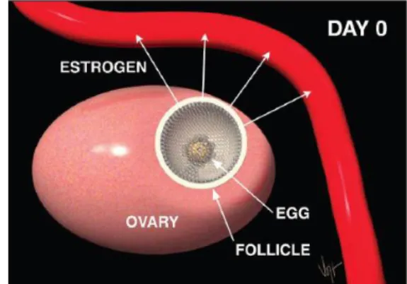
Figure 2. Uterine horn diameter measured by ultrasonography in the cows (Emma, 2010) In addition, the ovaries must resume normal cyclical activity for inseminating or mating a cow shortly after calving, the aim being one calf born per cow per year, so it is important that the postpartum proceeds normally. The estrous cycle normally repeat every 18 to 21 days (Figure 3). The cycle works starting with a cow in heat on day zero. One ovary has a large follicle approximately 15 to 20 mm in diameter. This follicle has a mature egg inside and ready to be released. The cell lining the follicle are producing the hormone estrogen (Figure 4).
Estrogen is transported in the blood stream to all parts of the cow’s body. It makes the uterus more sensitive to stimulation and aids in the transport of semen at the time of insemination. It causes the cervix to secrete viscous mucus that flows and lubricates the vagina. Estrogen is also
4
responsible for all signs of heat including a red swollen vulva, allowing other cows to mount her, going off feed, bellowing considerably and holding her ears erect are but a few of many signs. One day one, the follicle ruptures or ovulates releasing the egg to the waiting infundibulum. Several hours prior to ovulation estrogen production declines. As a result, the cow no longer displays the familiar signs of heat. After ovulation, new types of cell called, luteal cells, grow in the void on the ovary where the follicle was located. Quite rapidly over the next five to six days these cells grow to form the corpus luteum (CL). The CL produces another hormone, progesterone. Progesterone prepares the uterus for pregnancy. Under the influence of progesterone, the uterus produces a nourishing substance of the embryo called uterine milk. At the same time, progesterone causes a thick mucous plug to form in the cervix, preventing access of bacteria or viruses into the uterus. Progesterone also prevents the animal from returning to estrus by regulating the release of gonadotropins from the pituitary gland in the brain. There are two important gonadotropins produced, stores and released from the pituitary gland. The first is follicle stimulating hormone (FSH) which stimulates the growth of small follicles.
Luteinizing hormone (LH) is the other gonadotropin also produced. In addition to supporting progesterone production by the CL, LH ca also stimulate estrogen production in large follicles.
High levels of estrogen would bring the animal back into heat and make life difficult for a new embryo if she were pregnant. Thus, progesterone’s regulation of FSH and LH is very important aspect of maintaining the pregnancy (Figure 5).
Days 16 through 18 of the estrus cycle are referred to as “the period of maternal recognition”. During this time, the uterus sarches itself for the presence of a growing embryo.
If no embryo is detected, the uterus begins to produce prostaglandin which begins to detroy the CL. When the CL is destroyed, no more progesterone is produced and the pituitary gland begins to increase secretion of gonadotropins. Increased secrection of LH stimulate the dominant folicle to produce estrogen and bring the animal back into estrus.
Figure 3. The bovine estrus cycle (www.selectsires.com)
Figure 4. Estrogen from growing follicle is transported through the blood stream to all parts of the body (www.selectsires.com)
5
The average total time is about 21 days. The estrous cycle is subdivided into two phases based on the dominant hormone or ovarian struture during each phase (Figure 6). The luteal phase begind when the corpus luteum is formed, about 5 to 6 days after cow was in heat, and ands when the CL regresses, abnout day 17 to 19 of the cylce. Progesterone levels are high during this phase of the cycle and estrogen levels are low. The other pahse of the cycle is the follicular phase, begins when CL of one cycle is regressed and and when the new CL of the following cylcle is formed. Thus, the follicular phase encompasses the period of time surroungding estrus. During this phase of the cycle estrogen levels are typically high while progesterone level are low. Although it is typical for much follicular growth to occur throughtout the estrus cycle, low levels of LH during the luteal phase, prevents these follicles from producing high levels of estrogen which would bring the animal back to heat. It is only the domiant fillicle present at the time the CL regresses, when progesterone levels are low, that is permitted to produce enough estrogen to bring the animal back into estrus and to continue on to ovulation.
After calving, the muscles in the uterus begin to contract and eventually expel the calf and membranes through a dilated cervix and vagina. Several hormones including progesterone, estrogen, prolactin, relaxin and corticoids produces by the mother cow, the fetus and the placenta, interact to bring about the parturition. Calving in a clean environment and proper treatment of the cow after a difficult calving will help prevent reproductive problems.
The microbiota in uterus after calving
The vulva, vestibule, vagina and cervix function as anatomical barriers that protect the uterus from being contaminated by bacteria. These structures enable a normal uterus to remain sterile during pregnancy. During and after calving, however, the relaxed vulva and the dilated cervix allow the entrance of bacteria in to the uterus (Sheldon, 2004). Bacterial contamination of the uterus p.p is common and 33% of the animals show bacterial growth during the first week
Figure 5. Follicular growth occurs throughout the estrous cycle (www.selectsires.com)
Figure 6. Estrus cycle is divided into two phases (www.selectsires.com)
6
after calving. By the second week the number of cows positive for bacterial culture has increased to 44%.
In approximately 10-17% of the cows bacteria persist and cause uterine disease (Borsberry and Dobson, 1989). Whether or not the bacteria are eliminated depends on the involution, uterine contractions, endometrial regeneration and defence mechanisms such as leukocyte migration, phagocytosis and inflammatory mediators. Due to this inflammatory response an elevated rectal temperature is often seen in dairy cattle within the first ten days after calving (Sheldon, 2004). Factors that delay the elimination of bacterial contamination are the size of the bacterial load, the character of the bacterial flora, retained fetal membranes (RFM), and depression in the cow’s immune status (Sheldon, 2006 and Noakes, 2009).
The most common species of bacteria isolated from the uterine lumen p.p are Escherichia coli, Streptococci, Arcanobacterium pyogenes, Bacillus licheniformis, Prevotella spp and Fusobacterium necrophorum. Among bacteria that are associated with uterine infection (pathogenic species) the most common findings are E.coli, A. Pyogenes, F. necrophorum and Prevotella species (Table 1)
Table 1. List of bacteria according to their expected pathogenic potential (1-3) in the uterus
1 2 3
A. pyogenes Bacillus licheniformis Clostridius perfringens P. melaninogenicus Enterococcus faecalis Klebsiella pneumoniae
E. coli M. haemolytica Micrococcus species
F. necrophorum Pasteurella multocida Providencia stuartii Peptostreptococcus species Proteus species
Staphylococcus aureus Staphylococcus species, coagulase negative Non-haemolytic Streptococci α-Haemolytic Streptococci
Streptococcus acidominimus Aspergillus species
Data: (1) uterine pathogens associated with uterine endometrial lesions; (2) potential pathogens frequently isolated from the bovine uterine lumen and cases of endometritis but not commonly associated with endometrial lesions;
(3) opportunist contaminants transiently isolated from the uterine lumen and not associated with endometritis (Sheldon, 2005).
The appearance and odour of vaginal mucus reflects the bacteria in the uterus, A.
pyogenes, Proteus species and F. necrophorum are related with purulent or mucupurulent vaginal mucus. Furthermore, A.pyogenes, E.coli, non-haemolytic streptococci and M.
haemolytica are associated with a foul mucus odour (Williams et al., 2005).
Epithelial regeneration is complete by about 25 days after parturition, but the deeper layers of tissues are not fully restored until 6–8 weeks after calving. The postpartum environment of the uterine lumen supports the growth of a variety of aerobic and anaerobic bacteria. Many of these bacteria are contaminants in the uterine lumen and are removed by the mechanisms of defending from the uterus.
The uterine diseases after parturition
7
Bacterial contamination of the uterine lumen is common in cattle after parturition, often leading to infection and uterine disease. Clinical disease can be diagnosed and scored by examination of the vaginal mucus, which reflects the presence of pathogenic bacteria such as Escherichia coli and Arcanobacterium pyogenes (Sheldon et al., 2008). The infertility associated with uterine disease is caused by damage to the endometrium and disruption of ovarian cyclic activity.
Uterine disease is commonly associated with Escherichia coli, Arcanobacterium pyogenes, Fusobacterium necrophorum and Prevotella species. Indeed, A. pyogenes, F.
necrophorum and Prevotella species have been shown to enhance the likelihood of uterine disease, and increase the risk of clinical endometritis and its severity (Olson et al., 1984).
Numerically the most prevalent pathogens are E. coli (37% of pathogenic bacteria isolated) and A. pyogenes (49%) (Williams et al., 2005).
The placenta is normally expelled within 6 h of expulsion of the calf but if still present by 24 h, it is defined as a retained placenta. The incidence of retained placenta (RP) is between 2% and 5% of animals in a herd, but can be increased in cows with abortion, twins, dystocia, effects of diet and where infectious agents are endemic. Furthermore, retained placenta is associated with a substantial reduction in milk yield that persists even after resolution of the problem and in one study affected animals produced milk less than normal cows during the first 60 days of lactation (Sheldon et al., 2004).
The expression of clinical uterine infection depends on the balance between factors such as the animal, immunity, the number and pathogenicity of the microbes, and the uterine environment. Typically, 25–40% of animals have clinical metritis in the first 2 weeks after calving, and disease persists in up to 20% of animals as clinical endometritis (Figure 7).
Clinical metritis (CM) was diagnosed in 18.6% (inter farm range; 15.2–23.5%) and 30%
(19.4–42.3%) of cows and heifers, respectively. RP was diagnosed in 13.1% (9.4–18.1%) and 9.2% (3.6–13.8%) of cows and heifers, respectively (Goshen et al., 2006).
Puerperal metritis is defined as an animal with an abnormally enlarged uterus and a fetid watery red-brown uterine discharge, associated with signs of systemic illness (decreased milk yield, dullness or other signs of toxaemia) and fever >39.50C, within 21 days after parturition.
Animals that are not systemically ill, but have an abnormally enlarged uterus and a purulent uterine discharge detectable in the vagina, within 21 days after calving, may be classified as having clinical metritis.
LeBlanc et al. (2002) defined endometritis was the presence of purulent uterine discharge or cervical diameter >7.5 cm, or the presence of muco-purulent discharge after 26 DIM.
Clinical endometritis is characterized by the presence of purulent (>50% pus) uterine discharge detecting in the vagina 21 days or more after parturition, or mucopurulent (approximately 50%
pus, 50% mucus) discharge detectable in the vagina after 26 days. In the absence of clinical endometritis, a cow with subclinical endometritis is defined by >18% neutrophils in uterine cytology samples collected 21–33 days after calving, or >10% neutrophils at 34–47 days.
8
Pyometra is defined as the accumulation of purulent material within the uterine lumen in the presence of a persistent corpus luteum and a closed cervix.
Figure 7. Almost all cows have bacteria within the cavity of the uterus during the first 2 weeks of calving and uterine disease is very common. Each marker (♦) indicates the percent of animals with bacteria isolated from the uterine cavity; data are from four different studies, with animals sampled at various times between calving and 60 days after parturition (Elliot et al., 1968; Griffin et al., 1974; Sheldon et al., 2002b; Williams et al., 2005). The shaded areas represent the proportion of animals with metritis ( ) within 2 weeks of calving and endometritis ( ) 3–5 weeks after calving. The solid line ( ) indicates the percent of animal with histological evidence of inflammation of the endometrium (Gilbert et al., 2005).
The risk factors for uterine infection include retention of the placenta, the calving environment, twins, dystocia, and diet. There is bacterial contamination, clearance and re- contamination of the uterine lumen during the first few weeks after calving, not just infection around the time of parturition. Retained placenta, uterine bacterial infection and uterine disease are common after parturition in cattle and cause considerable infertility. Uterine bacterial infection stimulates a robust immune response but also modulates normal reproductive physiology. The interaction between the environment, uterine infection, immunity and reproduction are useful to improved control and treatment strategies for cattle infertility.
Therapy of the uterine diseases
Goshen et al. (2006) evaluated the intrauterine antibiotic treatment of clinical metritis (CM) and retained placenta (RP) with 5 g Chlortetracycline by intrauterine installation, twice weekly for 2 weeks. The results showed that Chlortetracycline treatment was proven to prevent the detrimental effect of CM on reproductive performance in heifers and cows and on milk production in cows only, while treatment of RP had no effect on reproductive performance or milk production.
In dairy cows diagnosed with clinical endometritis between 20 and 33 days in milk (DIM), LeBlanc et al. (2002) compared the effect of intrauterine with antibiotic 500 mg of cephapirin benzathine or intramuscular with prostaglandin (PGF2α) 500 μg cloprostenol. The results showed that there was no benefit of treatment of endometritis before 4 weeks postpartum.
9
Administration of between 20 and 26 DIM to cows with endometritis that did not have a palpable corpus luteum was associated with a significant reduction in pregnancy rate. Between 27 and 33 DIM, cows with endometritis treated with cephapirin had a significantly shorter time to pregnancy than untreated cows, and there was no difference in pregnancy rate between PGF2α and untreated cows. The difference in pregnancy rate between cows treated with cephapirin and with PGF2α was not statistically significant. In addition, Haimer et al. (2013) indicate the meta-analysis did not reveal an improvement of reproductive performance of cows with endometritis after treatment with PGF2α. Therefore, PGF2α was reconsidered as a routine treatment for cows with chronic endometritis. This is particularly important because a blanket treatment of PGF2α could be perceived by the public as unjustified hormone use.
Administration of PGF2α is the treatment of choice for cases of endometritis in which a corpus luteum is present. Whereas, in the absence of a corpus luteum, a range of intrauterine treatments have been administered including antiseptics and antibiotics. And Estradiol at doses of 5-10 mg per animal has been used therapeutically for postpartum endometritis and is as effective as PGF2α. However, the interval from treatment to conception was longer with estradiol treatment than PGF2α or intrauterine antibiotic (Sheldon and Noakes, 1998).
THE MILK MICROBIOTA Physiology of lactation
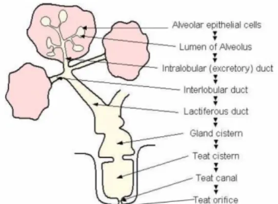
The bovine mammary gland (udder) grossly consists of four glands, commonly referred to as quarters. A schematic representation of a longitudinal cross section of bovine udder is shown in Figure 8. The glands are separated from one another by connective tissue and median suspensory ligaments. Each gland has a single teat that opens to the exterior through the streak canal through which the mammary secretions are drained. The teat canal is lined with skin-like epidermis known as keratin that acts as the main physical barrier against pathogens that can invade the mammary gland. The cavity within the teat seen above the teat canal is the teat cistern and is continuous with the gland cistern of the mammary gland. The secretory tissue and associated connective tissue constitute the interior of the mammary gland. Secretory tissue in the udder is organized into lobes, each of which is subdivided into lobules. Each lobule contains 150-200 microscopic alveoli. Each alveolus is a sac-like structure lined with a single layer of secretory epithelial cells, where milk is synthesized and then secreted. The epithelial lining of the alveolus is surrounded by myoepithelial cells. In response to the release of oxytocin a hormone from the pituitary gland, myoepithelial cells by their contractile nature assist in ejection of milk from each alveolus into the mammary duct. Outside the myoepithelial cells, the alveolus is surrounded with connective tissue, which consists of fibroblasts, adipocytes, capillaries and extracellular matrix components. Milk synthesized in alveoli is drained to the gland cistern through tubular structures known as ducts.
10
Figure 8. Schematic representation of bovine mammary gland
Nutrient composition of whole and whole, dry milk analyze in table 2.
Table 2. Nutrient composition of whole and whole, dry milk (Jenkins et al., 2006)
Whole Whole, dry
Water, % 88.32 2.47
Protein, % 3.22 26.32
Fat, % 3.25 26.71
Ash, % 0.69 6.08
Carbohydrate, % (by difference) 4.52 38.42
Energy, kcal/100 g 60 496
Cholesterol, mg/100 g 10 97
Fatty acids, % of total
Total saturated 64.9 66.1
Total monounsaturated 28.3 31.3
Total polyunsaturated 6.8 2.6
Figure 9. Schematic representation of relationships among lipid metabolism in adipose tissue, liver, and mammary gland. Plus signs (+) indicate stimulatory effects, minus signs (−) indicate inhibitory effects. Dashed lines indicate processes that occur at low rates or only during certain physiological states. Abbreviations: epi = epinephrine, TG
= triglyceride, VLDL = very-low-density lipoproteins, CPT-1 = carnitine palmitoyltransferase 1 (Drackley, 1999).
The seasonal pattern across the year has been known to effect the milk yield and milk composition (Baul et al., 2014; Salfer et al., 2019). Protein and solid-none-fat (SNF) percentage
11
were observed higher in winter than these in summer (Gernand et al. 2019), whilst Salfer et al (2019) showed the highest milk fat and protein concentration were during the winter and lowest occurring in the summer. Extreme rates of lipid mobilization lead to increased uptake of NEFA by liver and increased triglyceride (TG) accumulation (Figure 9). If this lipid infiltration becomes severe, the syndrome of hepatic lipidosis or fatty liver may result, which can lead to prolonged recovery for other disorders, increased incidence of health problems. Increased lipid accumulation and decreased glycogen in the liver were associated with an increased susceptibility to induction of ketosis (Drackley et al., 1992). Moreover, the primiparous cows produced less milk than the multiparous group (Oikonomou et al., 1989; Meikle et al., 2004), so milk composition of first-parity cows was higher than it in the second-, third-, and forth- parity cows.
The microbiota in milk
The microbiota in milk can play many roles, such as facilitating dairy fermentations (e.g.
Lactococcus, Lactobacillus, Streptococcus, Propionibacterium and fungal populations), causing spoilage (e.g. Pseudomonas, Clostridium, Bacillus and other spore-forming or thermoduric microorganisms), promoting health (e.g. lactobacilli and bifidobacteria) or causing disease (e.g. Listeria, Salmonella, Escherichia coli, Campylobacter and mycotoxin- producing fungi) (Quigley et al., 2013)
Microorganisms can bring about the fermentation of milk through the production of lactate and have a variety of different impacts on the sensory, texture, flavor and organoleptic properties of resultant products (Wouters et al., 2002). Lactic acid bacteria (LAB) is a group of bacteria that ferment lactose to lactate, which are a dominant population in dairy cow milk prior to pasteurization. The most common LAB genera in milk include Lactococcus, Lactobacillus, Leuconostoc, Streptococcus and Enterococcus. Other strains of non-LAB genera are also encountered in milk, as well as various yeasts and molds (Quigley et al., 2011).
Microorganisms can also have the negative impacts of milk quality, such as psychrotolerant bacteria can proliferate during refrigeration, which is a major component and frequently include Pseudomonas and Acinetobacter spp, and through the production of extracellular lipases and proteases, result in spoilage (Hantsis-Zacharov and Halpern, 2007).
The population of thermoduric bacteria (resisting pasteurisation) includes clostridia, Listeria monocytogenes, Salmonella, coagulase-positive staphylococci, Escherichia coli, Enterobacteriaceae, coliforms and Bacillus cereus. Masoud et al. (2012) compared the bacterial population present in cows’ milk pre- and postpasteurisation. The milk was from a variety of commercial producers, dominated by Lactococcus, Pseudomonas and Leuconostoc. The number of anaerobic taxa was detected, including Bacteroides, Faecalibacterium, Prevotella and Catenibacterium, which are more typically associated with the gut microbiota and may be entering the milk through fecal contamination. The results indicate that the bacterial population of pasteurised milk is more diverse than previously appreciated, but the nonthermoduric
12
bacteria that are present within these populations are likely to be in a damaged, nonculturable form.
The microbial composition of milk can also influence on health of consumers when the raw milk contaminated with pathogenic bacteria, in some cases, severe illness such as Staphylococcus aureus, Campylobacter spp., Salmonella, Listeria monocytogenes, Shiga toxin- producing E. coli (STEC). Staphylococcus aureus is one of major agents of mastitis in dairy cows and relate to the ingestion of milk and dairy products as a clinical food poisoning problem in humans characterized by vomiting and dehydration (Oliver et al., 2009; Gonzales-Barron et al., 2017). Campylobacter spp. comprises several species, the most important is Campylobacter jejuni which may be contaminated during milking from the intestinal tract of cattle and feces (Del Collo et al., 2017). However, Salmonella and STEC are few outbreaks of the ingestion of raw milk or milk products, in this case, the fecal contamination is their main source, thus hygienic practices should be taken to prevent fecal contact with milk (Gonzales-Barron et al., 2017). And the prevention of L. monocytogenes in raw milk is the application of hygienic practices involving the use of detergents and sanitizers (Kumar et al., 2017).
Mastitis
Bovine mastitis is an important disease in the dairy industry, causing economic losses as a result of milk reduces and treatment costs. Mastitis is inflammation of the mammary gland detected by increased somatic cell count (SCC) or visible abnormalities in milk and is the most common bacterial disease of dairy cattle. Although numerous bacteria can cause mastitis, historically, most mastitis was caused by Streptococcus spp. or Staphylococcus spp. To ensure accurate diagnosis, standardized culturing methods for milk samples have been developed (Ruegg, 2017)
Several studies have suggested milk amyloid A (MAA) as a promising biomarker in the diagnosis of mastitis, especially subclinical mastitis is difficult to detect because there are no clinical signs to identify by visual inspection and palpation of the udder. Jaeger et al. (2016) estimated the diagnostic test accuracy of a commercial MAA-ELISA, somatic cell count (SCC), and bacteriological culture for subclinical mastitis. The results showed that MAA-ELISA result of 3.9μg/mL to detect mastitis caused by major pathogens (e.g., Escherichia coli, Staphylococcus aureus, streptococci, and lacto-/enterococci) and 1.6 μg/mL to detect mastitis caused by all pathogens (major pathogens plus Corynebacterium bovis, coagulase-negative staphylococci, Bacillus spp., Streptomyces spp.). The optimal SCC threshold for identification of subclinical mastitis was 150,000 cells/mL. With the bacteriological culture, 63.5% milk samples contained at least 1 colony of any bacterial species; among them, 20.4% contained major pathogens and 79.6% contained minor pathogens. The MAA-ELISA is a valuable addition to existing tools for the diagnosis of subclinical mastitis.
Haptoglobin (Hp) was also indicated as one of the promised biomarkers of mastitis and the correlation between Hp and SCC has been related with the udder inflammation and mastitis
13
(Åkerstedt et al., 2011; Thomas et al., 2015). Beside that, Aspartate aminotransferase (AST) and alanine aminotransferase (ALT) were the liver functional parameters to indicate the degree of hepatocyte damage (Steen et al., 1997; Sun et al., 2015). The activities of ALT and AST were seen higher in winter that suggested the risk factor occurred more relate to liver function in winter. And ALT was found at 2 months higher than at 1 month after calving which followed the days of lactation period (Stojevie et al., 2005). Other factors, the higher values of non- esterified fatty acids (NEFA) was reported the relationship with mastitis by Melendez et al.
(2009), but some researches showed that urea nitrogen (BUN), serum albumin (Alb), total cholesterol (T-Cho), and calcium (Ca) were not increased in mastitic cows (Sharma et al., 2016;
Sadek et al., 2017; Zhong et al., 2018).
Treatment and prevention of mastitis
The bovine mammary gland is a difficult target for antimicrobial treatment. Penetration of substances into milk when administered parenteral or absorption and distribution throughout the udder when infused intramammary (IMM) depends on the pharmacokinetic characteristics.
These are lipid solubility, degree of ionization, extent of binding to serum and udder proteins, and the type of vehicle. Antimicrobial treatment of dairy cows creates residues into milk, and residue avoidance is an important aspect of mastitis treatment (Wagner and Erskine 2006).
Several antimicrobial classes are approved for use in food animals for the treatment of mastitis including β-lactams (e.g., penicillins, cephalosporins, ampicillin), tetracycline, macrolides (e.g., erythromycin), linocosamides (e.g., pirlimycin), and novobiocin (Mitchell et al., 1998).
The activity of macrolides, tetracyclines and trimethoprim-sulphonamides has been shown to be reduced in milk (Louhi et al. 1992). Selecting a substance with a low minimum inhibitory concentration (MIC) value for the target pathogen is preferable, particularly when the antimicrobial is administered systemically. The antimicrobial should have bactericidal rather than bacteriostatic action, because phagocytosis is impaired in the mammary gland.
Treatment of mastitis should be targeted towards the causative bacteria if possible, but in some acute situations, treatment is initiated based on herd data and personal experience. Rapid or on-farm bacteriological diagnosis would facilitate the selection of the most appropriate antimicrobial. Treatment protocols and medicine selection for each farm should be made by veterinarians familiar with the farm. The use of on-farm written protocols for mastitis treatment also can promote judicious use of antimicrobials. Therapeutic response of the cows can be monitored using individual somatic cell count data if available, or with bacteriological samples in herds with contagious mastitis. However, the use of narrow-spectrum antimicrobials is preferable.
THE ENVIRONMENT MICROBIOTA
The relationship between uterine and environmental microbiota
14
During parturition, the physical barriers of the cervix, vagina and vulva are compromised providing the opportunity for bacteria to ascend the genital tract from the environment as well as the animal’s skin and feces. The microbial sources could contaminate into the uterus from the environment such as their feces (Sheldon and Dobson, 2004) or fecal contamination directed to the angle of the vulva (Potter et al., 2010). However, according to Potter et al. (2010), the risk factors of uterine inflammation were the trauma of reproductive tract rather than fecal contamination.
The relationship between milk and environmental microbiota
The contaminated sources of milk microbiota were considered from environment including feces, air-borne dust, and bedding (Vacheyrou et al. 2011). Ruminococcaceae were present in milk and feces of healthy lactating cows (Young et al. 2015). Wu et al. (2018) found Staphylococcaceae and Aerococcaceae were the most abundance taxa in the air-borne dust associated with milk microbiota, while milk microbiota were associated with the microbiota from the bedding more than from the air-born dust (Metzger et al. 2018). The milk microbiota were not only affected by the environmental microbiota, but also varied by seasons. Indeed, during summer, the milk microbiota were associated with the microbiota from the bedding more than from the air-born dust (Metzger et al. 2018). However, both of the air-born dust and bedding microbiota could be suggested to become the contaminated sources of the milk microbiota in winter. Interestingly, the fecal microbiota were clearly separated from the milk, air-born dust and bedding microbiota (Wu et al. 2018).
Moreover, Vacheyrou et al. (2011) also showed some microbiota presented in milk, but not detected in the farm environment; and similarly, some microbiota taxa were detected in the farm environment, but not in milk. Most of the fungal species and environmental bacteria found in the milk were also found in the stable and milking parlour environments. While Lactobacilli and propionic acid bacteria were frequently identified in the milk, but were rarely found in other environments.
15 CHAPTER 3
THE RELATIONSHIP BETWEEN UTERINE, FECAL, BEDDING, AND AIRBORNE DUST MICROBIOTA FROM DAIRY COWS AND
THEIR ENVIRONMENT
ABSTRACT
A total of 98 samples were analyzed to characterize uterine, fecal, bedding, and airborne dust microbiota in a dairy farm managed by the free-stall housing system. Samples for the microbiota analysis were collected during summer and winter at one and two months postpartum. Blood samples were also collected to determine the levels of serum haptoglobin (Hp) and biochemical components. Uterine microbiota varied between seasons but not between one and two months postpartum; the five most prevalent taxa were Enterobacteriaceae, Moraxellaceae, Ruminococcaceae, Staphylococcaceae, and Lactobacillaceae during summer, and Ruminococcaceae, Lachnospiraceae, Bacteroidaceae, Moraxellaceae, and Clostridiaceae during winter. Although one cow showed a great abundance (19.2%) of Actinomycetaceae, the family Trueperella pyogenes belongs to, and four cows were considered to have uterine disease based on high Hp levels, the relationship between uterine microbiota and serum metabolite concentrations was unclear. The fecal microbiota was stable regardless of the season, whereas bedding and airborne dust microbiota varied between summer and winter. With regards to uterine, bedding, and airborne dust microbiota, Enterobacteriaceae, Moraxellaceae, Staphylococcaceae, and Lactobacillaceae were more abundant during summer, and Ruminococcaceae, Lachnospiraceae, Bacteroidaceae, and Clostridiaceae were more abundant during winter. Canonical analysis of principal coordinates confirmed the relationship between uterine and cowshed microbiota. Although our findings were derived mostly from healthy cows in a dairy farm, this study clarified that uterine microbiota may vary with changes in the microbiota from cowshed environments.
Keywords: airborne dust, bedding, dairy cow, microbiota, uterus
INTRODUCTION
The postpartum uterus of dairy cows has been shown to be contaminated with diverse bacterial species, including pathogens associated with uterine disease (Sheldon et al., 2006;
Ghanem et al., 2015). Although one-third to two-thirds of cows remain healthy, others may develop metritis and endometritis; this reduces their food intake and milk production ability
16
and renders them less likely to become pregnant (Lewis, 1997; LeBlanc et al., 2002).
Escherichia coli, Trueperella pyogenes, Fusobacterium necrophorum, and Bacteroides spp.
(e.g., Prevotella melaninogenica) are representative pathogens determined by isolation from the uteri of cows with postpartum uterine disease using the plate-culture method (Sheldon et al., 2006). The understanding of these pathogens has been expanded by employing culture- independent microbiota analyses; several bacterial families, e.g., Porphyromonadaceae, Mycoplasmataceae, Bacteroidaceae, and Leptotrichiaceae, have been recently considered as pathogens associated with uterine disease (Santos et al., 2011; Machado et al., 2012; Knudsen et al., 2016).
Restoration of the cervix diameter and regeneration of the uterine epithelium requires three to four weeks (LeBlanc et al., 2002; Sheldon et al., 2008; Gautam et al., 2009); hence, even in cows with no uterine disease, diverse bacterial species including anaerobes and facultative anaerobes can be detected after parturition. The bacteria contaminating the uteri of postpartum dairy cows are thus considered to originate from feces and the environment.
However, data for microbiota in cows from dairy farm environments are limited, and evidence showing the relationship between uterine, fecal, and cowshed microbiota is lacking. Because both Trueperella spp. and Fusobacterium spp. can be found in the uteri of virgin heifers and pregnant cows (Moore et al., 2017; Karstrup et al., 2017), the belief that the pregnant uterus is sterile until contamination with the environmental bacteria at calving, and metritis-causing bacteria gain access to the uterus when cows calve should be reconsidered. Regardless, factors affecting uterine microbiota need to be clarified to help prevent uterine disease, improve fertility, and ensure high milk production from the dairy cows.
In this study, a total of 98 samples of uterine mucus, feces, bedding, and air-borne dust collected in a dairy farm were analyzed by high-throughput amplicon sequencing. The cows were managed by the free-stall housing system, and samples for microbiota assessment were collected during summer and winter when the cows were at one and two months postpartum.
Blood samples were also collected to determine the serum levels of haptoglobin (Hp) and biochemical components. The objective was to characterize the uterine microbiota of postpartum dairy cows during different seasons, and examine if the uterine microbiota was related with the fecal, bedding, and air-borne dust microbiota.
MATERIALS AND METHODS Sampling
17
A total of 98 samples were collected from cows at the Okayama Prefecture Livestock Research Institute (Okayama, Japan). The cows were housed in a free stall barn and fed total mixed ration silage, which was formulated to contain 500–600 g/kg of dry matter (DM), 160–
180 g/kg DM of crude protein (N × 6.25), and 720–740 g/kg DM of total digestible nutrients.
The sampling was performed from 6 June to 22 August and from 17 November to 2 March in 2018; hereafter, the former series is referred to as the sampling during summer and the latter, as the sampling during winter. Uterine mucus and feces samples were collected from nine cows during summer and from eight cows during winter, with two sampling times each at one and two months after the calving. Uterine and fecal sampling from cows at one and two months postpartum was conducted occasionally on the same day. Accordingly, samples of bedding and air-borne dust were collected six times during summer and nine times during winter.
Uterine mucus was collected by using a cytobrush (Fujihira Industry Co. Ltd., Tokyo).
The cow was restrained, the tail was held, and the perineum area, especially the vulva, was cleaned by wiping with 70% ethanol. The cytobrush instrument was covered with a sanitary plastic sheath and then inserted into the cervix. Inside the cervix, the plastic sheath was ruptured, and the instrument was further passed through the cervix toward the base of the larger horn, at which point the stainless-steel tube was retracted to expose the cytobrush. Uterine mucus was collected by rotating the cytobrush while in contact with the uterine wall (Jeon et al., 2015);
then, the cytobrush was pulled back into the stainless-steel tube to avoid bacterial contamination from the vagina, vulva, and feces. The cytobrush was cut and placed into an Eppendorf tube and stored.
Fecal samples were collected from the rectum and blood samples were taken from the caudal vein. Airborne dust samples were collected by placing three petri dishes approximately 1.0 m above the ground for five minutes; they were then gathered into a tube using sterile physiological saline. Bedding samples were collected from three separate places in a cowshed.
In the free stall system, cows could move and rest freely, and determining their resting place was difficult. Thus, a composite sample prepared from three separate samples was regarded as a representative means of assessing the bedding and airborne dust microbiota at the time of sampling. All the samples were kept on ice during their transportation to the laboratory and stored at -20°C until further analyses. Procedures and protocols for the animal experiments were approved by the Animal Care and Use Committee, Okayama University, Japan.
Blood analyses
18
The levels of serum albumin (Alb), urea nitrogen (BUN), total cholesterol (T-Cho), non- esterified fatty acids (NEFA), aspartate aminotransferase (AST), alanine aminotransferase (ALT), calcium (Ca), and phosphate were determined by using the respective commercial kits (FUJIFILM Wako Pure Chemicals Co., Tokyo, Japan). The concentration of serum haptoglobin (Hp) was determined by the quantitative sandwich enzyme-linked immunoassay technique using the bovine Hp ELISA kit (Life Diagnostics, Inc., West Chester).
DNA extraction
For uterine samples, the thawed cytobrush was soaked in 1 mL of sterile physiological saline for 30 minutes to release the uterine microbiota. Bacterial pellets were obtained by centrifugation at 16,000 g for two minutes, and then washed with 500 µL of solution I containing 0.05 M D-glucose, 0.025 M Tris-HCl (pH 8.0), and 0.01 M sodium EDTA (pH 8.0).
After further centrifugation at 16,000 g for two minutes, the bacterial cells were lysed with 180 µL of lysozyme solution (20 g/L lysozyme, 0.02 M Tris-HCl [pH 8.0], 0.002 M sodium EDTA [pH 8.0], and 1.2 g/L Triton X-100) at 37°C for one hour. Air-borne dust samples dissolved in sterile physiological saline were processed in the same way. Bacterial DNA was purified by using the DNeasy blood & tissue kit (Qiagen, Germantown, MD, USA), according to the manufacturer’s instructions. In case of the fecal and bedding samples, bacterial DNA was extracted following the procedure for the repeated bead beating plus column method (Yu and Morrison, 2004) and purified using the mini DNeasy stool kit (Qiagen, Germantown, MD, USA).
Illumina MiSeq sequencing
Bacterial DNA was amplified by two-step polymerase chain reaction (PCR) to generate amplicon libraries for next-generation sequencing. The primers targeting the V4 region of 16S
ribosomal RNA (rRNA) genes (forward: 5′-
ACACTCTTTCCCTACACGACGCTCTTCCGATCTGTGCCAGCMGCCGCGGTAA-3′;
reverse: 5′-
GTGACTGGAGTTCAGACGTGTGCTCTTCCGATCTGGACTACHVGGGTWTCTAAT-3′;
tail sequences are underlined) (Tang et al., 2017) were used for the first round of PCR, with the following protocol: initial denaturation at 94°C for two minutes, followed by 35 cycles of denaturation at 94°C for 30 seconds, annealing at 50°C for 30 sec, elongation at 72°C for 30 sec, and an elongation step at 72°C for five minutes. The PCR products were purified by electrophoretic separation on a 2.0% agarose gel using a Fast Gene Gel/PCR Extraction Kit
19
(NIPPON Genetics Co., LTD., Tokyo, Japan). The second round of PCR, with adapter-attached primers, followed the protocol of initial denaturation at 94°C for two minutes, 10 cycles of denaturation at 94°C for 30 sec, annealing at 59°C for 30 sec, elongation at 72°C for 30 sec, and an elongation step at 72°C for five minutes. The second-round PCR products were purified in the same way as that in case of the first-round PCR products.
The purified amplicons were pair-end sequenced (2 × 250 bp) on an Illumina MiSeq platform at FASMAC Co., Ltd. (Kanagawa, Japan). Raw sequence data were analyzed using the Quantitative Insights Into Microbial Ecology (QIIME version 1.9.0). The 250-bp reads were truncated at any site receiving an average quality score under 20. Truncated reads that were shorter than 225 bp were discarded. In primer matching, sequences showing overlaps longer than 200 bp were assembled. The final reads obtained after pair-end joining were grouped into operational taxonomic units (OTUs) using a 97% similarity threshold. The sequence data were analyzed and categorized from the phylum to the family level using the default settings of the Ribosomal Database Project classifier.
Statistical analysis
Data for milk yield, blood metabolites concentration, and relative abundances of major bacterial families (proportion of the family in at least one sample is >1.0%) of uterine and fecal microbiota were analyzed by two-way analysis of variance (ANOVA). Models included the fixed effects of season, sampling time (1 and 2 months postpartum), and interaction between season and sampling time. Data for relative abundances of bedding and air-borne dust microbiota were analyzed by one-way ANOVA to examine the effect of season. Microbiota data were also subjected to canonical analysis of principal coordinates (CAP) to define the assignment and clustering that explained variations in the microbiota. Discriminant vectors with a Pearson correlation >0.7 were considered significant. Two-way ANOVA and t-test were performed using JMP (version 11; SAS Institute, Tokyo, Japan) and CAP was carried out using Primer version 7 with the Permanova+ add-on (Primer-E, Plymouth Marine Laboratory, Plymouth, UK).
RESULTS
Time of calving for the cows examined during summer and winter were 2.0±0.7 and 2.3±1.3, respectively. Milk yield of the cows was 35–40 kg, and no differences were seen between two seasons and between one and two months after the calving (Table 1). The content of milk protein was greater, and those of fat and SNF were numerically higher during winter
20
than during summer. The average somatic cell count (SCC) of the milk at two months postpartum during summer was as high as 429×103 cells/mL, because one cow showed a substantially high SCC (2.8×10⁶ cells/mL). The level of serum Alb was higher and that of serum ALT was lower during summer than during winter (Table 1). Numerical differences were also seen for the levels of serum NEFA (summer > winter) and AST (summer < winter). Likewise, the levels of serum T-Cho and ALT were higher at two months than at one month postpartum.
The Illumina MiSeq sequencing revealed that the microbiota showed a huge taxonomic diversity, including 34 phyla and 213 families, of which 10 phyla and 41 families were shared among all samples. Firmicutes, Proteobacteria, and Bacteroidetes were the 3 major phyla, and accounted for >90% of the total abundance regardless of the sampling time. The abundance of Proteobacteria was higher and those of Firmicutes and Bacteroidetes were lower during summer than during winter.
Differences owing to sample collection at one and two months postpartum were not found in any families of the uterine microbiota (Table 2). The five most abundant taxa of the uterine microbiota during summer were Enterobacteriaceae (12.4%), Moraxellaceae (12.1%), Ruminococcaceae (11.0%), Staphylococcaceae (7.6%), and Lactobacillaceae (3.9%), and those during winter were Ruminococcaceae (22.3%), Lachnospiraceae (7.3%), Bacteroidaceae (5.5%), Moraxellaceae (4.5%), and Clostridiaceae (3.9%). Season-to-season differences were seen for these families; relative abundances of Enterobacteriaceae, Moraxellaceae, Staphylococcaceae, and Lactobacillaceae were greater and those of Ruminococcaceae, Lachnospiraceae, Bacteroidaceae, and Clostridiaceae were lower during summer than during winter. The abundances of Actinomycetaceae and Mycoplasmataceae were low, except that the abundance of Actinomycetaceae in one cow was 19.2%, and that of Mycoplasmataceae in another cow was 11.6% during summer. The relative abundance of Fusobacteriaceae was substantially low (0.00–0.43%) in the uterine samples in this study.
The five most prevalent taxa of the fecal microbiota during summer were Ruminococcaceae (33.9%), Bacteroidaceae (11.3%), Lachnospiraceae (9.2%), Rikenellaceae (3.2%), and Clostridiaceae (3.2%), and those during winter were Ruminococcaceae (34.5%), Lachnospiraceae (10.1%), Bacteroidaceae (9.2%), Rikenellaceae (3.4%), and Clostridiaceae (3.0%). Except for Bacteroidaceae, the relative abundance of which was greater during summer than during winter, differences owing to season were not seen. The relative abundance of Mycoplasmataceae was 0.01% and those of Actinomycetaceae and Fusobacteriaceae were both
<0.005% in the fecal microbiota.
21
The five most abundant taxa of the bedding microbiota during summer were Aerococcaceae (13.8%), Ruminococcaceae (10.8%), Moraxellaceae (8.3%), Corynebacteriaceae (7.3%), and Staphylococcaceae (6.6%), and those during winter were Ruminococcaceae (17.0%), Aerococcaceae (13.3%), Lachnospiraceae (6.6%), Staphylococcaceae (6.3%), and Bacteroidaceae (6.0%). The relative abundance of Moraxellaceae was higher and those of Ruminococcaceae, Lachnospiraceae, and Bacteroidaceae were lower during summer than during winter. The abundances of Actinomycetaceae (0.17%), Fusobacteriaceae (0.02%), and Mycoplasmataceae (0.02%) were low in the bedding microbiota.
The five most abundant taxa of the air-borne dust microbiota during summer were Staphylococcaceae (13.4%), Moraxellaceae (7.2%), Corynebacteriaceae (7.0%), Pseudomonadaceae (6.7%), and Streptococcaceae (6.1%), and those during winter were Ruminococcaceae (17.4%), Aerococcaceae (7.3%), Bacteroidaceae (7.1%), Lachnospiraceae (6.8%), and Staphylococcaceae (4.7%). Season-to-season differences were found for the relative abundances of these families, except for Aerococcaceae and Peptostreptococcaceae.
The abundances of Actinomycetaceae (0.20%), Fusobacteriaceae (0.12%), and Mycoplasmataceae (0.02%) were also low in the air-borne dust microbiota.
Based on the CAP analysis, during summer, the uterine microbiota was grouped with the air-borne dust microbiota, whereas bedding and fecal microbiota formed two other (separate) groups. During winter, most of the uterine microbiota was grouped with the air-borne dust and bedding microbiota, whereas some of the uterine microbiota formed the same group with the fecal microbiota.
DISCUSSION
Milk yield and milk composition have been shown to follow a seasonal pattern across the year, typically being the greatest during winter and reaching a nadir during summer; thus, the higher protein and SNF contents of the milk obtained during summer were regarded to be normal (Salfer et al., 2019). It has been demonstrated that the level of blood metabolites can help describe postpartum uterine health and the resumption of postpartum cyclicity. Cows with high milk-producing ability may face a severe negative energy balance after calving, and cows with uterine disease may show lower levels of serum Alb, T-Cho, and Ca, and higher levels of serum NEFA, BUN, and AST (LeBlanc et al., 2011; Shin et al., 2018; Kawashima et al., 2018).
In this study, although the results revealed that the cows showed lower serum Alb and higher serum ALT levels, suggesting a greater risk of uterine disease during winter, they may have had
22
an appropriate protein-energy balance during both seasons, because the level of serum T-Cho was sufficiently high and those of serum BUN and NEFA were acceptably low. No differences between samples collected during different seasons were seen with regards to the serum concentrations of Hp, an acute-phase protein that reflects the severity of inflammatory responses in cattle with metritis (Chan et al., 2010; Nightingale et al., 2015; Pohl et al., 2015), thereby supporting this finding.
In this study, 24 out of 34 cow samples showed a baseline level (<20 μg/L) of serum Hp.
Four out of 10 other samples showed a high Hp level, at >130 μg/L, which was indicated as an upper cut-off value to differentiate between healthy and metritis-affected cows (Chan et al., 2010). Regardless, the cows with elevated Hp concentration were indicated to have a normal liver function, i.e., their serum AST and ALT levels were similar to those of others. Likewise, the cows with high Hp levels apparently showed healthy uterine microbiota, i.e., low abundances of typical pathogens such as members of Actinomycetaceae, Fusobacteriaceae, and Mycoplasmataceae (detailed data are not shown).
In the uterine microbiota examined in this study, Firmicutes was the most abundant phylum, followed by Proteobacteria and Bacteroidetes. Machado et al. (2012) and Wang et al.
(2018) also found that Firmicutes was the most abundant phylum, showing abundances of 52.3 and 76.7%, respectively, in the uterine microbiota of healthy cows at around one month postpartum. The predominant taxa of Firmicutes in the healthy uterine microbiota, however, is yet to be defined; Geobacillus spp. (Bacillaceae, Machado et al., 2012), Lactococcus spp.
(Streptococcaceae, Wang et al., 2018), and Ruminococcaceae (Knudsen et al., 2016) were reported as the most abundant components of healthy uterine microbiota. Although standard diagnosis methods, such as cytological assessment using polymorphonuclear neutrophils percentage (PMN%) were not performed in this study, most of the cows examined can be regarded to have healthy uterine microbiota, because the relative abundances of Actinomycetaceae (the family T. pyogenes belongs to), Fusobacteriaceae (the family F.
necrophorum belongs to), and Mycoplasmataceae were substantially small.
Santos et al. (2011) and Wang et al. (2018) reported that Proteobacteria was the most abundant phylum in the uterine microbiota of healthy cows; however, in this study, Proteobacteria was the second and third most abundant phylum in the samples obtained during summer and winter, respectively. The predominant taxa of Proteobacteria in the healthy uterine microbiota are also unclear; Santos et al. (2011) have indicated Pasteurella spp., and Bicalho et al. (2017) have demonstrated Escherichia spp. (Enterobacteriaceae), Shigella spp.
(Enterobacteriaceae), and Pseudomonas spp. (Pseudomonadaceae) as the major taxa of